REVIEW

Effects and Regulatory Mechanisms of Trace Element Selenium on Animal Intestinal Health

  • FANG Manxin , 1, 2, 3 ,
  • HU Wei 1, 2, 3 ,
  • LIU Ben 1, 2, 3
Expand
  • 1 College of Life Science and Resources and Environment, Yichun University, Yichun 336000, China
  • 2 Jiangxi Lvke Agriculture and Animal Husbandry Technology Co., Ltd., Yichun 336000, China
  • 3 Engineering Technology Research Center of Jiangxi Universities and Colleges for Selenium Agriculture, Yichun 336000, China

FANG Manxin, lecturer, E-mail: manxinfang@jxycu.edu.cn

Received date: 2023-12-25

  Online published: 2024-06-07

Abstract

The intestinal tract is an important digestive, absorption and immune organ of animals, and it is also an important defense line against pathogenic microorganisms and harmful substances. Selenium is an essential trace element for maintaining animal health. In livestock and poultry production, dietary selenium is of great significance for improving intestinal health of livestock and poultry. In this paper, the research progress of different selenium sources on the regulation of intestinal health in livestock and poultry was reviewed in order to provide reference for further understanding and scientific use of trace element selenium.

Cite this article

FANG Manxin , HU Wei , LIU Ben . Effects and Regulatory Mechanisms of Trace Element Selenium on Animal Intestinal Health[J]. Chinese Journal of Animal Nutrition, 2024 , 36(6) : 3425 -3437 . DOI: 10.12418/CJAN2024.293

肠道不仅是动物消化和吸收食物的器官,而且是对抗有害微生物及化合物的免疫器官,机体健康的维持在很大程度上依赖于肠道屏障功能。肠道屏障可以有效分隔肠腔内外的物质,抵制毒素、病原微生物的进入,在维护动物健康安全等方面发挥着重要的防御保护作用,尤其是肠道健康关乎动物综合生产机能与水平,优化肠道结构及调理肠道功能对促进健康养殖具有重要意义。
硒作为一种微量元素,最初被认为具有毒性,1957年,Schwarz等[1]证明,硒对动物机体发挥着有益作用。硒通过参与碳水化合物、蛋白质和脂质代谢[2]及在维持抗氧化防御等功能中发挥重要作用[3]。NRC建议每天硒摄入量肉牛为100 μg/kg DM,奶牛为300 μg/kg DM[4],家禽为150~200 μg/kg DM(在某些情况下可达到300 μg/kg DM),猪分别为150(母猪和育肥猪)和300 μg/kg DM(断奶仔猪)[5]。一般而言,在某地生产的饲料饲草中硒含量低于0.05 mg/kg标准的地区属于缺硒地区,我国有2/3面积属于缺硒地区,动物饲料硒含量偏低,因此补硒在我国动物生产上有重要意义。此外,大量相关研究显示,硒可通过多方面途径影响维护肠道健康,本文聚焦于动物肠道健康,归纳探讨硒在促进改善肠道屏障方面的功能和作用机制,以期为硒的深入研究及促进其在动物养殖生产中广泛应用提供参考。

1 硒的存在、使用及代谢

微量元素硒是瑞典化学家Berzelius于1817年发现的[6]。硒以有机和无机形式存在于自然界中,在土壤(0.1~0.7 mg/kg)、植物(0.02~0.40 mg/kg)、动物性食物来源(0.03~0.34 mg/kg)、水(通常≤10 mg/L)和空气(1~10 ng/m3)中都含有微量浓度硒。硒的主要无机形式包括亚硒酸盐(Se O 3 2 -)、硒酸盐(Se O 4 2 -)、硒化物(Se2-)和单质硒,有机形式包括硒代蛋氨酸(SeMet)、硒代半胱氨酸(SeCys)、甲基硒代半胱氨酸(MeSeCys)和2-羟基-4-甲基硒代丁酸(HMSeBA)等。目前,硒作为一种必需微量营养元素,经常被添加到动物饲粮中以维持重要生理功能。在养殖生产中,硒以亚硒酸钠(Na2SeO3)无机硒形式或酵母硒有机硒形式添加到饲粮中,其中无机硒易与维生素结合,稳定性差,生物利用度低,毒性较高且难以控制剂量;而有机硒稳定性较高,易于被机体吸收和利用。近年来,纳米颗粒因其物理和化学特性如较大比表面积、较多表面活性中心、较高表面活性和催化能力等引起人们极大兴趣,目前硒纳米颗粒(SeNPs)具有广泛应用价值,并且在所有硒补充剂中毒性最小,比有机硒低3倍,比无机硒低7倍[7]
硒酸盐可通过钠介导的载体吸收,亚硒酸盐通过被动扩散进入肠道细胞,在吸收后,亚硒酸盐在硫氧还蛋白还原酶(thiore-doxin reductase,TXNRD)和硫氧还蛋白的作用下可直接还原成硒氢基(HSe-)。在还原型谷胱甘肽(glutathione,GSH)存在时,亚硒酸盐与GSH反应生成硒代谷胱甘肽(selenodiglutathione,GSSeSG);GSSeSG在谷胱甘肽还原酶及还原型辅酶Ⅱ(NADPH)作用下还原成谷胱甘肽硒基硫醚(selenenylsulfide,GSSeH);GSSeH与GSH反应产生HSe-。硒酸盐通过相同途径还原成硒化合物从而被蛋白质同化。与无机硒相反,SeCys和SeMet通过氨基酸主动转运载体机制被吸收后,利用转硫途径转化为中间产物游离SeCys,进而分解为硒化氢(H2Se)。H2Se最终转化为硒代磷酸盐并以活性形式提供硒用于合成硒蛋白。此外,一些SeMet掺入硒酶或代替蛋氨酸(Met)参与硒蛋白合成。硒在动物机体内主要以硒蛋白形式存在,一般将以SeCys形式合成的蛋白称为硒蛋白(sele-noproteins,SePs)。随着研究的深入,目前哺乳动物中已发现并分离出硒蛋白有35种,包括谷胱甘肽过氧化物酶(glutathione peroxidase,GSH-Px)、TXNRD、碘甲腺原氨酸脱碘酶以及硒蛋白P和硒蛋白W等[8]

2 硒对肠道黏膜屏障的调控及其作用机制

动物健康生长和良好的生产效益离不开肠道功能的健全,肠道屏障受损则会导致肠道机能紊乱。研究表明,硒通过维持肠道黏膜上皮完整、促进抗菌肽分泌、增强肠道免疫功能、提高抵抗力、降低肠道炎症水平和维持肠道菌群平衡来保护提高动物肠道黏膜屏障功能。下文主要从肠道黏膜物理屏障、化学屏障、微生物屏障及免疫屏障4方面来阐述硒在保护动物肠道方面作用及机制。

2.1 硒对肠道黏膜物理屏障的影响

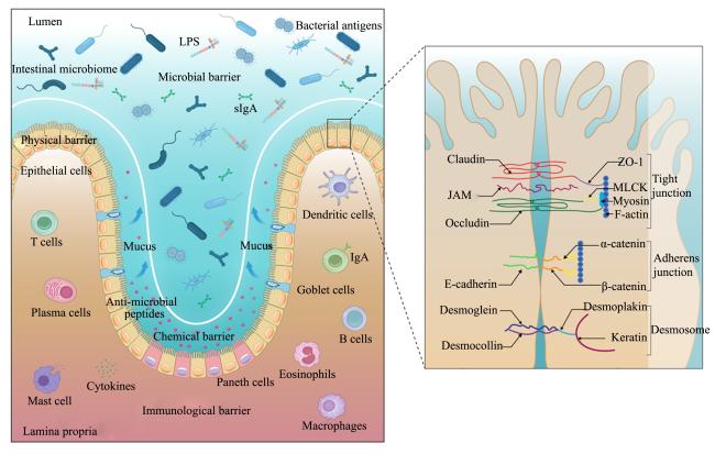
肠道黏膜屏障主要由物理屏障、化学屏障、微生物屏障和免疫屏障有机构成(图1),它们之间互相联系,共同维持肠道完整性和稳定性。肠道物理屏障是畜禽肠腔和内环境之间阻止有害物质入侵的第一道防线,主要由包括潘氏细胞、杯状细胞等多种肠上皮细胞及细胞间连接组成。肠上皮细胞间存在紧密连接(tight junction,TJ)、黏附连接(adherens junction,AJ)、缝隙连接(gapjunction,GJ)和桥粒(desmosome)等连接形式,其中TJ由上皮间紧密连接密封蛋白(claudin,CLDN)、闭合蛋白(occludin,OCLN)和闭锁小带蛋白(zonula occludens,ZO)组成。OCLN对紧密连接蛋白在肠道上皮细胞内的定位有重要影响,ZO-1能将CLDN和OCLN等跨膜蛋白与细胞骨架结合起来,协助建立肠道紧密连接;AJ主要由E-钙黏附蛋白(E-cadherin,E-Cad)组成,参与维持上皮细胞极性、组织器官发育和损伤修复等;GJ则参与细胞间物质及信息交换、细胞增殖分化等功能。
图1 肠道黏膜屏障结构

Lumen:肠腔;Intestinal microbiome:肠道微生物;LPS:脂多糖 lipopolysaccharide;Microbial barrier:微生物屏障;Bacterial antigens:细菌抗原;sIgA:分泌型免疫球蛋白A secretory immunoglobulin A;Physical barrier:物理屏障;Epithelial cells:上皮细胞;T cells:T细胞;Plasma cells:浆细胞;Mucus:黏蛋白;Anti-microbial peptides:抗菌肽;Chemical barrier:化学屏障;Mast cell:肥大细胞;Cytokines:细胞因子;Immunological barrier:免疫屏障;Paneth cells:潘氏细胞;Macrophages:巨噬细胞;Eosinophils:嗜酸性粒细胞;B cells:B细胞;Goblet cells:杯状细胞;IgA:免疫球蛋白A immunoglobulin A;Dendritic cells:树突状细胞;Lamina propria:固有层;Tight junction:紧密连接;Claudin:密封蛋白;JAM:连接黏附分子 junctional adhesion molecule;Occludin:闭合蛋白;ZO-1:闭锁小带蛋白-1 zonula occludens-1;MLCK:肌球蛋白轻链激酶 myosin light-chain kinase;Myosin:肌球蛋白;F-actin:F-肌动蛋白;Adherens junction:黏附连接;E-cadherin:E-钙黏附蛋白;α-catenin:α-连环蛋白;β-catenin:β-连环蛋白;Desmosome:桥粒;Desmoglein:桥粒芯蛋白;Desmocollin:桥粒芯胶黏蛋白;Desmoplakin:桥粒斑蛋白;Keratin:角蛋白。

Fig.1 Structure of intestine mucosal barrier

2.1.1 硒对肠上皮细胞稳态的影响

小肠绒毛是肠道物理屏障的重要组成部分,由一层单上皮细胞组成,绒毛高度/隐窝深度(V/C)值常用于评估肠道形态功能完整性。多项研究显示,添加不同硒源可以显著提高鸡肠道绒毛高度和V/C值[9-17]。Ahmed等[18]和程连平等[19]2个试验分别观察到,补充硒后对小山羊和湖羊羔羊十二指肠和空肠绒毛高度均有显著影响。此外,补充硒后显著提高了鱼及猪肠道绒毛高度和V/C值[20-21]。硒缺乏试验显示,缺硒组蛋鸡绒毛高度、隐窝深度和黏膜厚度都有不同程度下降,严重影响蛋鸡小肠黏膜结构,损伤小肠物理屏障功能[22]。在维持肠上皮更新过程中,肠道上皮细胞增殖与凋亡保持平衡状态,细胞凋亡过度会破坏肠上皮细胞稳态。内质网是蛋白质合成、折叠、运输和钙储存的重要部位,在各种应激源、内质网功能障碍、钙稳态失衡以及内质网腔内错误折叠或未折叠蛋白质累积作用下,细胞发生内质网应激(endoplasmic reticulum stress,ERS),持续和过度ERS可通过未折叠蛋白反应诱导自噬,同时内质网会启动相应信号通路诱导细胞凋亡。Song等[23]研究表明,呕吐毒素(DON)可抑制猪肠上皮细胞系IPEC-J2细胞GSH-Px和TXNRD活性,导致氧化应激并激活ERS相关激酶(PERK)通路,导致细胞质钙离子浓度提高;而SeNPs可以显著缓解DON引起的IPEC-J2细胞活性氧(ROS)含量增加,增强GSH-Px和TXNRD活性,并抑制PERK信号通路的激活,减轻DON诱导的IPEC-J2细胞凋亡,有效维持细胞钙离子稳态和肠上皮屏障完整性。
此外,研究显示,补充硒可显著降低猪及山羊肠上皮细胞凋亡率[21,24]。线粒体是细胞有氧呼吸的主要部位,线粒体受损是ERS过程中ROS的主要来源,线粒体自噬选择性清除受损线粒体。Yan等[25]研究表明,SeNPs可以通过调节哺乳动物雷帕霉素靶蛋白(mTOR)/磷酸酶及张力蛋白同源物诱导的激酶1(PINK1)介导的线粒体自噬,有效缓解过氧化氢(H2O2)诱导的肠上皮屏障功能障碍。Qiao等[26]研究显示,SeNPs有效减轻H2O2诱导的猪空肠上皮细胞内质网和线粒体结构损伤,其对肠屏障功能障碍的保护作用与ERS相关的PERK信号通路和线粒体自噬相关的单磷酸腺苷活化蛋白激酶(AMPK)信号通路密切相关。SeNPs也通过抑制ERS并抑制细胞凋亡,保护肠道上皮细胞免受双酚A(BPA)损伤,从而增强肠上皮屏障功能[27]。更进一步研究显示,SeNPs对肠上皮屏障损伤的保护作用与TBC1D15/Rab7介导的线粒体-溶酶体串扰信号通路密切相关[28]。在硒缺乏试验中,硒缺乏组与ERS细胞凋亡途径相关的B淋巴细胞瘤-2相关X蛋白(Bax)、B淋巴细胞瘤-2(Bcl-2)、胱天蛋白酶(Caspase)-3、Caspase-8、C/EBP同源蛋白(CHOP)、葡萄糖调节蛋白78(GRP78)等的蛋白或mRNA的表达水平在体内和体外均显著高于对照组,表明硒缺乏诱导了ERS并进而触发内源性和外源性细胞凋亡通路,导致猪小肠和IPEC-J2细胞凋亡[29]。另外的研究也显示,硒缺乏可通过氧化应激诱导的线粒体凋亡通路(内在通路)和炎症信号诱导的死亡受体通路(外在通路)引起十二指肠细胞凋亡[30]。BPA暴露和硒缺乏通过诱导氧化应激、线粒体功能障碍、线粒体凋亡和细胞周期停滞导致鸡空肠损伤,具体机制研究显示BPA暴露和硒缺乏诱导过量ROS释放以激活P53,P53与线粒体调节蛋白过氧化物酶体增殖物激活受体γ辅激活因子-1α(PGC-1α)发生蛋白互作,增加线粒体分裂进而诱导线粒体凋亡;此外,P53通过调节P21通路触发细胞周期停滞,最终导致空肠损伤[31]。除细胞凋亡外,细胞坏死是细胞死亡的方式之一,硒缺乏可以诱导IPEC-J2细胞坏死[32]。此外,添加SeNPs后,肿瘤坏死因子-α(TNF-α)、受体相互作用蛋白激酶(RIPK)1、RIPK3、混合谱系激酶结构域样假激酶(MLKL)、FAS相关的死亡结构域蛋白(FADD)和FAS表达下降,Caspase-8表达升高,表明SeNPs可以减轻十溴二苯醚BDE-209诱导的鸡肠道坏死[33]

2.1.2 硒对肠上皮紧密连接蛋白的影响

肠上皮紧密连接蛋白是维持肠道黏膜屏障功能平衡的基础。大量研究表明,硒可通过调节增强紧密连接蛋白表达和分布定位来维护物理屏障结构与功能的完整性。通过观察敌草快诱导氧化应激后猪的肠道变化发现,添加酵母硒改善了断奶猪肠道紧密连接蛋白ZO-1在空肠上皮中的分布和丰度[21]。右旋葡聚糖硫酸钠(DSS)诱导后大鼠结肠紧密连接蛋白水平下降,杜仲多糖修饰的SeNPs处理使紧密连接蛋白水平显著提高[34]。T-2毒素降低兔空肠ZO-1和OCLN表达,SeMet预处理后空肠中OCLN和ZO-1表达显著增多[35]。研究显示,酵母硒通过调节Toll样受体4(TLR4)/髓样分化因子88(MyD88)信号通路抑制核因子-κB(NF-κB)表达并增强紧密连接相关基因CLDN-1、OCLNZO-1表达,保护赭曲霉毒素A(OTA)暴露引起的肉鸡肠道屏障损伤[36]。BDE-209诱导CLDN-1、CLDN-3和OCLN蛋白表达分别降低了60.6%、82.7%和50.7%,在添加SeNPs后相应提高,表明SeNPs可以减轻BDE-209诱导的蛋鸡肠道屏障损伤,且这种保护作用与丝裂原活化蛋白激酶(MAPK)/NF-κB信号通路有关[33]。除此之外,猪肠上皮细胞、生长猪和断奶仔猪分别在BPA、热应激、产肠毒素大肠杆菌(enterotoxigenic Escherichia coli,ETEC)诱导情况下,肠道紧密连接相关蛋白如CLDN-1、OCLD和ZO-1等分别受到影响而下调,补充硒则增强了这些蛋白的表达[27,37-38]。研究显示,硒缺乏促进长链非编码RNA(lncRNA)-microrchidia 3(MORC3)和炎症因子表达,激活NOD样受体热蛋白结构域相关蛋白3(NLRP3)-Caspase-1/白细胞介素-1β(IL-1β)信号通路并破坏猪肠道紧密连接,降低紧密连接因子ZO-1、ZO-2、OCLD、E-Cad和锌指E盒结合同源蛋白-1(ZEB-1)表达[39]。更深入研究显示,肉鸡在28日龄时,补充硒提高了CLDN-1、OCLDZO-1表达,降低了CLDN-5和ZO-2表达;42日龄时,补充硒提高了OCLDZO-1和ZO-2表达,降低了CLDN-5表达[9]。尽管紧密连接蛋白上调增强了肠道完整性并抑制了细胞旁通透性,有助于上皮屏障发挥阻止病原体进入的防御作用,但在某些情况下,病原体如产气荚膜梭菌肠毒素会将紧密连接蛋白作为细胞表面蛋白或毒素受体,触发肠细胞损伤[40]。因此,对紧密连接蛋白上调或下调的影响需要综合评估肠道多种因素,如微生物群落等。

2.2 硒对肠道黏膜化学屏障的影响

动物肠道化学屏障包括由黏膜下腺体和上皮细胞分泌的防御素、抗菌肽等化学物质以及由肠道上皮杯状细胞分泌产生的糖基化黏蛋白(mucin,MUC)组成。MUC是肠黏液中的核心结构成分,包括MUC2、MUC5AC、MUC5B、MUC6及MUC19等,其中MUC2含量最多,在肠道发挥保护肠上皮细胞、表面清洁和免疫协同等作用。

2.2.1 硒对肠道杯状细胞及MUC的调节

杯状细胞是一种特殊的上皮细胞,硒通过不同信号通路促进杯状细胞增殖并改善肠道MUC的结构,从而保护肠道屏障功能免受氧化损伤[41]。研究显示,通过纳米硒调节自噬对增强肠道黏膜免疫和杯状细胞发育至关重要,在纳米硒组自噬会增强MUC2分泌,阻断自噬会导致杯状细胞生物学和生理功能受损,MUC2分泌减少[42]。艾美耳球虫寄生会入侵并破坏杯状细胞,在用SeNPs处理的小鼠中肠道杯状细胞特异性基因MUC2表达上调,表明杯状细胞活性的改善可影响调节MUC的产生[43-44]。此外,SeNPs缓解了DSS诱导的小鼠结肠炎,并增加了空肠杯状细胞数量和改善了MUC的表达[45]。Khajeh Bami等[13]研究发现,喂食添加0.3 mg/kg SeNPs的肉鸡回肠和空肠具有更高密度的杯状细胞。添加SeMet可以逆转T-2毒素诱导的杯状细胞凋亡,并使得MUC2转录增加[35]。三黄鸡生长过程中缺硒会增加杯状细胞胞质空泡化和溶解[30]。Krüppel样因子4(KLF4)是一种锌指转录因子,可促进细胞分化和组织稳态;c-Myc是Wnt/β-连环蛋白(β-catenin)通路诱导细胞增殖的关键下游效应器,对维持肠道稳态和再生至关重要;核因子E2相关因子2(Nrf2)是调控抗氧化应激的一种关键转录因子,通过Kelch样ECH关联蛋白1(Keap1)-Nrf2途径调节肠道细胞分化。试验表明,添加0.1 mg/kg SeNPs可显著增加空肠杯状细胞数量和提高MUC2 mRNA表达;且在添加0.4 mg/kg SeNPs后,空肠KLF4、c-MycNrf2 mRNA的表达显著降低,表明低剂量SeNPs可有效增加杯状细胞数量,高剂量SeNPs可能对肠道细胞有毒性并抑制肠道杯状细胞分化[14]

2.2.2 硒调节肠道防御肽的产生

动物肠道上皮细胞可产生大量抗菌肽,其中最多的是防御素,β-防御素(BD)1抗菌能力在所有BD中最强,参与诱导其他BD如BD2、BD3、BD4、BD5等分泌。研究显示,富硒枯草芽孢杆菌组肉鸡肠道BD1蛋白高于枯草芽孢杆菌yb-114246组,表明硒提高了肉鸡的免疫力[46]。研究表明,饲粮添加亚硒酸钠可以增强肉鸡对坏死性肠炎的保护性免疫反应,并上调促炎细胞因子、GSH-Px和禽β-防御素(AvBD)表达[47]。AvBD是存在于家禽体内的内源性抗菌肽,研究表明,缺硒组空肠中AvBD表达呈下降趋势,其中空肠中AvBD1、AvBD2和AvBD13表达分别下降了53%、65%和39%,表明硒缺乏会下调肉鸡空肠中AvBD mRNA的表达并破坏免疫屏障[48]。另外研究显示,硒缺乏通过激活ROS/NF-κB信号通路增加鸡十二指肠通透性并降低抗菌肽表达[49]。抗菌肽水平降低可能会损害肠道免疫屏障并导致微生物群失衡,从而影响肠道屏障功能并加重肠道损伤[50]

2.3 硒对肠道黏膜微生物屏障的影响

肠道微生物屏障是由数以亿计的肠道常驻细菌形成的一层生物防线,动物肠道细菌可分为有害菌(梭菌属、变形杆菌属、葡萄球菌属)、有益菌(乳杆菌属和双歧杆菌属)以及中性菌(拟杆菌属、肠球菌属和大肠杆菌属),后2类构成了肠道生物屏障。Al-Quwaie[51]研究发现,细菌SeNPs显著降低了肉鸡肠道细菌总数、酵母霉菌总数以及大肠杆菌和沙门氏菌数量,显著提高了乳酸菌数量,显示细菌SeNPs在减少病原体和增强有益细菌方面发挥重要作用,并有助于改善肉鸡生长性能。Dalia等[52]研究观察到肉鸡饲粮中添加ADS18有机硒可增加盲肠有益菌数量,ADS18有机硒组乳杆菌属丰度显著高于亚硒酸钠组和无硒组;此外,补充ADS18有机硒与最高的双歧杆菌属丰度和最低的大肠杆菌和沙门氏菌丰度有关。另外,相比于未添加硒组,添加硒组的肉鸡回肠大肠杆菌和沙门氏菌数量也显著减少[9]。补充酵母降低了蛋鸡肠道兼性厌氧和潜在致病性微生物丰度,调节了盲肠微生物群组成,使肠道更健康[53]。Alagawany等[54]和Reda等[55]研究发现,补充硒显著影响鹌鹑盲肠微生物群落,表现为更低的细菌总数、酵母和霉菌总数、大肠杆菌群、大肠杆菌、肠球菌和沙门氏菌定植,而乳酸菌数高于对照组。Pereira等[56]观察到,喂食有机硒的犬粪便中大肠杆菌DNA拷贝数较低,乳酸杆菌DNA拷贝数较高。同样,喂食富硒益生菌的仔猪也有类似结果[57]。此外,纳米硒通过提高幼鱼肠道有益菌(如梭杆菌)丰度,使高脂饮食(HFD)引起的幼鱼肠道微生物群失衡正常化[58]。另外研究显示,硒缺乏则显著降低GSH-Px活性并破坏肠道菌群,表现为罗伊氏乳杆菌数量减少,而通过补充硒、粪便微生物群移植或罗伊氏乳杆菌则逆转了这些减少,尤其是硒缺乏导致脂多糖(LPS)由于细菌失衡而增加,Toll样受体(TLR)信号相关基因表达水平显著升高,通过TLR4信号通路诱导炎症性肝损伤[59]
短链脂肪酸(SCFA)是肠道微生物发酵膳食纤维的主要产物,其中乙酸、丁酸和丙酸含量最高。SCFA能降低肠腔内pH,从而抑制致病菌生长并促进营养物质吸收。研究发现,饲喂纳米硒的鸡肠道显示高SCFA含量,尤其是SCFA含量最高的组生长性能表现最佳[60]。另有研究表明,硒显著促进山羊结肠发酵并提高SCFA含量[61]。用纳米硒替代亚硒酸钠改善了早期断奶仔猪生长性能和肠道健康,SeNPs提高了霍尔德曼氏菌丰度和乙酸、丙酸含量;相关分析表明,霍尔德曼氏菌参与脂质代谢与SeNPs对早期断奶仔猪的调节作用密切相关[62]。在纳米硒对HFD饲养的草鱼幼鱼肠道健康保护作用的相关研究中发现,纳米硒提高了肠道SCFA尤其是丁酸和己酸含量,且这与肠道通透性和炎症增加呈负相关[58]。在饲喂SeMet对减轻镉暴露引起的克氏原螯虾肠道炎症具体作用的研究中显示,一方面,硒通过提高硒依赖性酶活性有效抑制镉暴露引起的ROS产生,进而激活磷脂酰肌醇-3-羟激酶(PI3K)/蛋白激酶B(Akt)通路,同时抑制其下游调节通路NF-κB;另一方面,硒维持了肠道细菌丰富性和多样性,并显着提高乙酸和丁酸含量,SCFA进一步通过调节PI3K/Akt和NF-κB通路,减轻肠上皮细胞损伤和凋亡[63]。上述研究显示,硒在维系肠道微生态平衡方面具有良好的效果,其作用体现在其抑制有害菌定植、优化肠道菌群组成及代谢等方面,从而维护动物肠道乃至整个机体健康。硒对动物肠道微生物的影响见表1[16,64-71]
表1 硒对动物肠道微生物的影响

Table 1 Effects of selenium on intestinal microorganisms in animals

动物
Animals
硒添加
Selenium addition
结果
Results
参考文献
Reference
肉仔鸡
Broilers
硒纳米颗粒(SeNPs)负载
壳聚糖(200 mg/kg)
与对照组相比,SeNPs负载壳聚糖组回肠乳
杆菌属数量增加,同时大肠杆菌数量减少
[64]
罗斯肉鸡
Ross broilers
SeNPs(0.1、0.2 mg/kg) SeNPs对回肠细菌总数、酵母和霉菌总数、
大肠杆菌群、大肠杆菌、肠球菌和沙门氏菌
表现出剂量依赖性抗菌活性,对回肠乳酸菌
呈剂量依赖性增加
[65]
小鼠
Mice
SeNPs(0.6 mg/kg) SeNPs逆转了百草枯诱导的肠道微生物组成紊乱,
降低了厚壁菌门/拟杆菌门比例,提高了有益菌
丰度,如阿克曼菌属、Muribaculaceae、
拟杆菌属和副拟杆菌属
[66]
小母猪
Gilts
2-羟基-4-甲基硒代丁酸
(HMSeBA,0.3 mg/kg)
HMSeBA提高了肠道瘤胃球菌科和
考拉杆菌属丰度,并选择性降低副拟杆
菌属和普雷沃氏菌科丰度
[67]
公鸡
Roosters
纳米硒(0.9 mg/kg) 包括致病性盲肠肠球菌在内的肠球菌属操作分类
单元(OTU)因纳米硒完全减少,而乳杆菌属和
链球菌属被重构
[68]
蛋鸡
Laying hens
酵母硒(0.3 mg/kg)和
细菌有机硒ADS18
(0.3 mg/kg)
与无机硒和未添加硒相比,有机硒(细菌
有机硒ADS18或酵母硒)显著增加乳杆菌
属和双歧杆菌属数量,显著降低大肠
杆菌和沙门氏菌数量
[16]
蛋鸡
Laying hens
纳米硒(1 mg/kg) 毛螺菌科丰度在赭曲霉毒素A(OTA)-硒组
最高,相反梭状芽胞杆菌丰度在OTA组最高
[69]
蛋鸡
Laying hens
亚硒酸钠、酵母硒和富
硒酵母培养物(0.3 mg/kg)
补充亚硒酸钠提高毛螺菌科和克里斯滕森菌科
丰度,酵母硒提高巨单胞菌属和鞘氨醇单胞菌属丰度,
富硒酵母培养物提高梭杆菌属和乳球菌属丰度
[70]
小鼠
Mice
SeNPs(0.3 mg/kg) 呕吐毒素暴露显著降低了异杆菌属丰度,
SeNPs显著降低了螺杆菌属丰度
[71]

2.4 硒对肠道黏膜免疫屏障的影响

肠道黏膜免疫屏障主要由肠道相关淋巴组织(gut-associated lymphoid tissues,GALT)、肠系膜淋巴结等肠道组织和免疫细胞及因子构成。GALT是肠道免疫的主要诱导部位和效应部位,能摄取和转运抗原并分泌免疫球蛋白(immunoglobulin,Ig)和细胞因子。在肠道抗原刺激下产生的Ig、干扰素(interferon,IFN)、白细胞介素(interleukin,IL)等物质,可调节发挥肠道黏膜免疫功能。肠道浆细胞分泌的分泌型免疫球蛋白A(secretory immunoglobulin A,sIgA)是肠道免疫系统的关键指标。

2.4.1 硒直接增强动物肠道免疫力

动物免疫力的发挥依赖于补体因子、Ig和抗菌肽等一系列免疫物质。SeNPs对DON诱导的肠道屏障功能障碍有保护作用,研究显示,SeNPs可显著抑制DON暴露诱导的十二指肠sIgA含量下降,增强小鼠免疫力[71]。研究表明,低硒会减少肉鸡肠道sIgA分泌并通过激活NF-κB信号通路减弱十二指肠黏膜免疫,从而降低肠道免疫功能[72]。一种新的有机硒源HMSeBA在十二指肠、空肠和回肠中改善了仔猪肠道sIgA含量[67]。禽类体内主要存在3种免疫球蛋白:IgA、IgY(IgG)或IgM,与对照组相比,黄曲霉毒素B1(AFB1)组回肠IgA+细胞数量以及sIgA、IgA、IgG和IgM含量均降低;同时单纯加硒组(0.4 mg/kg硒)回肠IgA+细胞数量显著减少,回肠sIgA、IgA、IgG和IgM含量均低于对照组,说明单纯过量添加硒可降低鸡黏膜体液免疫功能[73],Khan等[74]也得到类似的研究结果;此外,雄性肉鸡在饲喂含有0.3 mg/kg AFB1的饲粮时添加0.4 mg/kg硒,AFB1+硒组回肠IgA+细胞数量和sIgA、IgA、IgG和IgM含量提高,表明硒对AFB1引起的黏膜体液免疫功能损害具有保护作用[73]。位于盲肠近端区域的鸡盲肠扁桃体属于鸡的周围淋巴组织,T淋巴细胞负责细胞介导的免疫并根据其细胞表面蛋白的表达进一步分为几个亚群,研究表明,AFB1可显著降低科宝(Cobb)肉鸡盲肠扁桃体CD3+、CD3+CD4+、CD3+CD8+ T细胞百分比和CD4+/CD8+值,然而,饲粮中添加硒使AFB1组T细胞亚群百分比、CD4+/CD8+值和细胞因子mRNA表达恢复到接近于对照组水平,表明硒对AFB1影响的Cobb肉鸡盲肠扁桃体功能有保护作用[75]。上皮内淋巴细胞(IELs)是肠道相关淋巴组织的一部分,低硒组蛋鸡小肠中sIgA分泌显著降低,十二指肠和回肠中IELs数量也不同程度减少,显示硒缺乏引起小肠黏膜免疫屏障损伤并降低鸡的生长性能[22]

2.4.2 硒通过缓解炎症反应增强动物肠道免疫力

研究显示,对肠道炎症反应的拮抗调节能增强动物免疫力[76]。促炎细胞因子含量升高和抗炎细胞因子含量的降低能够介导恶化炎症反应并损伤肠道健康。IL-6、TNF-α、IFN-γ、IL-1β和IL-8是促炎细胞因子,IL-10、TGF-β则是抗炎性细胞因子。研究表明,BDE-209引起蛋鸡肠道IL-1β、IL-2、IL-6和IL-17含量升高,IL-10和IFN-γ含量降低,激活MAPK/NF-κB信号通路并引起肠道炎症,而SeNPs可通过MAPK/NF-κB信号通路抑制BDE-209诱导的肠道炎症[33]。鼠乳状艾美耳球虫感染提高小鼠空肠炎性细胞因子(IL-6和TNF-α)和凋亡基因(Caspase-3和Bcl-2)表达,生物合成SeNPs处理显著降低空肠组织炎症和凋亡指标[44]。研究显示,补充HMSeBA能够通过调节炎症基因表达减少热应激诱导的生长猪空肠炎症[37]。母体在妊娠期补充HMSeBA后通过抑制NF-κB和细胞外信号调节激酶(ERK)/苄氯素-1(Beclin-1)信号传导来提高后代肠道抗氧化能力并降低炎症水平,并抑制新生仔猪回肠IL-1βIL-6和NF-κB基因表达[77]。研究显示,SeNPs通过NLRP3/Caspase-1/IL-1β信号通路减轻鸡肠道炎症反应[14]。在HFD诱导的幼鱼肠道中,炎症细胞因子(IL-8、IL-1β、IFN-γ、TNF-α和IL-6)、信号分子(TLR4、p38 MAPK和NF-κB p65)以及NF-κB p65和TNF-α的蛋白表达在补充纳米硒后被显著抑制[58]。硒缺乏则通过氧化应激诱导鸡空肠炎症反应并降低抗菌肽表达,通过激活NF-κB信号通路并诱导IL-1βIL-6、IFN-γTNF-α表达,引起肠道炎症[48]。NF-κB通路在炎症和免疫中发挥重要作用,是炎症反应中心介质,通过调节各种促炎基因的表达和转录参与免疫反应。MAPK通路包括ERK1/2通路、Jun氨基末端激酶(JNK)通路和p38通路,调节几乎所有关键细胞功能,包括凋亡、坏死和炎症。研究发现,缺硒小牛肠道NF-κB通路和MAPK通路被激活并上调了氧化应激相关蛋白表达,氧化应激和炎症相互加剧,形成恶性循环[78]。在仔猪硒缺乏时会促进lncRNA-MORC3和炎症因子表达,以激活NLRP3-Caspase-1/IL-1β信号通路并最终导致仔猪小肠组织炎症损伤[39]。以上资料说明,硒可以通过调节促炎细胞因子和抗炎细胞因子表达,缓解减轻机体肠道炎症反应。

2.4.3 硒通过提高抗氧化能力增强动物肠道免疫力

氧化应激被定义为ROS过量积累导致丙二醛(MDA)和其他代谢产物产生。研究发现,氧化应激会损害降低动物肠道免疫力[79]。抗氧化酶GSH-Px和超氧化物歧化酶(SOD)、过氧化氢酶(CAT)可以减轻ROS对组织细胞的过氧化损伤。研究显示,母体补充HMSeBA显著提高回肠GSH-Px2和磷酸化mTOR(p-mTOR)蛋白表达,且新生仔猪回肠磷酸化NF-κB(p-NF-κB)、Beclin-1和磷酸化ERK(p-ERK)蛋白表达降低,表明妊娠期补充HMSeBA可通过抑制NF-κB和ERK/Beclin-1信号传导以提高后代肠道抗氧化能力[77]。母体接受HMSeBA处理显著提高新生仔猪和断奶仔猪空肠中GSH-Px2和硒蛋白S(SELS)表达,同时降低LPS攻毒仔猪空肠中ERS标志物的基因表达和蛋白丰度[80]。在暴露于热应激时,虹鳟及生长猪肠道MDA含量上调,GSH-Px1a、GSH-Px1b1和硫氧还蛋白(Trx)表达水平在补充硒后上调,MDA含量在补充硒后降低,表明饲粮补硒提高了热应激条件下动物肠道黏膜抗氧化能力并减少氧化损伤[37,81]。T-2毒素和DON等霉菌毒素引起MDA积累,SOD活性降低,补充硒后则明显抑制上述现象,GSH-Px和TXNRD活性升高[23,35,36]。其他在不同诱导情况下的动物肠道也有类似结果,补充硒添加后降低了肠道MDA含量并提高了总抗氧化能力(T-AOC)以及SOD、GSH-Px、CAT和诱导型一氧化氮合酶(iNOS)等活性[21,24,33]。硒缺乏试验显示,GSH-PxGSHTXNRD转录水平和活性下调,肠黏膜T-AOC降低,并且ROS激活了NF-κB信号通路[22,30,78]。研究表明,在肉鸡十二指肠中,硒缺乏可降低25种硒蛋白表达水平和内源性抗氧化酶活性,包括SOD、CAT、GSH和GSH-Px等,同时提高MDA含量并导致氧化应激[49]

3 小结

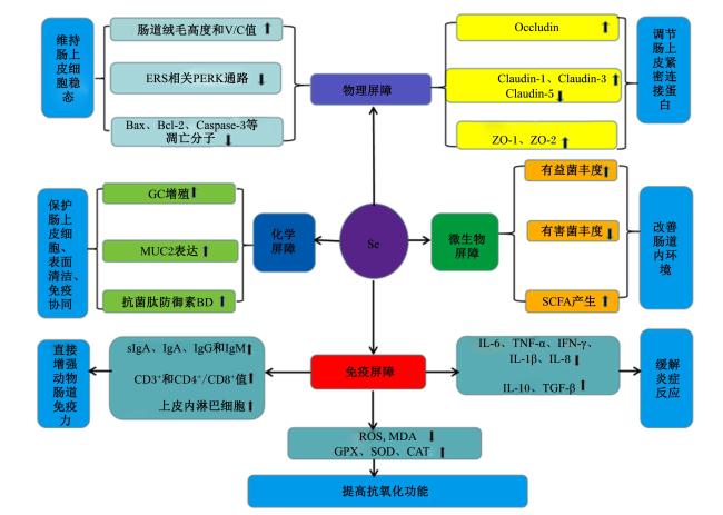
在畜禽养殖生产中,由于养殖环境、饲粮构成以及饲养方式不当等极易损伤肠道黏膜屏障功能并危害动物机体健康。综合本文所述,硒在保护和改善肠道黏膜屏障功能方面发挥了全面作用(图2)。今后,关于硒在畜禽生产中的应用尤其是在动物肠道健康保护方面还有待加强研究,如硒在不同动物类型、不同生长阶段及不同生理条件下对肠道黏膜屏障保护具体调控作用机制等;同时,由于硒与动物健康的关系十分复杂,硒在缺乏和毒性之间的范围非常窄,不同群体的基础硒水平也不同,硒摄入的安全方法和剂量以及适合补充硒的基础硒范围仍有待确定。未来随着研究的不断深入,在畜禽的营养研究方面开展更多体内外试验,以及对于动物品种的选取更加多样化,这将为硒作为畜禽饲料添加剂的开发及应用提供更加全面的科学依据。
图2 硒调节动物肠道的作用机制

Se:硒 selenium;V/C:绒毛高度/隐窝深度 villus height/crypt depth;ERS:内质网应激 endoplasmic reticulum stress;PERK:内质网应激相关激酶 endoplasmic reticulum stress-related kinase;Bax:B淋巴细胞瘤-2相关X蛋白 B cell lymphoma 2-associated X protein;Bcl-2:B淋巴细胞瘤-2 B cell lymphoma-2;Caspase:胱天蛋白酶 cysteine-aspartic protease;GC:杯状细胞 goblet cell;MUC2:黏蛋白2 mucin 2;BD:β-防御素 β-defensin;sIgA:分泌型免疫球蛋白A secretory immunoglobulin A;IgA:免疫球蛋白A immunoglobulin A;IgG:免疫球蛋白G immunoglobulin G;IgM:免疫球蛋白M immunoglobulin M;Occludin:闭合蛋白;Claudin:密封蛋白;ZO:闭锁小带蛋白 zonula occludens;SCFA:短链脂肪酸 short chain fatty acid;IL:白细胞介素 interleukin;TNF-α:肿瘤坏死因子-α tumor necrosis factor-α;IFN-γ:干扰素-γ interferon-γ;TGF-β:转化生长因子-β transforming growth factor-β;ROS:活性氧 reactive oxygen species;MDA:丙二醛 malonaldehyde;GSH-Px:谷胱甘肽过氧化物酶 glutathione peroxidase;SOD:超氧化物歧化酶 superoxide dismutase;CAT:过氧化氢酶 catalase。

⬆表示升高,⬇表示降低。⬆ indicated increase, and ⬇ indicated decrease.

Fig.2 Mechanism of selenium regulating animal intestines

[1]
SCHWARZ K, BIERI J, BRIGGS G, et al. Prevention of exudative diathesis in chicks by factor 3 and selenium[J]. Proceedings of the Society for Experimental Biology and Medicine, 1957, 95(4):621-625.

PMID

[2]
STAPLETON S R. Selenium:an insulin mimetic[J]. Cellular and Molecular Life Sciences, 2000, 57(13):1874-1879.

[3]
YE X Q, ZHU Y R, YANG Y Y, et al. Biogenic selenium nanoparticles synthesized with alginate oligosaccharides alleviate heat stress-induced oxidative damage to organs in broilers through activating Nrf2-mediated anti-oxidation and anti-ferroptosis pathways[J]. Antioxidants (Basel,Switzerland), 2023, 12(11):1973.

[4]
National Academies of Sciences,Engineering, and Medicine. Nutrient requirements of beef cattle[M]. 8th ed. Washington,D.C.:The National Academies Press, 2016.

[5]
National Research Council. Nutrient requirements of swine[S]. 11th ed.Washington,D.C.:The National Academies Press, 2012.

[6]
PRABHU K S, LEI X G. Selenium[J]. Advances in Nutrition, 2016, 7(2):415-417.

DOI PMID

[7]
ABDELNOUR S A, ALAGAWANY M, HASHEM N M, et al. Nanominerals:fabrication methods,benefits and hazards,and their applications in ruminants with special reference to selenium and zinc nanoparticles[J]. Animals:an Open Access Journal From MDPI, 2021, 11(7):1916.

[8]
GLADYSHEV V N, ARNÉR E S, BERRY M J, et al. Selenoprotein gene nomenclature[J]. Journal of Biological Chemistry, 2016, 291(46):24036-24040.

PMID

[9]
ALI F, SAEED K, FATEMEH H. Nano-bio selenium synthesized by Bacillus subtilis modulates broiler performance,intestinal morphology and microbiota,and expression of tight junction’s proteins[J]. Biological Trace Element Research, 2022, 200(4):1811-1825.

[10]
DALIA A M, LOH T C, SAZILI A Q, et al. Influence of bacterial organic selenium on blood parameters,immune response,selenium retention and intestinal morphology of broiler chickens[J]. BMC Veterinary Research, 2020, 16(1):365.

[11]
ZAMANI MOGHADDAM A K, MEHRAEI HAMZEKOLAEI M H, KHAJALI F, et al. Role of selenium from different sources in prevention of pulmonary arterial hypertension syndrome in broiler chickens[J]. Biological Trace Element Research, 2017, 180(1):164-170.

DOI PMID

[12]
TONG C, LI P, YU L, et al. Selenium-rich yeast attenuates ochratoxin A-induced small intestinal injury in broiler chickens by activating the Nrf2 pathway and inhibiting NF-κB activation[J]. Journal of Functional Foods, 2020,66:103784.

[13]
KHAJEH BAMI M, AFSHARMANESH M, ESPAHBODI M, et al. Effects of dietary nano-selenium supplementation on broiler chicken performance,meat selenium content,intestinal microflora,intestinal morphology,and immune response[J]. Journal of Trace Elements in Medicine and Biology, 2022,69:126897.

[14]
CHEN Y H, WU W, ZHOU H J, et al. Selenium nanoparticles improved intestinal health through modulation of the NLRP3 signaling pathway[J]. Frontiers in Nutrition, 2022,9:907386.

[15]
ZAINUDIN N N, HEMLY N I M, MUHAMMAD A I, et al. Effects of supplementing organic- and inorganic-based selenium with vitamin E on intestinal histomorphology,caecal bacterial proliferation,and short-chain fatty acid profile in layer hens[J]. Tropical Animal Health and Production, 2023, 55(2):90.

[16]
MUHAMMAD A I, MOHAMED D A, CHWEN L T, et al. Effect of selenium sources on laying performance,egg quality characteristics,intestinal morphology,microbial population and digesta volatile fatty acids in laying hens[J]. Animals:an Open Access Journal From MDPI, 2021, 11(6):1681.

[17]
ZHOU W T, MIAO S S, ZHU M K, et al. Effect of glycine nano-selenium supplementation on production performance,egg quality,serum biochemistry,oxidative status,and the intestinal morphology and absorption of laying hens[J]. Biological Trace Element Research, 2021, 199(11):4273-4283.

[18]
AHMED Z, MALHI M, SOOMRO S, et al. Dietary selenium yeast supplementation improved some villi morphological characteristics in duodenum and jejunum of young goats[J]. Journal of Animal & Plant Sciences, 2016,26:382-387.

[19]
程连平, 舒迎霜, 贺濛初, 等. 富硒酵母和枯草芽孢杆菌对湖羊羔羊小肠黏膜形态和直肠菌群的影响[J]. 动物营养学报, 2018, 30(11):4660-4669.

CHENG L P, SHU Y S, HE M C, et al. Effects of selenium-enriched yeast and Bacillus subtilis on intestinal mucosal morphology and rectum microflora of Hu lambs[J]. Chinese Journal of Animal Nutrition, 2018, 30(11):4660-4669. (in Chinese)

[20]
SAFFARI S, KEYVANSHOKOOH S, ZAKERI M, et al. Effects of dietary organic,inorganic,and nanoparticulate selenium sources on growth,hemato-immunological,and serum biochemical parameters of common carp (Cyprinus carpio)[J]. Fish Physiology and Biochemistry, 2018, 44(4):1087-1097.

[21]
LIU L, WU C M, CHEN D W, et al. Selenium-enriched yeast alleviates oxidative stress-induced intestinal mucosa disruption in weaned pigs[J]. Oxidative Medicine and Cellular Longevity, 2020,2020:5490743.

[22]
HE X J, LIN Y C, LIAN S, et al. Selenium deficiency in chickens induces intestinal mucosal injury by affecting the mucosa morphology,sIgA secretion,and GSH-Px activity[J]. Biological Trace Element Research, 2020, 197(2):660-666.

[23]
SONG X F, QIAO L, DOU X N, et al. Selenium nanoparticles alleviate deoxynivalenol-induced intestinal epithelial barrier dysfunction by regulating endoplasmic reticulum stress in IPEC-J2 cells[J]. Toxicology, 2023,494:153593.

[24]
SAMO S P, MALHI M, KACHIWAL A B, et al. Supranutritional selenium level minimizes high concentrate diet-induced epithelial injury by alleviating oxidative stress and apoptosis in colon of goat[J]. BMC Veterinary Research, 2020, 16(1):462.

DOI PMID

[25]
YAN S, QIAO L, DOU X, et al. Biogenic selenium nanoparticles by Lactobacillus casei ATCC 393 alleviate the intestinal permeability,mitochondrial dysfunction and mitophagy induced by oxidative stress[J]. Food & Function, 2021, 12(15):7068-7080.

[26]
QIAO L, YAN S Q, DOU X N, et al. Biogenic selenium nanoparticles alleviate intestinal epithelial barrier damage through regulating endoplasmic reticulum stress-mediated mitophagy[J]. Oxidative Medicine and Cellular Longevity, 2022,2022:3982613.

[27]
PAN Z Z, HUANG J Q, HU T, et al. Protective effects of selenium nanoparticles against bisphenol A-induced toxicity in porcine intestinal epithelial cells[J]. International Journal of Molecular Sciences, 2023, 24(8):7242.

[28]
DOU X N, QIAO L, SONG X F, et al. Biogenic nanoparticles alleviate intestinal epithelial barrier injury by regulating mitochondria-lysosome crosstalk[J]. Food & Function, 2023, 14(10):4891-4904.

[29]
ZHENG Y Y, ZHANG B, GUAN H Y, et al. Selenium deficiency causes apoptosis through endoplasmic reticulum stress in swine small intestine[J]. BioFactors, 2021, 47(5):788-800.

DOI PMID

[30]
WANG J F, LIU Z, HE X J, et al. Selenium deficiency induces duodenal villi cell apoptosis via an oxidative stress-induced mitochondrial apoptosis pathway and an inflammatory signaling-induced death receptor pathway[J]. Metallomics, 2018, 10(10):1390-1400.

DOI PMID

[31]
LUO D L, TANG X Y, WANG Y X, et al. Selenium deficiency exacerbated bisphenol A-induced intestinal toxicity in chickens:apoptosis and cell cycle arrest mediated by ROS/P53[J]. Science of the Total Environment, 2024,913:169730.

[32]
ZHANG Y, ZHANG J L, BAO J, et al. Selenium deficiency induced necroptosis,Th1/Th2 imbalance,and inflammatory responses in swine ileum[J]. Journal of Cellular Physiology, 2021, 236(1):222-234.

[33]
ZHANG Z Q, SHAN J H, SHI B D, et al. SeNPs alleviates BDE-209-induced intestinal damage by affecting necroptosis,inflammation,intestinal barrier and intestinal flora in layer chickens[J]. Ecotoxicology and Environmental Safety, 2023,262:115336.

[34]
YE R H, GUO Q Y, HUANG J Q, et al. Eucommia ulmoides polysaccharide modified nano-selenium effectively alleviated DSS-induced colitis through enhancing intestinal mucosal barrier function and antioxidant capacity[J]. Journal of Nanobiotechnology, 2023, 21(1):222.

[35]
LIU Y M, YANG Y X, DONG R Q, et al. Protective effect of selenomethionine on intestinal injury induced by T-2 toxin[J]. Research in Veterinary Science, 2020,132:439-447.

[36]
YANG S, LI L, YU L, et al. Selenium-enriched yeast reduces caecal pathological injuries and intervenes changes of the diversity of caecal microbiota caused by ochratoxin-A in broilers[J]. Food and Chemical Toxicology, 2020,137:111139.

[37]
HE Y, LIU Y, TANG J Y, et al. Selenium exerts protective effects against heat stress-induced barrier disruption and inflammation response in jejunum of growing pigs[J]. Journal of the Science of Food and Agriculture, 2022, 102(2):496-504.

[38]
ZHA A D, TU R Q, QI M, et al. Mannan oligosaccharides selenium ameliorates intestinal mucosal barrier,and regulate intestinal microbiota to prevent Enterotoxigenic Escherichia coli-induced diarrhea in weaned piglets[J]. Ecotoxicology and Environmental Safety, 2023,264:115448.

[39]
XUE Y, WANG H H, TIAN B W, et al. Selenium deficiency promotes the expression of lncRNA-MORC3,activating NLRP3-Caspase-1/IL-1β signaling to induce inflammatory damage and disrupt tight junctions in piglets[J]. Biological Trace Element Research, 2023, 201(5):2365-2376.

[40]
EICHNER M, PROTZE J, PIONTEK A, et al. Targeting and alteration of tight junctions by bacteria and their virulence factors such as Clostridium perfringens enterotoxin[J]. Pflugers Archiv-European Journal of Physiology, 2017, 469(1):77-90.

[41]
WEN Z S, DU M, TANG Z, et al. Low molecular seleno-aminopolysaccharides protect the intestinal mucosal barrier of rats under weaning stress[J]. International Journal of Molecular Sciences, 2019, 20(22):5727.

[42]
YIN Y X, AHMED N, HASSAN M F, et al. Effect of nano-selenium on biological mechanism of goblet cells of the small intestine within laying hen[J]. Biological Trace Element Research, 2024, 202(4):1699-1710.

[43]
ALKHUDHAYRI A A, DKHIL M A, AL-QURAISHY S. Nanoselenium prevents eimeriosis-induced inflammation and regulates mucin gene expression in mice jejunum[J]. International Journal of Nanomedicine, 2018,13:1993-2003.

[44]
ABDEL-GABER R, HAWSAH M A, AL-OTAIBI T, et al. Biosynthesized selenium nanoparticles to rescue coccidiosis-mediated oxidative stress,apoptosis and inflammation in the jejunum of mice[J]. Frontiers in Immunology, 2023,14:1139899.

[45]
ZHU C H, ZHANG S M, SONG C W, et al. Selenium nanoparticles decorated with Ulva lactuca polysaccharide potentially attenuate colitis by inhibiting NF-κB mediated hyper inflammation[J]. Journal of Nanobiotechnology, 2017, 15(1):20.

[46]
YANG J J, WANG J, HUANG K H, et al. Selenium-enriched Bacillus subtilis yb-114246 improved growth and immunity of broiler chickens through modified ileal bacterial composition[J]. Scientific Reports, 2021, 11(1):21690.

[47]
XU S Z, LEE S H, LILLEHOJ H S, et al. Dietary sodium selenite affects host intestinal and systemic immune response and disease susceptibility to necrotic enteritis in commercial broilers[J]. British Poultry Science, 2015, 56(1):103-112.

DOI PMID

[48]
HE Y J, PENG L, ZHAO X C, et al. Selenium deficiency induces inflammatory response and decreased antimicrobial peptide expression in chicken jejunum through oxidative stress[J]. Biological Trace Element Research, 2023, 201(7):3461-3473.

[49]
YU J, XIN Y, XUE F, et al. Selenium deficiency increased duodenal permeability and decreased expression of antimicrobial peptides by activating ROS/NF-κB signal pathway in chickens[J]. BioMetals, 2023, 36(1):137-152.

[50]
BERKOWITZ L, PARDO-ROA C, SALAZAR G A, et al. Mucosal exposure to cigarette components induces intestinal inflammation and alters antimicrobial response in mice[J]. Frontiers in Immunology, 2019,10:2289.

[51]
AL-QUWAIE D A. The influence of bacterial selenium nanoparticles biosynthesized by Bacillus subtilus DA20 on blood constituents,growth performance,carcass traits,and gut microbiota of broiler chickens[J]. Poultry Science, 2023, 102(9):102848.

[52]
DALIA A M, LOH T C, SAZILI A Q, et al. Effects of vitamin E,inorganic selenium,bacterial organic selenium,and their combinations on immunity response in broiler chickens[J]. BMC Veterinary Research, 2018, 14(1):249.

[53]
LI R, LIU J, LIU M, et al. Effects of selenium-enriched yeast dietary supplementation on egg quality,gut morphology and caecal microflora of laying hens[J]. Animal Biotechnology, 2024, 35(1):2258188.

[54]
ALAGAWANY M, QATTAN S Y A, ATTIA Y A, et al. Use of chemical nano-selenium as an antibacterial and antifungal agent in quail diets and its effect on growth,carcasses,antioxidant,immunity and caecal microbes[J]. Animals:an Open Access Journal From MDPI, 2021, 11(11):3027.

[55]
REDA F M, ALAGAWANY M, SALAH A S, et al. Biological selenium nanoparticles in quail nutrition:biosynthesis and its impact on performance,carcass,blood chemistry,and cecal microbiota[J/OL]. Biological Trace Element Research.(2023-12-18)[2023-12-20]. https://link.springer.com/article/10.1007/s12011-023-03996-3#citeas.

[56]
PEREIRA A M, PINNA C, BIAGI G, et al. Supplemental selenium source on gut health:insights on fecal microbiome and fermentation products of growing puppies[J]. FEMS Microbiology Ecology, 2020, 96(11):fiaa212.

[57]
LV C H, WANG T, REGMI N, et al. Effects of dietary supplementation of selenium-enriched probiotics on production performance and intestinal microbiota of weanling piglets raised under high ambient temperature[J]. Journal of Animal Physiology and Animal Nutrition, 2015, 99(6):1161-1171.

DOI PMID

[58]
LIU S, YU H, LI P, et al. Dietary nano-selenium alleviated intestinal damage of juvenile grass carp (Ctenopharyngodon idella) induced by high-fat diet:insight from intestinal morphology,tight junction,inflammation,anti-oxidization and intestinal microbiota[J]. Animal Nutrition, 2022, 8(10):235-248.

[59]
WANG G D, JIANG Z H, SONG Y W, et al. Gut microbiota contribution to Selenium deficiency-induced gut-liver inflammation[J]. BioFactors:1-15.(2023-09-07)[2023-11-10].https://iubmb.onlinelibrary.wiley.com/doi/10.1002/biof.2006.

[60]
GANGADOO S, DINEV I, CHAPMAN J, et al. Selenium nanoparticles in poultry feed modify gut microbiota and increase abundance of Faecalibacterium prausnitzii[J]. Applied Microbiology and Biotechnology, 2018, 102(3):1455-1466.

[61]
MOOLCHAND M, FARMAN S, BENAZIR A, et al. Influence of dietary selenium yeast on fermentation pattern mucosal growth and glutathione peroxidase (GSH-Px) activity in colon of goat[J]. Journal of Dairy Veterinary & Animal Research, 2018, 7(6):253-259.

[62]
QIAO L, DOU X, SONG X, et al. Replacing dietary sodium selenite with biogenic selenium nanoparticles improves the growth performance and gut health of early-weaned piglets[J]. Animal Nutrition, 2023, 15(12):99-113.

[63]
YANG H J, MO A J, YI L Y, et al. Selenium attenuated food borne cadmium-induced intestinal inflammation in red swamp crayfish (Procambarus clarkii) via regulating PI3K/Akt/NF-κB pathway[J]. Chemosphere, 2024,349:140814.

[64]
KHAN I, ZANEB H, MASOOD S, et al. Supplementation of selenium nanoparticles-loaded chitosan improves production performance,intestinal morphology,and gut microflora in broiler chickens[J]. Journal of Poultry Science, 2022, 59(3):272-281.

[65]
ABDEL-MONEIM A M E, SHEHATA A M, SELIM D A, et al. Spirulina platensis and biosynthesized selenium nanoparticles improve performance,antioxidant status,humoral immunity and dietary and ileal microbial populations of heat-stressed broilers[J]. Journal of Thermal Biology, 2022,104:103195.

[66]
QIAO L, DOU X N, SONG X F, et al. Protective effect of biogenic selenium nanoparticles against diquat-induced acute toxicity via regulation of gut microbiota and its metabolites[J]. Food and Chemical Toxicology, 2022,170:113480.

[67]
LI Z M, DONG Y P, CHEN S R, et al. Organic selenium increased gilts antioxidant capacity,immune function,and changed intestinal microbiota[J]. Frontiers in Microbiology, 2021,12:723190.

[68]
GANGADOO S, BAUER W B, BAJAGAI Y S, et al. In vitro growth of gut microbiota with selenium nanoparticles[J]. Animal Nutrition, 2019, 5(4):424-431.

[69]
FANG M X, HU W, LIU B. Effects of nano-selenium on cecum microbial community and metabolomics in chickens challenged with ochratoxin A[J]. Frontiers in Veterinary Science, 2023,10:1228360.

[70]
KANG R F, WANG W H, LIU Y F, et al. Dietary selenium sources alleviate immune challenge induced by Salmonella Enteritidis potentially through improving the host immune response and gut microbiota in laying hens[J]. Frontiers in Immunology, 2022,13:928865.

[71]
SONG X F, QIAO L, CHANG J J, et al. Dietary supplementation with selenium nanoparticles-enriched Lactobacillus casei ATCC 393 alleviates intestinal barrier dysfunction of mice exposed to deoxynivalenol by regulating endoplasmic reticulum stress and gut microbiota[J]. Ecotoxicology and Environmental Safety, 2022,248:114276.

[72]
LIU Z, QU Y P, WANG J F, et al. Selenium deficiency attenuates chicken duodenal mucosal immunity via activation of the NF-κB signaling pathway[J]. Biological Trace Element Research, 2016, 172(2):465-473.

DOI PMID

[73]
HE Y, FANG J, PENG X, et al. Effects of sodium selenite on aflatoxin B1-induced decrease of ileal IgA+ cell numbers and immunoglobulin contents in broilers[J]. Biological Trace Element Research, 2014, 160(1):49-55.

[74]
KHAN M Z I, AKTER S H, ISLAM M N, et al. The effect of selenium and vitamin E on the lymphocytes and immunoglobulin-containing plasma cells in the lymphoid organ and mucosa-associated lymphatic tissues of broiler chickens[J]. Anatomia Histologia Embryologia, 2008, 37(1):52-59.

DOI PMID

[75]
WANG F Y, ZUO Z C, CHEN K J, et al. Selenium rescues aflatoxin B1-inhibited T cell subsets and cytokine levels in cecal tonsil of chickens[J]. Biological Trace Element Research, 2019, 188(2):461-467.

[76]
ZHANG L, FENG L, JIANG W D, et al. Vitamin a deficiebcy suppresses fish immune function with difference in different intestinal segments:the role of transriotional factor NF-κB and p38 mitogen-activated protein kinase signalling pathways[J]. British Journal of Nutrition, 2017, 117(1):67-82.

[77]
MOU D L, DING D J, YANG M, et al. Maternal organic selenium supplementation during gestation improves the antioxidant capacity and reduces the inflammation level in the intestine of offspring through the NF-κB and ERK/Beclin-1 pathways[J]. Food & Function, 2021, 12(1):315-327.

[78]
LEI L, JING M, YINGCE Z, et al. Selenium deficiency causes oxidative stress and activates inflammation,apoptosis,and necroptosis in the intestine of weaned calves[J]. Metallomics, 2023, 15(6):mfad028.

[79]
SONG Z X, JIANG W D, LIU Y, et al. Dietary zinc deficiency reduced growth performance,intestinal immune and physical barrier functions related to NF-κB,TOR,Nrf2,JNK and MLCK signaling pathway of young grass carp (Ctenopharyngodon idella)[J]. Fish & Shellfish Immunology, 2017,66:497-523.

[80]
DING D J, MOU D L, ZHU H, et al. Maternal organic selenium supplementation relieves intestinal endoplasmic reticulum stress in piglets by enhancing the expression of glutathione peroxidase 4 and selenoprotein S[J]. Frontiers in Nutrition, 2022,9:900421.

[81]
LI L L, LIU Z, QUAN J Q, et al. Dietary nano-selenium alleviates heat stress-induced intestinal damage through affecting intestinal antioxidant capacity and microbiota in rainbow trout (Oncorhynchus mykiss)[J]. Fish & Shellfish Immunology, 2023,133:108537.

Outlines

/