RESEARCH PAPER

Screening Combination of Lactobacillus spp. and Prebiotics and Evaluating Their Impact on Intestinal Barrier Utilizing IPEC-J2 Cell Model

  • JING Xueyi , 1, 2 ,
  • YU Aibing 1 ,
  • CHENG Yufei 2 ,
  • GAO Yang 1, 2 ,
  • JIANG Nan 3 ,
  • HONG Liang , 1, 2, *
Expand
  • 1 Tianjin Key Laboratory of Green Ecological Feed, Tianjin Modern Tianjiao Agricultural Technology Co., Ltd., Tianjin 301800, China
  • 2 Tianjin Key Laboratory of Agricultural Animal Breeding and Healthy Husbandry, Department of Animal Science and Animal Medicine, Tianjin Agricultural University, Tianjin 300392, China
  • 3 Institute of Animal Husbandry and Veterinary, Tibet Autonomous Regional Academy of Agricultural Sciences, Lhasa 850000, China

Received date: 2024-02-06

  Online published: 2024-07-09

Abstract

In order to develop green additive products to improve the intestinal health of animals, this study aimed to screen the synbiotic combination of Lactobacillus spp. and prebiotics, and investigate its influence on the integrity of the intestinal barrier. Initially, the fermentation results using API 50 CHL kit were used to identify five strains of Lactobacillus spp. [Lactobacillus plantarum 177 (LP), Lactobacillus salivarius KLW001 (LSa), Lactobacillus reuteri KLR3004 (LRe), Lactobacillus paracaeei KCTC3510 (LPa), and Lactobacillus rhamnosus KCTC5033 (LRh)] corresponding to candidate prebiotics. Then, based on the fermentation ability of short-chain fatty acids (SCFA) by the five strains of Lactobacillus spp. and candidate prebiotics [sorbitol (So), mannitol (Ma), rhamnose (Rh), xylo-oligosaccharide (Xyo), trehalose (Tr), raffinose (Ra), and pullulan (Pu)], synbiotic combinations were selected. Three selected synbiotic combinations (LP+Pu, LSa+Pu, and LRe+Ra) were then applied to lipopolysaccharide (LPS)-induced “leaky gut” cell model. The effect of synbiotics on intestinal barrier function was validated by measuring changes in monolayer cell permeability, trans-epithelial electrical resistance (TEER) values, and the mRNA expression of intestinal tight junction proteins [Claudin-1, Occludin, and zonula occluden-1 (ZO-1)]. Results showed that the combination of LP+Pu exhibited enhanced fermentation capabilities, resulting in higher production of SCFA, particularly an increase in butyric acid content; in the “leaky gut” cell model, the synbiotic consisting of LP+Pu demonstrated an upregulation of mRNA relative expression for intestinal tight junction proteins, specifically Claudin-1 and ZO-1. Furthermore, this synbiotic combination contributed to the preservation of cell epithelial integrity. The results of this study confirm that the combination of LP+Pu can serve as a synbiotic formulation, effectively enhancing the production of SCFA and increasing the expression of intestinal tight junction proteins. These findings present a novel avenue for the treatment and prevention of intestinal barrier-related diseases in livestock and poultry.

Cite this article

JING Xueyi , YU Aibing , CHENG Yufei , GAO Yang , JIANG Nan , HONG Liang . Screening Combination of Lactobacillus spp. and Prebiotics and Evaluating Their Impact on Intestinal Barrier Utilizing IPEC-J2 Cell Model[J]. Chinese Journal of Animal Nutrition, 2024 , 36(7) : 4693 -4705 . DOI: 10.12418/CJAN2024.402

肠道屏障是调节物质进入机体的“一扇门”,在动物出生后逐渐建立并稳定[1]。在畜牧养殖中,肠道屏障功能异常会导致脂多糖(lpopolysaccharide, LPS)和毒素暴露,抑制食欲、消化和蛋白质的合成,使营养物质和能量不能被充分利用和转化,最终导致畜禽饲料转化率下降[2]。由于新生幼畜肠道黏膜和肠道菌群发育不完善,无法有效抵抗环境中的有害因素,易患肠道相关疾病,导致畜禽存活率和经济效益下降,增加养殖成本。在维持肠道屏障功能方面,相比治疗已经受损的肠道屏障,预防更为重要。因此,迫切需要寻求其他有效措施来预防肠道疾病并维持肠道健康和屏障功能。益生元是不能被机体吸收的碳水化合物,进入体内后能被益生菌发酵产生短链脂肪酸(SCFA),SCFA能直接调节肠道菌群结构,或与相应受体结合,通过调节肠道pH、为上皮细胞提供能量等方式参与肠道屏障完整性和功能的调控[3-4]。越来越多的研究证明,摄入益生元和益生菌能发酵产生更多SCFA,有利于改善肠道屏障功能,在治疗肠道疾病方面具有临床益处[5],特别是丁酸,不仅能抑制肠道炎症,还有助于肠黏膜紧密连接的组装、增强肠道的屏障功能[6-7]
乳酸菌是小肠中最常见的有益菌[8],可以和肠黏膜共同组成一道生物学屏障,防止有害菌在肠道表面附着和定植,维持动物机体的健康状态[9]。乳酸菌单独或复合用于动物生产中,可以提高畜禽机体免疫、生长性能,调节肠道菌群结构,保障动物健康[10]。因此,本研究中选取了乳杆菌作为益生菌。本研究目的是根据SCFA的发酵能力来筛选合生元组合,并验证其在LPS诱导的“肠漏”细胞模型中的作用,为今后在畜牧养殖中预防并保障动物肠道健康提供新方法,提高养殖效益。

1 材料与方法

1.1 试验材料

1.1.1 主要试剂

本研究所用材料和化学试剂,除非特别说明,均购自西格玛奥德里奇(上海)贸易有限公司。MRS肉汤/琼脂购自上海玉博生物科技有限公司,乳酸菌选择性培养基、鉴定培养基、噻唑蓝(MTT)、二甲基亚砜(DMSO)、SCFA标准品、总RNA提取试剂盒、反转录通用试剂盒、Real-time PCR荧光定量试剂盒(SYBR Green Ⅰ)均购自北京索莱宝科技有限公司。碳水化合物鉴定试剂条(API 50CH)购自上海欣中生物工程有限公司。

1.1.2 候选益生菌

植物乳杆菌177(Lactobacillus plantarum 177, LP)、唾液乳杆菌KLW001(Lactobacillus salivarius KLW001, LSa)和罗伊氏乳杆菌KLR3004(Lactobacillus reuteri KLR3004, LRe)从猪的粪便中分离。类干酪乳杆菌KCTC3510(Lactobacillus paracaeei KCTC3510, LPa)和鼠李糖乳杆菌KCTC5033(Lactobacillus rhamnosus KCTC5033, LRh)购自韩国典型菌种保存中心(KCTC),保存于-80 ℃待用。

1.1.3 候选益生元和IPEC-J2细胞系

本研究所用到的候选益生元中山梨醇(sorbitlol, So)和甘露醇(mannitol, Ma)购自生工生物工程(上海)股份有限公司,鼠李糖(rhamnose, Rh)、麦芽糖(maltose, Mal)、低聚木糖(xylo-oligo, Xyo)、海藻糖(trehalose, Tr)、棉子糖(raffinose, Ra)和普鲁兰多糖(pullulan, Pu)购自北京索莱宝科技有限公司。
IPEC-J2细胞系(45~60代)购自上海雅吉生物科技有限公司。

1.2 试验设计

1.2.1 候选益生菌和候选益生元的筛选

将5株益生菌菌株接种于MRS培养基中,37 ℃条件下培养12 h,将培养好的细菌混悬液加入到碳水化合物生化鉴定试剂条中,37 ℃培养24 h后观察培养物的变化。根据细菌对不同碳水化合物的利用情况来获取碳水化合物的发酵谱,再根据益生元的定义选择益生元。将不同组合的合生元培养上清液与标准品(丙酸2,2-d2)和甲醇混合,利用气相色谱仪(Agilent Technologies, 7890B)检测SCFA含量。通过比较不同合生元组合发酵SCFA的能力选择合生元。筛选出3种合生元组合(LP+Pu、LSa+Pu、LRe+Ra)用于后续试验。

1.2.2 体外“肠漏”细胞模型的构建

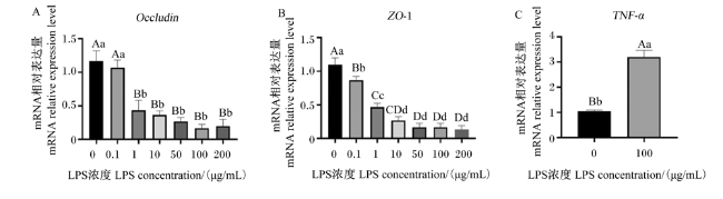
选用不同浓度的LPS(0.1、1、10、50、100和200 μg/mL)处理IPEC-J2细胞24 h。测定IPEC-J2细胞单层细胞渗透率、跨上皮细胞电阻(trans-epithelial electrical resistance, TEER)值的变化、肿瘤坏死因子-α(tumor necrosis factor-α, TNF-α)的mRNA表达,构建“肠漏”细胞模型。选择能破坏屏障且对细胞无毒性的LPS浓度用于后续研究。

1.2.3 体外合生元功能试验

采用3种试验方法:治疗试验、预防试验和保护试验来评估合生元的功能。每个试验分别设置了空白对照(C)、阴性对照(NC)和试验处理,每个处理3个重复。
在治疗试验中,先用100 μg/mL的LPS处理细胞24 h,之后用PBS洗涤细胞去除LPS,然后用10%(v/v)的益生菌或合生元的无细胞上清液(CFS)处理。在预防试验中,处理和治疗试验处理相反,先用10%(v/v)的益生菌或合生元的CFS处理细胞,然后再用100 μg/mL的LPS处理。在保护试验中,同时用10%(v/v)的益生菌或合生元的CFS和100 μg/mL LPS共同处理细胞。之后测定与肠屏障相关的紧密连接蛋白的mRNA表达和TEER值的变化,以评估3种合生元组合(LP+Pu、LSa+Pu、LRe+Ra)对屏障完整性的影响。

1.3 测定方法

1.3.1 SCFA的含量测定

将合生元在37 ℃条件下培养24 h,之后进行离心,取上清液经滤膜过滤后用气相色谱仪(Agilent Technologies, 7890B)进样检测。将SCFA的标准品稀释成不同浓度的工作液,进行进样检测。以质量浓度为横坐标,对应峰面积为纵坐标,制备不同SCFA浓度的回归曲线,测定合生元上清液中乙酸、丙酸和丁酸的含量,每个样品重复测定3次。色谱的条件如下:用Thermo ScientificTrace 1310系统,包括带有TG-5MS柱(30 mm×0.25 mm,5%苯基-甲基聚硅氧烷毛细血管柱,薄膜厚度为0.25 μm)的质量选择器。温度程序设定如下:初始温度为50 ℃,保持5 min,然后以每分钟4 ℃的速率升温至250 ℃。载气使用高纯度氦气,每次进样量为1 μL,电离能设定为70 eV。

1.3.2 IPEC-J2细胞培养及MTT法测定细胞活性

选取45~60代IPEC-J2细胞进行培养,培养基为添加了1%青霉素/链霉素(100×)以及1%胰岛素/转铁蛋白/硒(100×)和10%胎牛血清的DMEM/F12。培养条件为37 ℃,5% CO2和95%相对湿度,每2 d更换1次培养基。
MTT法测定细胞活性:活细胞可以将MTT还原成蓝紫色甲瓒结晶,而DMSO能溶解细胞中的甲瓒结晶,通过吸光度测定细胞存活率。使用前制备MTT贮备液(1 mg/mL),用1.5 mL DMEM/F12替代IPEC-J2的细胞培养基,并加入0.1 mL MTT储备液。37 ℃条件下孵育4 h,弃掉培养基并用1×PBS(pH 7.4)洗涤,再加入DMSO振荡使其溶解,之后用酶标仪(SpectraMax Absorbance Microplate Reader, CMax Plus)在540 nm处测定吸光度(OD)用来表示细胞活力。细胞存活率用相对于空白对照的百分比表示。
细胞存活率(%)=(OD试验/OD空白)×100。

1.3.3 TEER值测定

将IPEC-J2细胞以105个细胞的密度接种在0.33 cm2和0.4 μm孔径的聚对苯二甲酸乙二醇酯膜的培养基中,隔1 d更换1次培养基。使用细胞电阻仪(CellZscopeE)测定TEER值,判断细胞的完整性。在测量过程中,上皮的顶侧和基底侧都被浸泡在缓冲液中。在处理细胞24 h后,测量TEER值。

1.3.4 细胞通透性测定

当IPEC-J2细胞贴壁于Trans-well细胞培养板上时,按照1.3.3提到的方法处理后,测量TEER值。接下来,在细胞的顶侧加入荧光素异硫氰酸酯标记的右旋糖苷4 ku(FITC-D4),浓度为1 g/L,并将细胞置于37 ℃下孵育4 h。之后,在96孔细胞培养板中收集100 μL底部培养基,并使用紫外分光光度计(METASH,UV-5100B)在激发波长为498 nm、发射波长为540 nm处测定其OD值。细胞通透性通过FITC-D4从顶部渗透到底部的量来表示。

1.3.5 实时荧光定量PCR(qRT-PCR)

IPEC-J2细胞培养至融合,用PBS洗涤3次,用Trizol法提取总RNA,之后用通用反转录试剂盒将1 μg的RNA反转录成cDNA。再以cDNA为模板在实时定量荧光PCR仪(CFX96 Touch)上用SYBR-qPCR-Mix一步法对目的基因进行扩增。扩增体系15 μL:7.5 μL SYBR,0.75 μL上游引物,0.75 μL下游引物,1 μL cNDA,5 μL RNase-free water。扩增程序为:预变性95 ℃,10 min,变性95 ℃,10 s,退火60 ℃,30 s,延伸72 ℃,30 s,35个循环。以β-肌动蛋白(β-actin)为内参,通过2-ΔΔCt法计算目的基因的mRNA相对表达量。引物序列见表1
表1 引物序列

Table 1 Primer sequence

基因 Genes 引物序列 Primer sequence (5'—3') 退火温度 Tm/℃
β-肌动蛋白 β-actin F:TGCGGGACATCAAGGAGAAG
R:AGTTGAAGGTGGTCTCGTGG
58.8
58.6
闭合蛋白 Occludin F:ATCAACAAAGGCAACTCT
R:GCAGEAGCCATGTACTCT
50.5
56.1
闭锁小带蛋白-1 ZO-1 F:GAGTTTGATAGTGGCGTT
R:GTGGGAGGATGCTGTTGT
51.4
55.9
跨膜蛋白-1 Claudin-1 F:TTTCCTCAATACAGGAGG
R:CCCTCTCCCCACATTCGA
56.1
58
肿瘤坏死因子-α TNF-α F:ATTCAGGGATGTGTGGCCTG
R:CCAGATGTCCCAGGTTGCAT
59
59

1.4 统计分析

试验数据用Excel 2016软件进行初步处理,再通过SPSS 22.0软件进行统计分析,用单因素方差分析(one-way ANOVA)和Tukey's检验分析各组间的统计学显著性,P<0.05为差异显著,P<0.01为差异极显著。

2 结果与分析

2.1 益生元的筛选

使用碳水化合物鉴定试剂条(API 50CH),测定碳水化合物家族及其衍生物发酵情况。表2中列出了5种乳酸菌对49种底物的发酵情况。葡萄糖、果糖、半乳糖、甘露醇和核糖都能被这5种乳酸菌发酵,但是因其作为单糖,可以被机体吸收利用,不符合益生元的定义。在5种乳酸菌中,4种乳酸菌可以发酵麦芽糖。因此,选取了由α-(1-4)糖苷键连接的麦芽三糖构成的Pu作为候选益生元进行后续研究。木糖可以被LRe发酵,但其也能被动物机体吸收,因此,选择Xyo作为候选益生元进行筛选,其中Rh是已知的可以被LRh发酵的多糖;So和Ma互为同分异构体,进入机体之后不能被吸收利用;Tr是由2分子葡萄糖连接而成的双糖,本身不易被机体消化吸收;Ra可以被3种乳酸菌发酵,其本身不能被动物机体吸收利用。因此,选择Rh、So、Ma、Tr、Ra、Pu和Xyo作为候选益生元。
表2 5种乳酸菌的发酵能力

Table 2 Fermentation ability of five kinds of Lactobacillus spp.

糖类
Carbohydrates
乳酸菌 Lactobacillus spp.
LP LRe LSa LPa LRh
葡萄糖 Glucose + + + + +
果糖 Fructose + + + +
半乳糖 Galactose + + + + +
甘露糖 Mannose + + + +
核糖 Ribose + + +
鼠李糖 Rhamnose +
山梨醇 Sorbitlol + + +
葡萄糖苷 Glucopyranside +
甘露醇 Mannitol + +
乙酰葡萄糖胺 Acety-glucosamine + + + +
苦杏仁苷 Amygdalin +
熊果苷 Arbutin + +
水杨苷 Salicin + + + +
纤维二糖 Cellobiose + + +
麦芽糖 Maltose + + + +
塔格糖 Tagatose + +
木糖 Xylose +
乳糖 Lactose + + + +
蜜二糖 Melibiose + + + + +
蔗糖 Sucrose + + +
海藻糖 Trehalose + + +
棉子糖 Raffinose + + +
松二糖 Turanose + +
龙胆二糖 Gentiobiose + +

LP:植物乳杆菌177;LRe:罗伊氏乳杆菌KLR3004;LSa:唾液乳杆菌 KLW001;LPa:类干酪乳杆菌 KCTC3510;LRh:鼠李糖乳杆菌 KCTC5033。

“+”表示可以被对应的乳酸菌发酵。表中只列出了24种能被5种乳酸菌发酵的底物,没列出的25种底物不能被5种乳酸菌中任意一种发酵。

LP:Lactobacillus plantarum 177;LRe:Lactobacillus reuteri KLR3004;LSa:Lactobacillus salivarius KLW001;LRh:Lactobacillus rhamnose KCTC5033.

Symbol “+” means fermented by the corresponding Lactobacillus spp. The table only lists 24 substrates that can be fermented by the 5 Lactobacillus spp. mentioned. The 25 substrates not listed are incapable of being fermented by any of the 5 Lactobacillus spp.

根据碳水化合物发酵谱选择LP+So、LP+Tr、LP+Ra以及LP+Pu组合;LRe+Ra、LRe+Xyo和LRe+Pu组合;LPa+Ma、LPa+Tr组合;LSa+So、LSa+Ma、LSa+Pu和LSa+Ra组合;LRh+So、LRh+Pu、LRh+Rh和LRh+Tr组合。通过测定不同细菌和相应候选益生元组合发酵SCFA的能力选择用于后续试验的合生元。

2.2 合生元组合的筛选

使用气相色谱仪测定了合生元的SCFA发酵性能。如表3所示,LP可以将Pu发酵为乙酸、丙酸和丁酸。除了Pu和Tr可以被LP发酵成乙酸外,So和Ra也可被发酵成丙酸和丁酸。另外,从表4中可以看出,LRe+Ra组合相较于其他组合和单独的LRe组,极显著提高发酵产生的乙酸含量(P<0.01)。
表3 LP与潜在益生元组合发酵SCFA的能力

Table 3 Ferment SCFA ability of LP in combination with potential prebioticsmmol/L

项目
Items
处理 Treatments SEM P
P-value
LP LP+Tr LP+So LP+Ra LP+Pu
乙酸 Acetate 62.54BCc 64.58ABb 62.15Cc 62.19Cc 66.41Aa 0.47 <0.001
丙酸 Propionate 0.09Cc 0.25Cc 2.10Aa 1.96ABab 1.78Bb 0.24 <0.001
丁酸 Butyrate 0.00Dd 0.00Dd 0.98Cc 1.56Aa 1.37Bb 0.18 <0.001

LP:植物乳杆菌177;LP+Tr:植物乳杆菌177+海藻糖;LP+So:植物乳杆菌177+山梨醇;LP+Ra:植物乳杆菌177+鼠李糖;LP+Pu:植物乳杆菌177+普鲁兰多糖。

同行数据肩标无字母或相同字母表示差异不显著(P>0.05),不同小写字母表示差异显著(P<0.05),不同大写字母表示差异极显著(P<0.01)。下表同。

LP: Lactobacillus plantarum 177; LP+Tr: Lactobacillus plantarum 177+trehalose; LP+So: Lactobacillus plantarum 177+sorbitol; LP+Ra: Lactobacillus plantarum 177+raffinose; LP+Pu: Lactobacillus plantarum 177+pullulan.

In the same row, values with no letter or the same letter superscripts mean no significant difference (P>0.05), while with different small letter superscripts mean significant difference (P<0.05), and with different capital letter superscripts mean significant difference (P<0.01). The same as below.

表4 LRe与潜在益生元组合发酵SCFA的能力

Table 4 Ferment SCFA ability of LRe in combination with potential prebioticsmmol/L

项目
Items
处理 Treatments SEM P
P-value
LRe LRe+Ra LRe+Xyo LRe+Pu
乙酸 Acetate 80.05Bb 82.88Aa 77.13Cc 79.70Bb 0.64 <0.001
丙酸 Propionate 1.51Aa 1.29Bb 1.13Bb 0.75Cc 0.08 <0.001
丁酸 Butyrate 0.79a 0.40ab 0.19b 0.64ab 0.08 0.021

LRe:罗伊氏乳杆菌KLR3004;LRe+Ra:罗伊氏乳杆菌KLR3004+棉子糖;LRe+Xyo:罗伊氏乳杆菌KLR3004+低聚木糖;LRe+Pu:罗伊氏乳杆菌KLR3004+普鲁兰多糖。

LRe: Lactobacillus reuteri KLR3004; LRe+Ra: Lactobacillus reuteri KLR3004+raffinose; LRe+Xyo: Lactobacillus reuteri KLR3004+xylo-oligosacchaide; LRe+Pu: Lactobacillus reuteri KLR3004+pullulan.

表5所示,LPa不能将Ma和Tr发酵成SCFAs。Pu能被LSa发酵为乙酸(表6)。此外,已有研究表明Rh是LRh的益生元,能够被其发酵生成丁酸。如表7所示,除了Rh外,So和Tr也能被LRh发酵生成丁酸。根据LP、LRe、LSa、LPa以及LRh和不同候选益生元发酵能力分别选择LP+Pu、LRe+Ra和LSa+Pu组成的合生元用于后续试验。
表5 LPa与潜在益生元组合发酵SCFA的能力

Table 5 Ferment SCFA ability of LPa in combination with potential prebioticsmmol/L

项目
Items
处理 Treatments SEM P
P-value
LPa LPa+Ma LPa+Tr
乙酸 Acetate 58.96 58.88 58.77 0.13 0.875
丙酸 Propionate 1.35 1.30 1.44 0.03 0.248
丁酸 Butyrate 0.43Aa 0.27Bc 0.30Bb 0.45 <0.001

LPa:类干酪乳杆菌KCTC3510;LPa+Ma:类干酪乳杆菌KCTC3510+甘露醇;LPa+Tr:类干酪乳杆菌KCTC3510+海藻糖。

LPa: Lactobacillus paracasei KCTC3510; LPa+Ma: Lactobacillus paracasei KCTC3510+mannitol; LPa+Tr: Lactobacillus paracasei KCTC3510+trehalose.

表6 LSa与潜在益生元组合发酵SCFA的能力

Table 6 Ferment SCFA ability of LSa in combination with potential prebioticsmmol/L

项目
Items
处理 Treatments SEM P
P-value
LSa LSa+So LSa+Ma LSa+Pu LSa+Ra
乙酸 Acetate 57.82Bb 56.83Bbc 56.79BCbc 65.44Aa 55.97Cc 0.94 <0.001
丙酸 Propionate 1.60 1.63 1.70 1.65 1.61 0.01 0.099
丁酸 Butyrate 0.35b 0.65a 0.51ab 0.61ab 0.49ab 0.04 0.034

LSa:唾液乳杆菌KLW001;LSa+So:唾液乳杆菌KLW001+山梨醇;LSa+Ma:唾液乳杆菌KLW001+甘露醇;LSa+Pu:唾液乳杆菌KLW001+普鲁兰多糖;LSa+Ra:唾液乳杆菌KLW001+棉子糖。

LSa: Lactobacillus salivarius KLW001; LSa+So: Lactobacillus salivarius KLW001+sorbitol; LSa+Ma: Lactobacillus salivarius KLW001+mannitol; LSa+Pu: Lactobacillus salivarius KLW001+pullulan; LSa+Ra: Lactobacillus salivarius KLW001+raffinose.

表7 LRh与潜在益生元组合发酵SCFA的能力

Table 7 Ferment SCFA ability of LRh in combination with potential prebioticsmmol/L

项目
Items
处理 Treatments SEM P
P-value
LRh LRh+So LRh+Pu LRh+Rh LRh+Tr
乙酸 Acetate 61.96Aab 62.34Aa 60.30ABab 58.16Bd 59.20Bcd 0.45 <0.001
丙酸 Propionate 1.74ab 1.68b 1.77ab 1.87ab 1.91a 0.03 0.023
丁酸 Butyrate 0.49Bc 0.65ABab 0.52Bbc 0.73Aa 0.76Aa 0.03 <0.001

LRh:鼠李糖乳杆菌KCTC5033;LRh+So:鼠李糖乳杆菌KCTC5033+山梨醇;LRh+Pu:鼠李糖乳杆菌KCTC5033+普鲁兰多糖;LRh+Rh:鼠李糖乳杆菌KCTC5033+鼠李糖;LRh+Tr:鼠李糖乳杆菌KCTC5033+海藻糖。

LRh: Lactobacillus rhamnose KCTC5033; LRh+So: Lactobacillus rhamnose KCTC5033+sorbitol; LRh+Pu: Lactobacillus rhamnose KCTC5033+pullulan; LRh+Rh: Lactobacillus rhamnose KCTC5033+rhamnose; LRh+Tr: Lactobacillus rhamnose KCTC5033+trehalose.

2.3 体外“肠漏”细胞模型的构建

为了验证合生元对肠道屏障功能,尤其是肠上皮细胞紧密连接的有效作用,在体外选用IPEC-J2细胞系构建了一种“肠漏”细胞模型。LPS诱导细胞功能紊乱,可以通过Toll样受体-4(TLR-4)通路中TNF-α表达量来验证。通过与不同浓度的LPS处理IPEC-J2细胞24 h,之后测定单层细胞的渗透性、TEER值变化以及紧密连接相关蛋白的mRNA表达。如表8所示,随着LPS浓度的增加,IPEC-J2细胞的单层细胞渗透性、TEER值变化都变大,表明IPEC-J2细胞的完整性被破坏。MTT法测定结果表明,当LPS浓度为200 μg/mL时,IPEC-J2细胞存活率开始出现降低趋势。
表8 体外“肠漏”细胞模型的构建

Table 8 Construction of an in vitro “leaky gut” cell model

项目
Items
LPS浓度 LPS concentration/(μg/mL) SEM P
P-value
0 0.1 1 10 50 100 200
单层细胞渗透性
Monolayer permeability/μg
3.33Bb 3.37Bb 3.77ABb 4.13ABab 3.80ABb 5.03Aa 4.87Aa 0.16 <0.001
跨细胞上皮电阻值变化
TEER value change/%
11.33Aa 5.00Bb -3.00Cc -41.33Dd -47.67Ee -77.67Ff -80.33Ff 7.95 <0.001
细胞存活率
Cell survival rate/%
102.33Aa 100.00Aa 102.33Aa 103.33Aa 100.33Aa 102.00Aa 85.33Bb 1.34 <0.001
同时,肠道紧密连接相关蛋白闭合蛋白(Occludin)和闭锁小带蛋白-1(ZO-1)的mRNA相对表达量也随着LPS浓度升高而显著降低(P<0.05)(图1-A图1-B)。相关研究已证实,TNF-α是一种促炎细胞因子,可以用来检测LPS对IPEC-J2细胞的破坏作用,且LPS浓度为100和200 μg/mL(未列出)时的破坏作用最强,但二者之间无显著差异(图1-C)。因此,选用浓度为100 μg/mL的LPS进行后续试验。
图1 IPEC-J2细胞经LPS处理后OccludinZO-1和TNF-α的mRNA表达

Occludin:闭合蛋白;ZO-1:闭锁小带蛋白-1 zonula occluden-1;TNF-α: 肿瘤坏死因子-α tumor necrosis factor-α。
数据柱标注相同字母表示差异不显著(P>0.05),不同大写字母表示差异极显著(P<0.01),不同小写字母表示差异显著(P<0.05)。下图同。

Fig.1 mRNA expression of Occludin, ZO-1 and TNF-α in IPEC-J2 cells treated with LPS

Value columns with the same letters mean no significant difference (P>0.05), with different small letters mean significant difference (P<0.05), and with different capital letter superscripts mean significant difference (P<0.01). The same as below.

以上结果表明,LPS能诱导IPEC-J2细胞功能紊乱,选用100 μg/mL的LPS构建体外“肠漏”细胞模型。

2.4 合生元发酵能力测定

所有益生元均能被乳酸菌发酵成乙酸,且LRe发酵乙酸的能力最强。Pu可以被LP发酵为丙酸和丁酸。通过表9可以看出,LP+Pu组合能极显著提高发酵产生的丁酸含量(P<0.01),对肠道SCFA的产生具有最明显的效果。
表9 无细胞培养液中SCFAs含量的测定

Table 9 Determination of SCFAs content in cell-free culture solutionmmol/L

项目
Items
处理 Treatments SEM P
P-value
LP LP+Pu LSa LSa+Pu LRe LRe+Ra
乙酸 Acetate 62.54Dd 66.41Cc 57.82Ee 65.44Cc 80.05Bb 82.88Aa 2.22 <0.001
丙酸 Propionate 0.08Dd 1.78Aa 1.60ABab 1.65ABab 1.51BCb 1.29Cc 0.14 <0.001
丁酸 Butyrate 0.00Cc 1.37Aa 0.35BCb 0.61Bb 0.58Bb 0.40Bb 0.10 <0.001

LP:植物乳杆菌177;LP+Pu:植物乳杆菌177+普鲁兰多糖;LSa:唾液乳杆菌KLW001;LSa+Pu:唾液乳杆菌KLW001+普鲁兰多糖;LRe:罗伊氏乳杆菌KLR3004;LRe+Ra:罗伊氏乳杆菌KLR3004+棉子糖。

LP: Lactobacillus plantarum 177; LP+Pu: Lactobacillus plantarum 177+pulullan; LSa: Lactobacillus salivarius KLW001; LSa+Pu: Lactobacillus salivarius KLW001+pulullan; LRe: Lactobacillus reuteri KLR3004; LRe+Ra: Lactobacillus reuteri KLR3004+raffinose.

2.5 合生元对肠道屏障的作用

2.5.1 治疗试验

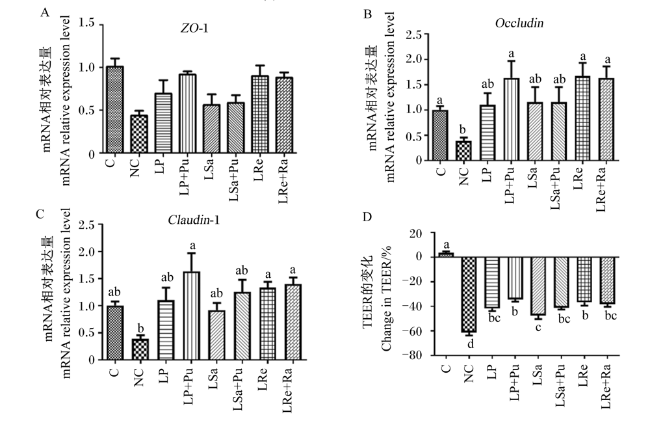
与单独的益生菌相比,在紧密连接蛋白mRNA表达和TEER值变化方面,未发现益生元对合生元组合的其他影响。但与阴性对照相比,LP+Pu与LRe+Ra组合显著提高了跨膜蛋白-1(Claudin-1)的mRNA相对表达量(P<0.05)(图2-C)。
图2 治疗试验IPEC-J2细胞中ZO-1、OccludinClaudin-1的mRNA表达和TEER值的改变

ZO-1: 闭锁小带蛋白-1 zonula occluden-1; Occludin:闭合蛋白;Claudin-1:跨膜蛋白-1;TEER: 跨上皮细胞电阻trans-epithelial electrical resistance。 下图同 the same as below。
C:空白对照;NC:阴性对照;LP:植物乳杆菌177;LP+Pu:植物乳杆菌177+普鲁兰多糖;LSa:唾液乳杆菌KLW001;LSa+Pu:唾液乳杆菌KLW001+普鲁兰多糖;LRe:罗伊氏乳杆菌KLR3004;LRe+Ra:罗伊氏乳杆菌KLR3004+棉子糖。下图同。

Fig.2 Changes in ZO-1, Occludin, Claudin-1 mRNA expression and TEER values in IPEC-J2 cells of curing test

C: blank control; NC: negative control; LP: Lactobacillus plantarum 177; LP+Pu: Lactobacillus plantarum 177+pullulan; LSa: Lactobacillus salivarius KLW001; LSa+Pu: Lactobacillus salivarius KLW001+pullulan; LRe: Lactobacillus reuteri KLR3004; LRe+Ra: Lactobacillus reuteri KLR3004+raffinose. The same as below.

2.5.2 预防试验

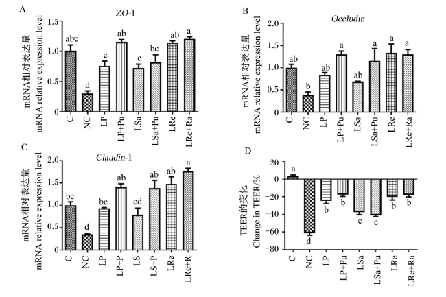
图3所示,与单独使用LP相比,LP+Pu组合显著上调紧密连接相关蛋白ZO-1 mRNA相对表达量(P<0.05),同时也有效缓解了TEER值的下降。而与LSa相比,LSa+Pu组合上调了Claudin-1 mRNA相对表达量(P<0.05)(图3-C),但对TEER值没有显著影响(P>0.05,图3-D)。
图3 预防试验IPEC-J2细胞中ZO-1、OccludinClaudin-1的mRNA表达和TEER值的改变

Fig.3 Changes in ZO-1, Occludin, Claudin-1 mRNA expression and TEER values in IPEC-J2 cells of preventing test

2.5.3 保护试验

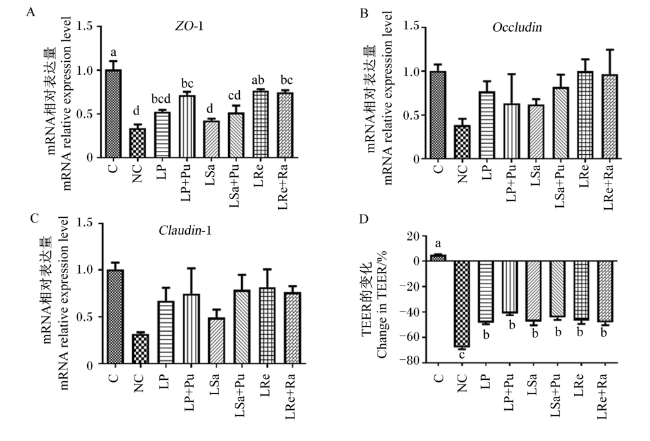
图4所示,在紧密连接蛋白mRNA表达和TEER值变化方面,并没有发现益生元对益生菌的显著作用。但与NC相比,LP+Pu、LRe和LRe+Ra组合能显著上调ZO-1的mRNA相对表达量(P<0.05)。
图4 保护试验IPEC-J2细胞中ZO-1、OccludinClaudin-1的mRNA表达和TEER值的改变

Fig.4 Changes in ZO-1, Occludin, Claudin-1 mRNA expression and TEER values in IPEC-J2 cells of protecting test

3 讨论

肠上皮屏障是由肠上皮细胞构成的物理屏障,它可以选择性地调节肠道上皮对肠道内容物和抗原的通透性。当暴露于毒素和病原体时,肠道屏障可能会受到破坏,进而加重肠道和消化系统的炎症[11]。紧密连接相关蛋白是构成肠上皮屏障完整性的主要结构蛋白,主要由3种类型的蛋白组成:ZO、Claudin和Occludin,这3类蛋白共同组成并维持肠道屏障[12]。当紧密连接相关蛋白表达下降时,意味着肠道屏障被破坏,导致肠道黏膜结构受损和肠道通透性增加[13]。常振宇等[14]研究发现,高剂量乳酸杆菌可以更好地抵御有害菌导致的肠道屏障损伤,维持肠道屏障完整性。朱涛[15]研究发现,罗伊氏乳杆菌ZJ617干预可以缓解仔猪因LPS引起的肠通透性增加、紧密连接蛋白表达的下降,恢复肠道屏障功能。本研究发现,合生元能增强物理肠道屏障完整性,缓解肠道紧密连接相关蛋白(ZO-1和Claudin-1)因LPS破坏引起的表达下调。特别是LP+P组合显示出对肠道屏障的保护作用,能够有效抵抗LPS引起的破坏,这很可能是通过上调Claudin-1的表达来实现的。
研究发现,益生菌可以抑制炎症反应,提高肠道黏膜屏障功能,抑制有害菌的侵入和定植,从而维护机体健康[16-17]。合生元是益生菌与益生元的组合,选择合适的益生元作用于益生菌,能提高益生菌的益生性能及抑菌作用,可以达到1+1>2的效果。研究发现,植物乳杆菌HW1能提高肉鸡的生长性能,显著增加抗炎因子白细胞介素-10(IL-10)的分泌增强机体免疫机能,上调紧密连接蛋白的表达,并调节肠道菌群组成[18]。杜丽欣等[19]用植物乳杆菌和罗伊氏乳杆菌组成复合菌株添加到断奶仔猪饲粮中,发现益生菌组可以降低断奶仔猪腹泻率,减少有害菌丰度。丁立人等[20]用低聚木糖、低聚壳聚糖、枯草芽胞杆菌和改性凹凸棒石组成合生元用于蛋鸡生产,发现合生元能提高鸡群抗氧化能力,增强肠道免疫力,抵御疾病侵袭。SCFA是细菌的发酵产物,包括乙酸、丙酸、丁酸等。研究证明,SCFA对肠道屏障完整性和功能有一定的改善作用[11];此外,SCFA还具有降低幼龄反刍动物腹泻率、促进肠道发育、改善因断奶引起的应激、提高生长性能和免疫力等功能[21-23]。特别是丁酸被证明是肠上皮细胞主要的能量来源,它可以通过降低编码紧密连接蛋白的DNA甲基化率、抑制蛋白激酶C(protein kinase C,PKC)和丝裂原激活的蛋白激酶(mitogen-activated protein kinase, MAPK)信号通路,进而增强紧密连接蛋白基因的表达[24-25]。Wang等[26]研究发现,在仔猪饲粮中添加丁酸钠,能降低肠黏膜中肥大细胞的百分比和促炎因子水平,这一过程可能与激活c-Jun氨基端激酶(c-Jun N-terminal kinase, JNK)信号通路有关。此外,丁酸通过抑制转录因子核因子-κB(NF-κB)信号通路的激活发挥对肠道炎症的缓解作用[27]。本研究的结果与丁酸盐对Caco-2细胞上皮屏障完整性的增强结果[28]一致。
IPEC-J2细胞系是猪小肠上皮细胞系,具有选择性吸收营养物质和矿物质的能力,并能有效阻止有毒有害物质通过上皮细胞进入体内,因此可作为研究上皮功能的细胞模型[29]。目前,IPEC-J2细胞广泛被用于研究肠屏障功能[30],因此本研究也选择采用IPEC-J2细胞模型来研究肠道屏障功能。LPS是革兰氏阴性菌细胞壁的成分。当阴性致病菌进入肠道之后,在肠上皮细胞定植,通过释放毒素或LPS与其同源受体TLR-4结合来激活炎症通路[31-32],从而导致肠黏膜损伤、肠上皮细胞通透性改变和肠道屏障破坏。因此,LPS被广泛用于“肠漏”模型的建立[33]。在本研究中,通过SCFA的发酵能力筛选合生元,并发现LP+Pu组合可以发酵产生更多的丁酸,增强肠道屏障完整性以抵抗LPS的破坏。丁酸抑制LPS的作用可能是下调TLR-4/NF-κB信号通路的表达,且LP+Pu组合预防肠道屏障破坏的效果最佳。

4 结论

本研究发现,LP+Pu组合作为一种合生元,在发酵过程中产生大量丁酸。LP+Pu组合通过促进紧密连接相关蛋白(特别是Claudin-1)的表达,恢复了LPS诱导的肠屏障损伤。这一发现有助于维护肠道健康,并为治疗和预防畜禽肠道屏障相关疾病提供了新的方法。
[1]
GILBERT J A, BLASER M J, CAPORASO J G, et al. Current understanding of the human microbiome[J]. Nature Medicine, 2018, 24(4):392-400.

DOI PMID

[2]
YOU X Y, ZHANG H Y, HAN X, et al. Intestinal mucosal barrier is regulated by intestinal tract neuro-immune interplay[J]. Frontiers in Pharmacology, 2021, 12:659716.

[3]
王海波, 占今舜, 霍俊宏, 等. 短链脂肪酸对反刍动物糖和脂代谢及肠道屏障的调控以及其在生产中的应用[J]. 动物营养学报, 2023, 35(7):4127-4137.

DOI

WANG H B, ZHAN J S, HUO J H, et al. Regulation of short chain fatty acids on glucose and lipid metabolism and intestinal barrier in ruminants and their application in production[J]. Chinese Journal of Animal Nutrition, 2023, 35(7):4127-4137. (in Chinese)

DOI

[4]
BOLOGNINI D, DEDEO D, MILLIGAN G. Metabolic and inflammatory functions of short-chain fatty acid receptors[J]. Current Opinion in Endocrine and Metabolic Research, 2021, 16:1-9.

DOI PMID

[5]
CABRÉ E, DOMÈNECH E. Impact of environmental and dietary factors on the course of inflammatory bowel disease[J]. World Journal of Gastroenterology, 2012, 18(29):3814-3822.

DOI PMID

[6]
SUZUKI T, YOSHIDA S, HARA H. Physiological concentrations of short-chain fatty acids immediately suppress colonic epithelial permeability[J]. British Journal of Nutrition, 2008, 100(2):297-305.

[7]
XU H, AGALIOTI T, ZHAO J, et al. The induction and function of the anti-inflammatory fate of TH17 cells[J]. Nature Communications, 2020, 11:3334.

[8]
WALTER J. Ecological role of Lactobacilli in the gastrointestinal tract:implications for fundamental and biomedical research[J]. Applied and Environmental Microbiology, 2008, 74(16):4985-4996.

[9]
黄明星, 陈艳, 虞龙飞, 等. 乳酸菌在家禽生产中的应用[J]. 饲料研究, 2021, 44(2):142-146.

HUANG M X, CHEN Y, YU L F, et al. Application of lactic acid bacteria in poultry production[J]. Feed Research, 2021, 44(2):142-146. (in Chinese)

[10]
孙笑非, 王文娟, 孙冬岩, 等. 益生菌在家禽饲料中应用的研究进展[J]. 饲料研究, 2021, 44(4):107-110.

SUN X F, WANG W J, SUN D Y, et al. Research progress of probiotics used in poultry feed[J]. Feed Research, 2021, 44(4):107-110. (in Chinese)

[11]
VON MARTELS J Z H, SADAGHIAN SADABAD M, BOURGONJE A R, et al. The role of gut microbiota in health and disease: in vitro modeling of host-microbe interactions at the aerobe-anaerobe interphase of the human gut[J]. Anaerobe, 2017, 44:3-12.

[12]
HAMMER A M, MORRIS N L, EARLEY Z M, et al. The first line of defense:the effects of alcohol on post-burn intestinal barrier,immune cells,and microbiome[J]. Alcohol Research, 2015, 37(2):209-222.

[13]
刘同岳, 安文东, 张文龙, 等. 博落回提取物对脂多糖刺激的肉仔鸡抗氧化功能、免疫功能及肠道紧密连接蛋白表达的影响[J]. 饲料工业, 2024, 45(1):67-72.

LIU T Y, AN W D, ZHANG W L, et al. Effects of Macleaya cordata extract on antioxidant function,immune function and mRNA expression of intestinal tight junction protein in lipopolysaccharide-challenged broilers[J]. Feed Industry, 2024, 45(1):67-72. (in Chinese)

[14]
常振宇, 董海龙, 叶炳涛, 等. 乳酸杆菌对牦牛小肠黏膜屏障的保护作用及机制[J]. 中国农业大学学报, 2024, 29(3):155-163.

CHANG Z Y, DONG H L, YE B T, et al. Protective effect and mechanism of Lactobacillus on the mucosal barrier of the small intestine in yaks[J]. Journal of China Agricultural University, 2024, 29(3):155-163. (in Chinese)

[15]
朱涛. 罗伊氏乳杆菌ZJ617对LPS致炎仔猪肠道和肝脏的保护效应及机制研究[D]. 硕士学位论文. 杭州: 浙江大学, 2021.

ZHU T.Protective effect and mechanism of Lactobacillus reuteri ZJ 617 on intestine and liver of piglets induced by lipopolysaccharides[D]. Master's Thesis. Hangzhou: Zhejiang University, 2021. (in Chinese)

[16]
KANG H S, KOO J S, LEE K M, et al. Two-year delay in ulcerative colitis diagnosis is associated with anti-tumor necrosis factor alpha use[J]. World Journal of Gastroenterology, 2019, 25(8):989-1001.

DOI PMID

[17]
WONG W Y, CHAN B D, LEUNG T W, et al. Beneficial and anti-inflammatory effects of formulated prebiotics, probiotics, and synbiotics in normal and acute colitis mice[J]. Journal of Functional Foods, 2022, 88:104871.

[18]
CHEN P, LV H M, LIU W Y, et al. Effects of Lactobacillus plantarum HW1 on growth performance,intestinal immune response,barrier function,and cecal microflora of broilers with necrotic enteritis[J]. Animals, 2023, 13(24):3810.

[19]
杜丽欣, 张峰瑞, 邹雪, 等. 复合乳酸菌制剂对预防断奶仔猪细菌性腹泻及生长性能的影响[J]. 天津农学院学报, 2023, 30(4):46-52.

DOI

DU L X, ZHANG F R, ZOU X, et al. Effect of compound lactic acid bacteria on prevention of bacterial diarrhea and growth performance of weaned piglets[J]. Journal of Tianjin Agricultural University, 2023, 30(4):46-52. (in Chinese)

[20]
丁立人, 杨一丁, 邵云, 等. 合生元对蛋鸡生产性能、抗氧化能力、肠道功能和蛋品质的影响[J]. 南京农业大学学报, 2023, 46(6):1161-1168.

DING L R, YANG Y D, SHAO Y, et al. Effect of symbiotic on laying performance,antioxidant capacity,intestinal function,and egg quality of laying hens[J]. Journal of Nanjing Agricultural University, 2023, 46(6):1161-1168.

[21]
吴东霖, 徐萍, 邵凯, 等. 丁酸在幼龄反刍动物中的作用机理及其产品应用的研究进展[J]. 动物营养学报, 2020, 32(3):1034-1046.

DOI

WU D L, XU P, SHAO K, et al. Mechanism of butyric acid and application of its products in young ruminants[J]. Chinese Journal of Animal Nutrition, 2020, 32(3):1034-1046. (in Chinese)

[22]
王可鑫, 左丽君, 陈想, 等. 包膜丁酸钠对脂多糖刺激下断奶羔羊血清炎性细胞因子、肠道通透性及肠道组织形态的影响[J]. 动物营养学报, 2021, 33(2):954-964.

DOI

WANG K X, ZUO L J, CHEN X, et al. Effects of coated sodium butyrate on serum inflammatory cytokines, intestinal permeability and intestinal morphology of weaned lambs challenged with lipopolysaccharide[J]. Chinese Journal of Animal Nutrition, 2021, 33(2):954-964. (in Chinese)

[23]
GÓRKA P, KOWALSKI Z M, ZABIELSKI R, et al. Invited review: use of butyrate to promote gastrointestinal tract development in calves[J]. Journal of Dairy Science, 2018, 101(6):4785-4800.

DOI PMID

[24]
YAN H, AJUWON K M. Butyrate modifies intestinal barrier function in IPEC-J2 cells through a selective upregulation of tight junction proteins and activation of the Akt signaling pathway[J]. PLoS One, 2017, 12(6):e0179586.

[25]
ZHANG K, MENG M J, GAO L P, et al. Sodium butyrate improves high-concentrate-diet-induced impairment of ruminal epithelium barrier function in goats[J]. Journal of Agricultural and Food Chemistry, 2018, 66(33):8729-8736.

DOI PMID

[26]
WANG C C, WU H, LIN F H, et al. Sodium butyrate enhances intestinal integrity,inhibits mast cell activation, inflammatory mediator production and JNK signaling pathway in weaned pigs[J]. Innate Immunity, 2018, 24(1):40-46.

[27]
WANG K X, CHEN X, ZUO L J, et al. Dietary sodium butyrate protects lipopolysaccharide-induced inflammatory response in lambs through inhibiting TLR4/NF-κB signalling pathway[J]. Italian Journal of Animal Science, 2021, 20(1):1398-1409.

[28]
VALENZANO M C, DIGUILIO K, MERCADO J, et al. Remodeling of tight junctions and enhancement of barrier integrity of the CACO-2 intestinal epithelial cell layer by micronutrients[J]. PLoS One, 2015, 10(7):e0133926.

[29]
XIAO Z P, LIU L J, TAO W J, et al. Clostridium tyrobutyricum protect intestinal barrier function from LPS-induced apoptosis via P38/JNK signaling pathway in IPEC-J2 cells[J]. Cellular Physiology and Biochemistry, 2018, 46(5):1779-1792.

[30]
BROSNAHAN A J, BROWN D R. Porcine IPEC-J 2 intestinal epithelial cells in microbiological investigations[J]. Veterinary Microbiology, 2012, 156(3/4):229-237.

[31]
SCHLEGEL N, LEWEKE R, MEIR M, et al. Role of NF-κB activation in LPS-induced endothelial barrier breakdown[J]. Histochemistry and Cell Biology, 2012, 138(4):627-641.

DOI PMID

[32]
GUO S H, NIGHOT M, AL-SADI R, et al. Lipopolysaccharide regulation of intestinal tight junction permeability is mediated by TLR4 signal transduction pathway activation of FAK and MyD88[J]. Journal of Immunology, 2015, 195(10):4999-5010.

DOI PMID

[33]
李保良, 庞胜美, 吕林芬, 等. 产肠毒素大肠杆菌诱导肠上皮细胞凋亡的研究进展[J]. 微生物学报, 2023, 63(6):2340-2349.

LI B L, PANG S M, LV L F, et al. Research progress in enterotoxigenic Escherichia coli-induced apoptosis of intestinal epithelial cells[J]. Acta Microbiologica Sinica, 2023, 63(6):2340-2349. (in Chinese)

Outlines

/