REVIEW

Research Progress on Protective Effects of Short Chain Fatty Acids on Intestinal Barrier

  • LI Xinke ,
  • YANG Xue ,
  • ZHANG Xuan ,
  • ZHENG Nan , *
Expand
  • Laboratory of Quality and Safety Risk Assessment for Dairy Products of Ministry of Agriculture and Rural Affairs (Beijing), Key Laboratory of Quality and Safety Control for Milk and Dairy Products of Ministry of Agriculture and Rural Affairs, Institute of Animal Sciences, Chinese Academy of Agricultural Sciences, Beijing 100193, China
*professor, E-mail:

Received date: 2024-02-20

  Online published: 2024-08-12

Abstract

The intestinal barrier is a crucial physiological defense mechanism that regulates and maintains homeostasis within the organism’s internal environment. Short chain fatty acids, emanating from the metabolism of intestinal microorganisms, not only serve as the primary energy source for intestinal epithelial cells but also play a critical role in alleviating tissue inflammation and sustaining the functionality of the intestinal barrier. This article aims to elucidate the protective effects of short chain fatty acids on the four major barriers of the intestine (mechanical, microbial, chemical, and immune barriers), offering a new research perspective for the treatment of diseases related to intestinal injury.

Cite this article

LI Xinke , YANG Xue , ZHANG Xuan , ZHENG Nan . Research Progress on Protective Effects of Short Chain Fatty Acids on Intestinal Barrier[J]. Chinese Journal of Animal Nutrition, 2024 , 36(8) : 4861 -4871 . DOI: 10.12418/CJAN2024.416

肠道主要负责吸收功能和屏障功能,是动物获得充足营养和抵抗疾病的关键。作为一个关键的营养传感器,肠道可以感知和调节氨基酸、脂肪酸和葡萄糖的吸收[1-2]。除了调节营养吸收外,肠道还发挥屏障保护作用,以防止病原体入侵并维持肠道的完整性。肠道屏障系统作为维持肠道稳态的关键组成部分,主要通过以紧密连接(tight junction,TJ)蛋白和肠上皮细胞(intestinal epithelial cells,IECs)为主的机械屏障、以免疫细胞和细胞因子为主的免疫屏障、以肠道菌群为主的微生物屏障和以黏蛋白(mucoprotein,MUC)和抗菌肽为主的化学屏障等四大屏障发挥作用。肠道屏障功能受损可能严重危害机体。首先,肠道屏障的破坏可能导致有害微生物的入侵,从而引发疾病,如炎症性肠病等[3-4]。其次,肠道屏障的破坏可能使毒素、抗生素和代谢产物等物质进入血液循环,影响器官功能,甚至影响中枢神经系统[5]。此外,肠道屏障的破坏还可能引发免疫系统异常,导致过敏或自身免疫性疾病的发生[6]。因此,维持肠道屏障的完整性对机体健康至关重要。近年来,利用微生物代谢物,如短链脂肪酸(short chain fatty acids,SCFAs),对恢复健康肠道屏障功能的研究逐渐增多[7-8]
SCFAs作为肠道微生物代谢的关键产物,不仅为肠道菌群和宿主IECs提供能量,同时有助于维持肠道pH平衡,抑制有害微生物的生长。此外,SCFAs在调节宿主肠道免疫系统方面发挥着重要作用,具有免疫调节的特性,有助于减轻炎症反应。与此同时,SCFAs还能加强肠道屏障功能,有效防止有害物质的渗透。这些生物学功能共同促使SCFAs在维持肠道微生态平衡、支持肠道健康以及对机体免疫系统的调控中发挥关键作用[9]

1 SCFAs概述

1.1 SCFAs的种类及作用

SCFAs是含有1~6个碳原子的小分子有机羧酸,在肠道中是难消化多糖(如抗性淀粉、菊粉、纤维素和果胶)厌氧发酵的主要产物。根据国际纯粹与应用化学联合会的标准,可以根据它们所含的碳原子数进行命名,即甲酸、乙酸、丙酸、丁酸、戊酸和己酸[10]。其中,乙酸、丙酸和丁酸是人类肠道中最常见的SCFAs,其比例约为3∶1∶1,而甲酸、戊酸和己酸的含量相对较少。
SCFAs在多种细胞类型中发挥作用,调控关键的生物过程,如宿主代谢、肠道功能和免疫应答等。甲酸作为最短碳链的SCFAs,通过参与葡萄糖代谢、调节酵母酶活性和抗氧化作用,调控氧化还原平衡。乙酸作为主要的SCFAs,可参与细胞代谢途径、维护肠道完整性、调节脂质和碳水化合物代谢等[11]。丙酸则被发现具有抗炎和抗菌特性,有助于保护肠道免受病原体的侵害。此外,丙酸还能减少肝脏胆固醇合成,改善脂质代谢[12]。丁酸作为结肠细胞的能量来源,可以调节多种肠道功能,包括免疫调节、肠道发育、细胞分化以及基因表达等,从而减少氧化应激和炎症[13]。尽管对于戊酸和己酸的研究关注相对较少,但已有研究表明它们具有积极的健康作用[14],例如抑制肝癌细胞的生长和分化[15]、防止辐射损伤[16]等。此外,SCFAs还在预防和治疗多种疾病等方面发挥着作用,如肥胖[17]、心血管疾病[18]、糖尿病[19]、神经性疾病[20]和牙周病[21]等。这些研究结果强调了SCFAs在调控肠道屏障功能和机体健康中的重要性。

1.2 肠道中SCFAs及其微生物来源

SCFAs是肠道中非消化性碳水化合物厌氧发酵的主要代谢产物,代表了从饮食通过微生物群传递到宿主的主要碳代谢途径。肠道中最主要的SCFAs包括乙酸、丙酸和丁酸,它们的总和大约占所有SCFAs的95%[22]。在膳食纤维向SCFAs的转化过程中,受到肠道特定微生物群的影响。
研究指出,乙酸作为肠道中含量最高的SCFAs,由多种结肠微生物,如嗜黏蛋白阿克曼菌、拟杆菌和双歧杆菌等,通过伍德-隆达尔代谢途径和丙酮酸氧化脱羧形成乙酰辅酶A途径产生乙酸[23-24]。此外,肠道菌群通过拟杆菌门介导的琥珀酸途径和厚壁菌门介导的乳酸途径,参与调控丙酸的产生[25]。丁酸作为第3种主要的SCFAs,由2分子乙酰辅酶A缩合并随后还原为丁酰辅酶A形成,丁酰辅酶A可通过磷酸转丁酰酶和丁酸激酶通过经典途径由特定微生物转化为丁酸。此外,丁酸也可以通过外源性乙酸盐的替代途径生产[26]。然而,很少有报道关注具有甲酸、戊酸以及己酸代谢特性的菌种,目前只有巨型球菌属被报道能同时产生戊酸和己酸。在未来的研究中,预期会发现更多能够产生甲酸、戊酸和己酸的细菌,从而使甲酸、戊酸和己酸对健康的影响更加清晰。综合研究数据表明,产SCFAs的菌株主要集中在厚壁菌门,该菌门展现出最高的物种多样性,其他参与SCFAs产生的菌株包括放线菌门、拟杆菌门、变形菌门和疣微菌门;研究重点则主要集中在产生乙酸、丙酸和丁酸的菌属上[25,27-35]

1.3 影响SCFAs产生的因素

多种因素如口腔微生物群对肠道微生物群的影响[36]、口服益生菌[37]、粪便微生物群移植[38]以及肠道微生物相互作用等[39]都可能扰乱肠道微生物群,影响产生SCFAs的细菌丰度和多样性,从而影响SCFAs生成。此外,宿主的生理状态也是一个重要因素,肠道环境[40]、微生物-宿主相互作用[41]甚至社会压力[42]等都可能对肠道微生物组成及其代谢产物SCFAs的生成造成影响。此外,食物组成也是影响肠道微生物群产SCFAs的关键因素。短期内饮食带来的宏量营养物质变化可能对肠道微生物群产生可重复性影响,从而影响SCFAs的生成[43]。抗生素的使用对肠道微生物群的影响同样不容忽视[44]。所有这些因素,从外部介入到宿主生理状态,再到食物因素和药物应用,都会影响肠道微生物群的丰度和多样性,最终影响肠道产生SCFAs菌群的代谢特征,从而导致内源性SCFAs水平的波动。

2 SCFAs和肠道屏障
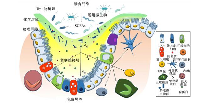
机体通过肠道屏障系统来防御外来病原体入侵,包括机械屏障、化学屏障、免疫屏障和微生物屏障(图1)。肠道屏障完整性的破坏可能导致机体代谢功能障碍,损害肠道健康,对动物的生产性能带来负面影响[45]。此外,肠道屏障破损可能导致机体对病原体的无效应答,加剧疾病风险。因此,维持肠道屏障完整性对维护生物体整体健康至关重要。
图1 肠道屏障的组成和解剖结构

SCFAs:短链脂肪酸 short chain fatty acids;IECs:肠上皮细胞 intestinal epithelial cells。

Fig.1 Composition and anatomical structure of gut barrier[46]

2.1 SCFAs与肠道机械屏障

2.1.1 肠道机械屏障

肠道机械屏障是肠道屏障的重要组成部分,由黏液、IECs以及IECs之间的TJ蛋白组成[47]。IECs如杯状细胞、潘氏细胞和扁平细胞,是肠道机械屏障的支柱,具有快速增殖和再生,分泌黏液、抗菌肽以及免疫球蛋白A(immunoglobulin A,IgA)的能力,是维持肠道屏障功能的结构基础[47]。黏液是一种由杯状细胞分泌的高度糖基化的MUC,是肠道物理屏障的重要组成部分。它覆盖在IECs表面,起到物理隔离的作用,能有效阻止肠道微生物群直接作用于肠黏膜[48]。此外,肠道物理屏障的连续性还取决于IECs之间TJ蛋白的存在。TJ蛋白由跨膜蛋白,如咬合蛋白(Occludin)、闭合蛋白(Claudins)、钙黏蛋白(Cadherin)以及连接黏附分子和细胞质支架蛋白,如闭合小环蛋白(zonula occluden,ZO)组成。这些蛋白在维持IECs结构完整性和生物学功能上发挥着重要作用,不仅能通过调节细胞间隙阻止肠道腔内的细菌和抗原等物质进入肠固有层,还能促进营养物质的运输[49]

2.1.2 SCFAs对机械屏障的调节作用

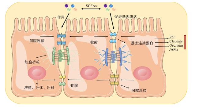
SCFAs能促进IECs增殖。相关研究发现,在无菌小鼠和经抗生素处理无特定病原体小鼠中,IECs的增殖活性和细胞更新率降低。通过补充SCFAs不仅能逆转这一现象,还能调节参与能量代谢(例如脂质代谢)的上皮基因表达,促进体外肠道类器官的发育,从而增强其支持IECs增殖的作用[50]。研究发现,丙酸盐能通过增强细胞扩散和极化,提高细胞速度和持久性,从而促进小鼠IECs和人Caco-2细胞的增殖和迁移。而丙酸盐的这种功能依赖于对Ⅰ类组蛋白去乙酰化酶和G蛋白偶联受体(G protein-coupled receptor,GPR)43的抑制以及信号转导子和转录激活子3(signal transducer and activator of transcription 3,STAT3)的激活[51]。肠道干细胞的增殖和分化是肠道更新和再生的基础。Xie等[52]研究发现,瘤胃球菌科产生的SCFAs可以通过激活Wnt/β-连环蛋白(β-catenin)信号通路介导α-亚麻酸,促进肠道干细胞增殖。
SCFAs可以提高TJ蛋白的表达。研究表明,4 mmol/L丁酸盐不仅能促进猪IECs中TJ蛋白OccludinZO-1的表达[53],还能促进小鼠IECs中TJ蛋白Claudin-1的表达,增加肠绒毛高度并降低小鼠肠道通透性[54]。此外还有研究报道,丙酸盐通过提高肠道TJ蛋白ZO-1、OccludinCadherin的表达,从而促进肠道功能[55]。Voltolini等[56]研究发现,SCFAs通过激活腺苷酸激活蛋白激酶(AMP-activated protein kinase,AMPK)信号通路,上调IECs中TJ蛋白ZO-1的表达,增强IECs的跨膜抵抗,从而保持肠屏障的完整性。此外,Wang等[57]研究发现,丁酸钠能改善断奶后仔猪的肠道形态,增强IECs的跨膜抵抗,降低异硫氰酸荧光素-葡聚糖4(fluorescein isothiocyanate-dextran 4,FD4)表达,改善断奶造成的肠道应激损伤,从而增强断奶仔猪的肠道屏障功能。研究指出,通过补充丙酸盐可以促进猪肠道绒毛高度和绒毛高度/隐窝深度,并选择性增强了空肠黏膜中Claudin-4的mRNA表达水平以及Claudin-1、Claudin-4和Occludin的蛋白表达水平[58]
综上所述,SCFAs主要通过以下2个机制实现对肠道机械屏障的保护作用:1)通过激活IECs表面受体如GPR,参与调节细胞增殖,从而促使肠道上皮的更新和修复;2)激活细胞信号通路如AMPK信号通路,提高肠道TJ蛋白如ClaudinOccludinZO等相关基因的表达,调节肠道通透性,进而改善肠道机械屏障功能(图2)。
图2 SCFAs对机械屏障的主要影响

SCFAs:短链脂肪酸 short chain fatty acids;ZO:闭合小环蛋白 zonula occluden;Claudins:闭合蛋白;Occludin:咬合蛋白;JAMs:连接黏附分子 unction adhesion molecules。

Fig.2 Main effects of SCFAs on mechanism barrier[59]

2.2 SCFAs与肠道微生物屏障

2.2.1 肠道微生物屏障

肠道微生物屏障是指定居在肠道表面的微生物群之间形成的一种相互依赖并与其他微生物互作的稳定微生物系统。由于共生微生物群在肠道中建立了平衡的生态位,导致致病微生物群必须竞争结合位点和营养才能在肠道中定植。因此,在大多数情况下,肠道微生态平衡被破坏是导致传染性肠病以及各种代谢疾病的主要原因。

2.2.2 SCFAs对微生物屏障的调节作用

SCFAs可通过多种机制抑制肠腔内细菌的生长和发育。首先,SCFAs进入肠道后,部分解离产生氢离子(H+),导致肠道环境中pH下降,从而在一定程度上抑制了有害微生物的定植[60]。相关试验也表明,通过补充SCFAs能显著降低肉鸡和仔猪肠道中鼠伤寒沙门菌和肠炎沙门菌的相对丰度[61-62]。其次,部分未解离的SCFAs通过自由扩散进入细菌内部,由于胞内pH为中性或碱性,高于SCFAs的解离常数,导致SCFAs在胞内解离生成酸根离子和 H + [ 63 ]。解离产生的H+在细菌内积累,导致其内部pH下降。为了维持胞内pH平衡,细菌通过主动运输消耗大量ATP释放胞内H+,从而与其正常的生长代谢产生能量竞争[64]。此外,肠道中的多种有益菌,如双歧杆菌、嗜黏蛋白阿克曼氏菌和普拉梭菌等,通过利用SCFAs,特别是从其他微生物发酵产生的SCFAs,来获得所需的能量和营养物质,从而在肠道内繁殖和维持其丰度[65]。这种共生关系对于维持肠道微生物群的稳定和促进肠道健康至关重要。SCFAs还可以影响肠道内微生物的组成和丰度。Lu等[66]研究发现,在高脂饮食诱导的肥胖小鼠中,添加乙酸盐或丙酸盐能显著降低肠道中拟杆菌门和厚壁菌门的比例。有研究指出,在补充了SCFAs的猪肠道中观察到乳酸杆菌属和双歧杆菌属数量增加的现象[67]。此外,补充SCFAs可改善慢性脑灌注不足引起的肠道微生物群失调,并通过调节SCFAs的微生物代谢,增加乙酸含量、提高线粒体电子传递链活性和促进氧化磷酸化,从而有效预防结肠炎症和线粒体能量代谢的紊乱[68]
综上所述,SCFAs主要通过释放H+降低肠道pH以及能量竞争的形式抑制病原微生物的生长繁殖、能量供应以及调控肠道微生物组成和丰度等方式,改善肠道微生物屏障功能,从而有助于维持肠道稳态。

2.3 SCFAs与肠道化学屏障

2.3.1 肠道化学屏障

肠道化学屏障是指由肠黏膜上皮细胞分泌的抗菌相关蛋白,如MUC和抗菌肽所构成的黏液层。该黏液层在肠道表面覆盖且功能复杂,其主要功能是防止肠道微生物与宿主IECs直接接触。其中,杯状细胞分泌的MUC是黏液层中最主要的高分子糖蛋白,它能有效隔离并控制肠道微生物的入侵和定植等,在肠道化学屏障中起重要作用。抗菌肽由IECs产生,包括α-防御素、β-防御素、C型凝集素和再生胰岛衍生蛋白3γ(REG3γ)等,其对细菌表现出广谱且高效的杀菌活性,能够有效清除绝大多数微生物,阻止抗原进入宿主细胞。作为肠道机械屏障的良好补充,肠道化学屏障在维持肠道微生态平衡中发挥关键作用。

2.3.2 SCFAs对化学屏障的调节作用

MUC相关基因的表达对于维持肠道化学屏障的稳定起着至关重要的作用[24]。先前的研究表明,丁酸盐对于调节肠道杯状细胞中MUC基因的表达具有特异性。具体而言,它能够通过抑制组蛋白去乙酰化酶(histone deacetylase,HDAC)途径来实现对MUC3基因表达的调控[69]。此外,丁酸盐能够提高人结肠细胞LS174T中目标基因MUC3、MUC4和MUC12的表达水平,随之而来的MUC含量提高增强了益生菌的黏附性,同时降低了大肠杆菌的贴壁能力,从而增强了黏膜层对抗肠道病原体的防御能力[70]。此外,Diao等[8]对断奶仔猪进行了SCFAs的干预研究,发现SCFAs通过丝裂原活化蛋白激酶(mitogen-activated protein kinase,MAPK)信号通路显著提高肠黏膜表面MUC1和MUC2的表达水平。同时,它还抑制了促凋亡基因B细胞淋巴瘤相关X蛋白(B-cell lymphoma-associated X protein,Bax)和半胱天冬蛋白酶-3(cysteinyl aspartate specific proteinase-3,Caspase-3)的表达水平,降低了细胞凋亡指数,从而增强肠道化学屏障功能。
相关研究报道,抗菌肽具有抗菌和免疫调节特性,可保护肠道免受感染。有证据表明,Zhao等[71]发现在GPR43基因敲除鼠中,REG3γ和β-防御素的表达受到抑制,而通过补充SCFAs则能够促进野生型小鼠体内REG3γ和β-防御素的生成。在机制上,这是由于GPR43通过激活哺乳动物雷帕霉素靶蛋白(mammalian target of rapamycin,mTOR)和STAT3途径,介导SCFAs对IECs抗菌肽的表达进行调控。此外研究还发现,SCFAs特别是丁酸盐,可以诱导离体小肠隐窝潘氏细胞分泌α-防御素,有效杀灭有害微生物,从而促进肠道稳态[72]。这些证据表明SCFAs可通过激活相关信号通路,提高抗菌肽的表达,从而增强肠道对感染和炎症性疾病的保护作用。
综上所述,SCFAs在肠道化学屏障的调控中主要通过以下途径发挥作用:HDAC抑制以激活MAPK信号通路等方式促进肠道MUC的表达,激活GPR43受体触发细胞信号转导途径影响抗菌肽的表达,降低肠道细胞的凋亡指数(图3)。
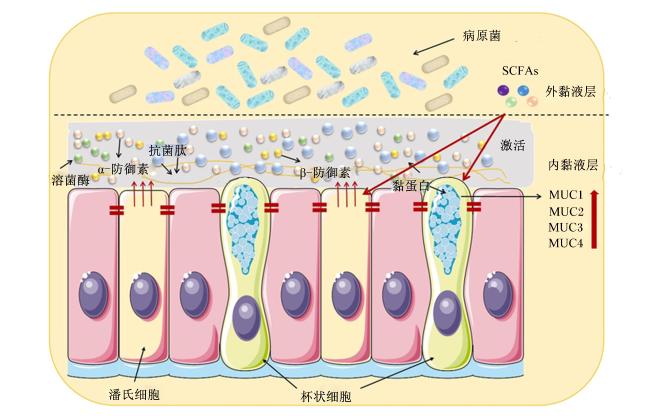
图3 短链脂肪酸对化学屏障的主要影响

SCFAs:短链脂肪酸 short chain fatty acids;MUC:黏蛋白 mucin。

Fig.3 Main effects of SCFAs on chemical barrier[59]

2.4 SCFAs与肠道免疫屏障

2.4.1 肠道免疫屏障

肠道免疫屏障是肠道组织不可缺少的组成部分,其主要由肠道黏膜层的免疫细胞和免疫细胞因子构成,通过参与先天性免疫和特异性免疫来维持局部和系统内的稳态。肠黏膜负责识别各类食物和微生物抗原,这使得肠道成为免疫细胞最为丰富的部位,主要包括T细胞、B细胞、巨噬细胞和自然杀伤细胞等[73]。按T细胞表面分化抗原的不同,可分为CD4+T细胞(如Th细胞、Treg细胞)和CD8+T细胞(如Tc细胞)两大亚群。在肠道环境中,CD4+T细胞和CD8+T细胞的协同作用是维持免疫平衡和有效对抗病原微生物的重要组成部分[74]。B细胞则可以通过分泌IgA和调控免疫应答等,参与维护肠道免疫屏障的完整性[75]。在肠道稳态条件下,肠道驻留型巨噬细胞扮演IECs的“守门人”,具有促进微生物降解、清除凋亡细胞、推动组织修复以及促进肠道蠕动等功能[76]。而自然杀伤细胞作为最早被鉴定的固有淋巴细胞亚型,可对病毒感染或转化的细胞做出反应,具备细胞杀伤和促炎细胞因子产生等多种效应子功能[77]。此外,这些免疫细胞产生的细胞因子,如肿瘤坏死因子-α(tumor necrosis factor-α,TNF-α)、γ-干扰素(interferon-γ,IFN-γ)和白细胞介素(interleukin,IL)-10等,可通过对应机制调控肠道通透性,在炎症性疾病中引起的屏障破坏中发挥关键作用[78]

2.4.2 SCFAs对免疫屏障的调节作用

SCFAs对T细胞和B细胞的分化及其介导的特异性免疫具有调节作用。研究表明,肠道SCFAs浓度与Treg细胞数量呈正相关,且丁酸在体内和体外均能诱导Treg细胞分化,并改善结肠炎症的发展。这可能是由于丁酸处理增强了叉头框蛋白P3(forkhead box protein P3,Foxp3)基因启动子和保守非编码序列区域的组蛋白H3乙酰化,从而调节Treg细胞分化[79]。此外,Sun等[80]研究发现,SCFAs可通过GPR43介导微生物抗原特异性Th1细胞产生IL-10,缓解肠道炎症,从而有助于维持肠道稳态。在机制上,这是由于SCFAs通过激活Th1细胞STAT3和mTOR信号通路,进而上调B淋巴细胞诱导成熟蛋白-1(B lymphocyte induced maturation protein-1,Blimp-1)转录因子来诱导IL-10的产生。另外,Yang等[81]研究发现,SCFAs可通过GPR41和抑制HDAC促进CD4+T细胞产生IL-22,从而维持肠道稳态。除此之外,通过补充SCFAs可以增加小鼠肠道中B细胞数量,并通过促进代谢调节(如线粒体能量代谢、糖酵解等过程)和相关基因表达以促进B细胞的活化和分化[82]。IgA是维持肠道稳态和防止炎症的重要因素。Wu等[83]研究发现,乙酸能够通过GPR43介导,促进肠道B细胞的IgA类别转换和产生,其中乙醛脱氢酶1家族成员A2(aldehyde dehydrogenase 1 family member A2,Aldh1A2)的表达调控了维生素A向视黄酸的转化,而阻断视黄酸信号则会抑制乙酸诱导的IgA产生。
SCFAs对先天性免疫细胞,如巨噬细胞、中性粒细胞、嗜酸性粒细胞和自然杀伤细胞的调节,对维持肠道内的免疫平衡至关重要。研究发现,SCFAs可调节巨噬细胞的分化和代谢,提高其抗菌活性和对肠道病原体的抵抗能力[84]。此外,SCFAs还能通过抑制HDAC减少单核巨噬细胞产生促炎性肿瘤坏死因子,抑制核转录因子-κB(nuclear factor-kappa B,NF-κB)的表达,减少促炎细胞因子如IL-2、IL-6和IL-8的分泌[85]。中性粒细胞被认为是骨髓和外周血中最丰富的先天免疫细胞。研究表明,丁酸盐可以显著抑制结肠炎患者肠道中性粒细胞分泌的促炎细胞因子和趋化因子的产生,从而减轻肠道炎症[86]。Theiler等[87]研究发现,经SCFAs处理的嗜酸性粒细胞表现出细胞大小和数量减少、双叶核丢失、线粒体去极化和效应Caspase激活,从而调节细胞内通路诱导细胞凋亡。此外,丁酸盐可通过介导哺乳动物雷帕霉素靶蛋白复合物1(mechanistic target of rapamycin complex 1,mTORC1)、c-Myc和细胞代谢,降低IL-12/IL-15联合激活后的自然杀伤细胞中细胞表面受体的表达,抑制TNF-α、IFN-γ、IL-22和可溶性Fas配体等促炎细胞因子的产生,进而维持免疫稳态[88]
综上所述,SCFAs对免疫屏障的重要意义主要是基于以下3点实现的:1)促进肠道免疫细胞分化,调节免疫细胞活性;2)激活STAT3和mTOR信号通路,促进免疫调节因子的产生,从而调节免疫细胞的活性和功能;3)影响免疫细胞的炎症介质的产生和释放,缓解肠道炎症。

3 小结

肠道屏障作为机体重要的防御系统,可以阻止病原体进入机体,维持机体免疫平衡。当肠道屏障功能受损时,可能会导致以下几种情况发生:1)肠道内有害微生物渗透,引发感染和炎症:2)毒素、抗生素和代谢产物等物质进入血液循环,对全身器官产生负面影响:3)引发免疫系统异常,导致过敏反应或自身免疫性疾病。作为肠道微生物最重要的代谢产物之一,SCFAs已被证明通过肠道屏障和免疫应答调节宿主的生理和健康。例如,通过调节肠道微生物群结构、MUC和抗菌物质的表达,促炎或抗炎细胞因子的分泌,IECs和免疫细胞的活性等,维持机体肠道健康。目前,研究SCFAs对肠道屏障的影响面临一些具有挑战性的任务,其中包括多因素交互影响、剂量效应以及机制解析等,综合解决这些问题将有助于更全面、深入地理解SCFAs对肠道屏障的影响,为未来的治疗策略和健康干预提供更有力的科学支持。
[1]
EVERARD A, GEURTS L, CAESAR R, et al. Intestinal epithelial MyD88 is a sensor switching host metabolism towards obesity according to nutritional status[J]. Nature Communications, 2014,5:5648.

[2]
DYER J, VAYRO S, SHIRAZI-BEECHEY S P. Mechanism of glucose sensing in the small intestine[J]. Biochemical Society Transactions, 2003, 31(Pt 6):1140-1142.

DOI PMID

[3]
CHEN Y Y, CUI W W, LI X, et al. Interaction between commensal bacteria, immune response and the intestinal barrier in inflammatory bowel disease[J]. Frontiers in Immunology, 2021,12:761981.

[4]
盛天骄, 闫思蒙, 兰威儒, 等. 肠道微生物对炎症性肠病影响研究进展[J]. 临床军医杂志, 2023, 51(9):904-907.

SHENG T J, YAN S M, LAN W R, et al. Research progress on the influence of intestinal microbes on inflammatory bowel disease[J]. Clinical Journal of Medical Officers, 2023, 51(9):904-907. (in Chinese)

[5]
罗文辉, 何兴祥. 肠道屏障功能在创伤性脑损伤中的研究进展[J]. 胃肠病学和肝病学杂志, 2021, 30(1):51-54.

LUO W H, HE X X. Research progress of intestinal barrier dysfunction in traumatic brain injury[J]. Chinese Journal of Gastroenterology and Hepatology, 2021, 30(1):51-54. (in Chinese)

[6]
GOPALAKRISHNAN V, HELMINK B A, SPENCER C N, et al. The influence of the gut microbiome on cancer,immunity,and cancer immunotherapy[J]. Cancer Cell, 2018, 33(4):570-580.

[7]
KELLY C J, ZHENG L, CAMPBELL E L, et al. Crosstalk between microbiota-derived short-chain fatty acids and intestinal epithelial HIF augments tissue barrier function[J]. Cell Host & Microbe, 2015, 17(5):662-671.

[8]
DIAO H, JIAO A R, YU B, et al. Gastric infusion of short-chain fatty acids can improve intestinal barrier function in weaned piglets[J]. Genes & Nutrition, 2019,14:4.

[9]
SADLER R, CRAMER J V, HEINDL S, et al. Short-chain fatty acids improve poststroke recovery via immunological mechanisms[J]. Journal of Neuroscience, 2020, 40(5):1162-1173.

DOI PMID

[10]
LAYDEN B T, ANGUEIRA A R, BRODSKY M, et al. Short chain fatty acids and their receptors:new metabolic targets[J]. Translational Research, 2013, 161(3):131-140.

[11]
MERLI G, BECCI A, AMATO A, et al. Acetic acid bioproduction:the technological innovation change[J]. The Science of Total Environment, 2021,798:149292.

[12]
MARKOWIAK-KOPEĆ P, ŚLIŻEWSKA K. The effect of probiotics on the production of short-chain fatty acids by human intestinal microbiome[J]. Nutrients, 2020, 12(4):1107.

[13]
BEDFORD A, GONG J. Implications of butyrate and its derivatives for gut health and animal production[J]. Animal Nutrition, 2018, 4(2):151-159.

DOI PMID

[14]
TAP J, FURET J P, BENSAADA M, et al. Gut microbiota richness promotes its stability upon increased dietary fibre intake in healthy adults[J]. Environmental Microbiology, 2015, 17(12):4954-4964.

DOI PMID

[15]
HAN R, NUSBAUM O, CHEN X Y, et al. Valeric acid suppresses liver cancer development by acting as a novel HDAC inhibitor[J]. Molecular Therapy Oncolytics, 2020,19:8-18.

[16]
LI Y, DONG J L, XIAO H W, et al. Gut commensal derived-valeric acid protects against radiation injuries[J]. Gut Microbes, 2020, 11(4):789-806.

DOI PMID

[17]
JIAO A R, YU B, HE J, et al. Sodium acetate,propionate,and butyrate reduce fat accumulation in mice via modulating appetite and relevant genes[J]. Nutrition, 2021,87/88:111198.

[18]
HAGHIKIA A, ZIMMERMANN F, SCHUMANN P, et al. Propionate attenuates atherosclerosis by immune-dependent regulation of intestinal cholesterol metabolism[J]. European Heart Journal, 2022, 43(6):518-533.

[19]
党崇舒, 赵可新, 李华文, 等. 肠道短链脂肪酸与2型糖尿病相关性研究进展[J]. 中国微生态学杂志, 2021, 33(12):1471-1475.

DANG C S, ZHAO K X, LI H W, et al. Current studies on relationship between short-chain fatty acids and type 2 diabetes mellitus[J]. Chinese Journal of Microecology, 2021, 33(12):1471-1475. (in Chinese)

[20]
LIU J M, LI H J, GONG T Y, et al. Anti-neuroinflammatory effect of short-chain fatty acid acetate against Alzheimer’s disease via upregulating GPR41 and inhibiting ERK/JNK/NF-κB[J]. Journal of Agricultural and Food Chemistry, 2020, 68(27):7152-7161.

[21]
DAHLSTRAND RUDIN A, KHAMZEH A, VENKATAKRISHNAN V, et al. Short chain fatty acids released by fusobacterium nucleatum are neutrophil chemoattractants acting via free fatty acid receptor 2 (FFAR2)[J]. Cellular Microbiology, 2021, 23(8):e13348.

[22]
RÍOS-COVIÁN D, RUAS-MADIEDO P, MARGOLLES A, et al. Intestinal short chain fatty acids and their link with diet and human health[J]. Frontiers in Microbiology, 2016,7:185.

[23]
RAGSDALE S W, PIERCE E. Acetogenesis and the wood-ljungdahl pathway of CO(2) fixation[J]. Biochimica et Biophysica Acta, 2008, 1784(12):1873-1898.

DOI PMID

[24]
SUN M M, WU W, LIU Z J, et al. Microbiota metabolite short chain fatty acids,GPCR,and inflammatory bowel diseases[J]. Journal of Gastroenterology, 2017, 52(1):1-8.

[25]
LOUIS P, HOLD G L, FLINT H J. The gut microbiota,bacterial metabolites and colorectal cancer[J]. Nature Reviews Microbiology, 2014, 12(10):661-672.

[26]
LOUIS P, DUNCAN S H, MCCRAE S I, et al. Restricted distribution of the butyrate kinase pathway among butyrate-producing bacteria from the human colon[J]. Journal of Bacteriology, 2004, 186(7):2099-2106.

DOI PMID

[27]
TERNES D, TSENKOVA M, POZDEEV V I, et al. The gut microbial metabolite formate exacerbates colorectal cancer progression[J]. Nature Metabolism, 2022, 4(4):458-475.

DOI PMID

[28]
HAYASHI H, SHIBATA K, SAKAMOTO M, et al. Prevotella copri sp.nov.and Prevotella stercorea sp.nov.,isolated from human faeces[J]. International Journal of Systematic and Evolutionary Microbiology, 2007, 57(Pt 5):941-946.

[29]
SCOTT K P, MARTIN J C, CAMPBELL G, et al. Whole-genome transcription profiling reveals genes up-regulated by growth on fucose in the human gut bacterium “Roseburia inulinivorans”[J]. Journal of Bacteriology, 2006, 188(12):4340-4349.

[30]
SHETTY S A, MARATHE N P, LANJEKAR V, et al. Comparative genome analysis of Megasphaera sp.reveals niche specialization and its potential role in the human gut[J]. PloS One, 2013, 8(11):e79353.

[31]
REY F E, FAITH J J, BAIN J, et al. Dissecting the in vivo metabolic potential of two human gut acetogens[J]. Journal of Biological Chemistry, 2010, 285(29):22082-22090.

[32]
FLINT H J, DUNCAN S H, SCOTT K P, et al. Links between diet,gut microbiota composition and gut metabolism[J]. Proceedings of the Nutrition Society, 2015, 74(1):13-22.

[33]
REICHARDT N, DUNCAN S H, YOUNG P, et al. Phylogenetic distribution of three pathways for propionate production within the human gut microbiota[J]. ISME Journal, 2014, 8(6):1323-1335.

DOI PMID

[34]
LOUIS P, YOUNG P, HOLTROP G, et al. Diversity of human colonic butyrate-producing bacteria revealed by analysis of the butyryl-CoA:acetate CoA-transferase gene[J]. Environmental Microbiology, 2010, 12(2):304-314.

DOI PMID

[35]
VAN DEN ABBEELE P, BELZER C, GOOSSENS M, et al. Butyrate-producing clostridium cluster ⅩⅣ a species specifically colonize mucins in an in vitro gut model[J]. ISME Journal, 2013, 7(5):949-961.

[36]
PRODAN A, LEVIN E, NIEUWDORP M. Does disease start in the mouth,the gut or both?[J]. eLife, 2019,8:e45931.

[37]
LINNINGE C, XU J, BAHL M I, et al. Lactobacillus fermentum and Lactobacillus plantarum increased gut microbiota diversity and functionality,and mitigated Enterobacteriaceae,in a mouse model[J]. Beneficial Microbes, 2019, 10(4):413-424.

[38]
WANG Y P, HE Y Y, LIANG Y, et al. Fecal microbiota transplantation attenuates Escherichia coli infected outgrowth by modulating the intestinal microbiome[J]. Microbial Cell Factories, 2023, 22(1):30.

[39]
GERBABA T K, GUPTA P, RIOUX K, et al. Giardia duodenalis-induced alterations of commensal bacteria kill Caenorhabditis elegans:a new model to study microbial-microbial interactions in the gut[J]. American Journal of Physiology:Gastrointestinal and Liver Physiology, 2015, 308(6):G550-G561.

[40]
SCOTT K P, GRATZ S W, SHERIDAN P O, et al. The influence of diet on the gut microbiota[J]. Pharmacological Research, 2013, 69(1):52-60.

DOI PMID

[41]
ELZINGA J, VAN DER OOST J, DE VOS W M, et al. The use of defined microbial communities to model host-microbe interactions in the human gut[J]. Microbiology and Molecular Biology Reviews:MMBR, 2019, 83(2):e00054-18.

[42]
MALTZ R M, KEIRSEY J, KIM S C, et al. Social stress affects colonic inflammation,the gut microbiome,and short-chain fatty acid levels and receptors[J]. Journal of Pediatric Gastroenterology and Nutrition, 2019, 68(4):533-540.

[43]
DAVID L A, MAURICE C F, CARMODY R N, et al. Diet rapidly and reproducibly alters the human gut microbiome[J]. Nature, 2014, 505(7484):559-563.

[44]
SERINO M. SCFAs—the thin microbial metabolic line between good and bad[J]. Nature Reviews Endocrinology, 2019, 15(6):318-319.

[45]
CAMILLERI M, MADSEN K, SPILLER R, et al. Intestinal barrier function in health and gastrointestinal disease[J]. Neurogastroenterology and Motility, 2012, 24(6):503-512.

DOI PMID

[46]
LIU W X, LUO X L, TANG J, et al. A bridge for short-chain fatty acids to affect inflammatory bowel disease,type 1 diabetes,and non-alcoholic fatty liver disease positively:by changing gut barrier[J]. European Journal of Nutrition, 2021, 60(5):2317-2330.

[47]
DOKLADNY K, ZUHL M N, MOSELEY P L. Intestinal epithelial barrier function and tight junction proteins with heat and exercise[J]. Journal of Applied Physiology, 2016, 120(6):692-701.

DOI PMID

[48]
JOHANSSON M E V, HANSSON G C. Immunological aspects of intestinal mucus and mucins[J]. Nature Reviews Immunology, 2016, 16(10):639-649.

DOI PMID

[49]
BUCKLEY A, TURNER J R. Cell biology of tight junction barrier regulation and mucosal disease[J]. Cold Spring Harbor Perspectives in Biology, 2018, 10(1):a029314.

[50]
PARK J H, KOTANI T, KONNO T, et al. Promotion of intestinal epithelial cell turnover by commensal bacteria:role of short-chain fatty acids[J]. PloS One, 2016, 11(5):e0156334.

[51]
BILOTTA A J, MA C Y, YANG W J, et al. Propionate enhances cell speed and persistence to promote intestinal epithelial turnover and repair[J]. Cellular and Molecular Gastroenterology and Hepatology, 2021, 11(4):1023-1044.

[52]
XIE J, LI L F, DAI T Y, et al. Short-chain fatty acids produced by Ruminococcaceae mediate α-linolenic acid promote intestinal stem cells proliferation[J]. Molecular Nutrition & Food Research, 2022, 66(1):e2100408.

[53]
MA X, FAN P X, LI L S, et al. Butyrate promotes the recovering of intestinal wound healing through its positive effect on the tight junctions[J]. Journal of Animal Science, 2012,90 (Suppl.4):266-268.

[54]
WANG H B, WANG P Y, WANG X, et al. Butyrate enhances intestinal epithelial barrier function via up-regulation of tight junction protein claudin-1 transcription[J]. Digestive Diseases and Sciences, 2012, 57(12):3126-3135.

[55]
TONG L C, WANG Y, WANG Z B, et al. Propionate ameliorates dextran sodium sulfate-induced colitis by improving intestinal barrier function and reducing inflammation and oxidative stress[J]. Frontiers in Pharmacology, 2016,7:253.

[56]
VOLTOLINI C, BATTERSBY S, ETHERINGTON S L, et al. A novel anti-inflammatory role for the short-chain fatty acids in human labor[J]. Endocrinology, 2012, 153(1):395-403.

[57]
WANG C C, WU H, LIN F H, et al. Sodium butyrate enhances intestinal integrity,inhibits mast cell activation,inflammatory mediator production and JNK signaling pathway in weaned pigs[J]. Innate Immunity, 2018, 24(1):40-46.

[58]
ZHANG Y N, CHEN H Z, ZHU W Y, et al. Cecal infusion of sodium propionate promotes intestinal development and jejunal barrier function in growing pigs[J]. Animals, 2019, 9(6):284.

[59]
MA J Y, PIAO X S, MAHFUZ S, et al. The interaction among gut microbes,the intestinal barrier and short chain fatty acids[J]. Animal Nutrition, 2022,9:159-174.

[60]
HSIAO C P, SIEBERT K J. Modeling the inhibitory effects of organic acids on bacteria[J]. International Journal of Food Microbiology, 1999, 47(3):189-201.

PMID

[61]
MCHAN F, SHOTTS E B. Effect of feeding selected short-chain fatty acids on the in vivo attachment of Salmonella typhimurium in chick ceca[J]. Avian Diseases, 1992, 36(1):139-142.

[62]
WILLAMIL J, CREUS E, PÉREZ J F, et al. Effect of a microencapsulated feed additive of lactic and formic acid on the prevalence of Salmonella in pigs arriving at the abattoir[J]. Archives of Animal Nutrition, 2011, 65(6):431-444.

[63]
RICKE S C. Perspectives on the use of organic acids and short chain fatty acids as antimicrobials[J]. Poultry Science, 2003, 82(4):632-639.

DOI PMID

[64]
GADDE U, KIM W H, OH S T, et al. Alternatives to antibiotics for maximizing growth performance and feed efficiency in poultry:a review[J]. Animal Health Research Reviews, 2017, 18(1):26-45.

[65]
KOH A, DE VADDER F, KOVATCHEVA-DATCHARY P, et al. From dietary fiber to host physiology:short-chain fatty acids as key bacterial metabolites[J]. Cell, 2016, 165(6):1332-1345.

[66]
LU Y Y, FAN C N, LI P, et al. Short chain fatty acids prevent high-fat-diet-induced obesity in mice by regulating G protein-coupled receptors and gut microbiota[J]. Scientific Reports, 2016,6:37589.

[67]
DIAO H, JIAO A R, YU B, et al. Stimulation of intestinal growth with distal ileal infusion of short-chain fatty acid:a reevaluation in a pig model[J]. RSC Advances, 2017, 7(49):30792-30806.

[68]
SU S H, WU Y F, LIN Q, et al. Fecal microbiota transplantation and replenishment of short-chain fatty acids protect against chronic cerebral hypoperfusion-induced colonic dysfunction by regulating gut microbiota,differentiation of Th17 cells,and mitochondrial energy metabolism[J]. Journal of Neuroinflammation, 2022, 19(1):313.

[69]
GAUDIER E, JARRY A, BLOTTIÈRE H M, et al. Butyrate specifically modulates MUC gene expression in intestinal epithelial goblet cells deprived of glucose[J]. American Journal of Physiology.Gastrointestinal and Liver Physiology, 2004, 287(6):G1168-G1174.

[70]
JUNG T H, PARK J H, JEON W M, et al. Butyrate modulates bacterial adherence on LS174T human colorectal cells by stimulating mucin secretion and MAPK signaling pathway[J]. Nutrition Research and Practice, 2015, 9(4):343-349.

[71]
ZHAO Y, CHEN F D, WU W, et al. GPR43 mediates microbiota metabolite SCFA regulation of antimicrobial peptide expression in intestinal epithelial cells via activation of mTOR and STAT3[J]. Mucosal Immunology, 2018, 11(3):752-762.

DOI PMID

[72]
TAKAKUWA A, NAKAMURA K, KIKUCHI M, et al. Butyric acid and leucine induce alpha-defensin secretion from small intestinal paneth cells[J]. Nutrients, 2019, 11(11):2817.

[73]
崇洁, 马继登, 张进威, 等. SCFAs对肠道免疫调控的研究进展[J]. 生命科学, 2023, 35(5):663-670.

CHONG J, MA J D, ZHANG J W, et al. Advances in the intestinal immune regulation by SCFAs[J]. Chinese Bulletin of Life Sciences, 2023, 35(5):663-670. (in Chinese)

[74]
LI G Q, XIA J L, ZENG W H, et al. The intestinal γδ T cells:functions in the gut and in the distant organs[J]. Frontiers in Immunology, 2023,14:1206299.

[75]
SABATINO J J Jr, PRÖBSTEL A K, ZAMVIL S S. B cells in autoimmune and neurodegenerative central nervous system diseases[J]. Nature Reviews Neuroscience, 2019, 20(12):728-745.

DOI PMID

[76]
WANG S, YE Q H, ZENG X F, et al. Functions of macrophages in the maintenance of intestinal homeostasis[J]. Journal of Immunology Research, 2019,2019:1512969.

[77]
LASKOWSKI T J, BIEDERSTÄDT A, REZVANI K. Natural killer cells in antitumour adoptive cell immunotherapy[J]. Nature Reviews Cancer, 2022, 22(10):557-575.

DOI PMID

[78]
MEYER F, WENDLING D, DEMOUGEOT C, et al. Cytokines and intestinal epithelial permeability:a systematic review[J]. Autoimmunity Reviews, 2023, 22(6):103331.

[79]
FURUSAWA Y, OBATA Y, FUKUDA S, et al. Commensal microbe-derived butyrate induces the differentiation of colonic regulatory T cells[J]. Nature, 2013, 504(7480):446-450.

[80]
SUN M M, WU W, CHEN L, et al. Microbiota-derived short-chain fatty acids promote Th1 cell IL-10 production to maintain intestinal homeostasis[J]. Nature Communications, 2018, 9(1):3555.

DOI PMID

[81]
YANG W J, YU T M, HUANG X S, et al. Intestinal microbiota-derived short-chain fatty acids regulation of immune cell IL-22 production and gut immunity[J]. Nature Communications, 2020, 11(1):4457.

DOI PMID

[82]
KIM C H. Immune regulation by microbiome metabolites[J]. Immunology, 2018, 154(2):220-229.

DOI PMID

[83]
WU W, SUN M, CHEN F, et al. Microbiota metabolite short-chain fatty acid acetate promotes intestinal IgA response to microbiota which is mediated by GPR43[J]. Mucosal Immunology, 2017, 10(4):946-956.

DOI PMID

[84]
SCHULTHESS J, PANDEY S, CAPITANI M, et al. The short chain fatty acid butyrate imprints an antimicrobial program in macrophages[J]. Immunity, 2019, 50(2):432-445.e7.

DOI PMID

[85]
LEVY M, BLACHER E, ELINAV E. Microbiome,metabolites and host immunity[J]. Current Opinion in Microbiology, 2017,35:8-15.

[86]
LI G F, LIN J, ZHANG C, et al. Microbiota metabolite butyrate constrains neutrophil functions and ameliorates mucosal inflammation in inflammatory bowel disease[J]. Gut Microbes, 2021, 13(1):1968257.

[87]
THEILER A, BÄRNTHALER T, PLATZER W, et al. Butyrate ameliorates allergic airway inflammation by limiting eosinophil trafficking and survival[J]. The Journal of Allergy and Clinical Immunology, 2019, 144(3):764-776.

DOI PMID

[88]
ZAIATZ-BITTENCOURT V, JONES F, TOSETTO M, et al. Butyrate limits human natural killer cell effector function[J]. Scientific Reports, 2023, 13(1):2715.

Outlines

/