RESEARCH PAPER

Effects of Clostridium butyricum on Protein Synthesis of Rumen Explants in Dairy Cows

  • SUN Siyu ,
  • WANG Qiuju , * ,
  • WAN Baoxia ,
  • WANG Jiaqi ,
  • ZHAO Yujie
Expand
  • Heilongjiang Province Key Laboratory of Exploration and Innovative Utilization of White Goose Germplasm Resources in Cold Region, College of Animal Science and Technology, Heilongjiang Bayi Agricultural University, Daqing 163319, China
* professor, E-mail:

Received date: 2024-03-25

  Online published: 2024-09-08

Abstract

This experiment takes Clostridium butyricum as the research object and utilizes rumen explants for in vitro studies. The purpose was to evaluate the effects of Clostridium butyricum on protein synthesis in rumen explants through molecular analysis and amino acid profiling analysis. Experiment 1: mucosal tissue from the rumen of three healthy Holstein calves was collected. Through pathological observation, as well as detection of the expression level of calcium adhesion protein E and the vitality of rumen explant cells, the optimal culture time was determined. Experiment 2: a total of 200 μL of living bacteria solution of Clostridium butyricum at concentrations of 3.0×10⁷, 3.0×10⁸, 3.0×10⁹, and 3.0×10¹⁰ CFU/mL, inactivated bacteria solution, and the corresponding inactivated bacteria culture supernatant were added to the culture medium of rumen explants. Each group was set with three repetitions. Western blot and real-time quantitative PCR (RT-qPCR) were used to detect the effects of Clostridium butyricum on the key proteins and genes of the phosphatidylinositol 3-kinase (PI3K)/protein kinase B (AKT)/mammalian target protein complex of rapamycin 1 (mTORC1) protein synthesis pathway in explants. The differences in the contents of 17 free amino acids in the living bacteria culture supernatant and inactivated bacteria culture supernatant were also examined. The results showed as follows: 1) the optimal culture time for rumen explants was 24 h. 2) Western blot results showed that when the living bacteria solution concentration of Clostridium butyricum was 3.0×10⁸ CFU/mL, the protein expression levels of phosphorylated ribosomal 70S small subunit S6 kinase (p-P70S6K), phosphorylated eukaryotic initiation factor 4E-binding protein 1 (p-4EBP1), and phosphorylated mammalian target of rapamycin (p-mTOR) were the highest. At living bacteria solution concentrations of 3.0×10⁸ and 3.0×10⁹ CFU/mL, the protein expression levels of p-PI3K and p-AKT were significantly higher than those in the control group (P<0.01). Phosphorylated programmed cell death factor 4 (p-PDCD4), which mainly inhibited protein synthesis, showed significantly lower protein expression levels in the groups with Clostridium butyricum living bacteria solution concentrations of 3.0×10⁸ and 3.0×10⁹ CFU/mL compared with the control group (P<0.01). The protein expression levels of p-PI3K, p-AKT, p-mTOR, p-P70S6K, and p-4EBP1 were highest at the inactivated bacteria solution concentration of 3.0×10⁹ CFU/mL, all significantly higher than those in the control group (P<0.01). 3) Western blot results indicated that there were significant differences in the protein expression levels of p-mTOR, p-P70S6K, p-4EBP1, and p-PDCD4 in the living bacteria group compared wiht the control group (P<0.01). In the inactivated bacteria group, the protein expression levels of p-AKT and p-PDCD4 showed significant or extremely significant differences compared with the control group (P<0.05 or P<0.01). Compared with the inactivated bacteria group, the protein expression levels of p-P70S6K, p-4EBP1, and p-PDCD4 in the living bacteria group were significantly higher (P<0.05). RT-qPCR results showed that the relative expression levels of AKT, mTOR, PI3K, P70S6K, 4EBP1, and PDCD4 genes in the living bacteria group were significantly different from the control group (P<0.01). The relative expression levels of P70S6K, AKT, mTOR, 4EBP1, and PDCD4 genes in the inactivated bacteria group also showed significant or extremely significant differences compared to the control group (P<0.05 or P<0.01). 4) Compared with the control group and the inactivated bacteria culture supernatant group, the contents of asparagine (Asp), threonine (Thr), alanine (Ala), valine (Val), isoleucine (Ile), leucine (Leu), and cysteine (Cys) in the living bacteria culture supernatant were significantly different (P<0.05). In contrast, the contents of glutamic acid (Glu), glycine (Gly), methionine (Met), tyrosine (Tyr), lysine (Lys), and proline (Pro) in the inactivated bacteria culture supernatant were significantly different compared with the control group and the living bacteria culture supernatant group (P<0.05). In summary, for Clostridium butyricum, both live and inactivated bacteria can promote protein synthesis in rumen tissue explants by changing the expression levels of related factors in the PI3K/AKT/mTORC1 pathway.

Cite this article

SUN Siyu , WANG Qiuju , WAN Baoxia , WANG Jiaqi , ZHAO Yujie . Effects of Clostridium butyricum on Protein Synthesis of Rumen Explants in Dairy Cows[J]. Chinese Journal of Animal Nutrition, 2024 , 36(9) : 5709 -5721 . DOI: 10.12418/CJAN2024.486

随着试验技术的发展,添加菌株与瘤胃外植体共培养成为一种新的研究微生物与反刍动物瘤胃相互作用的方式,而瘤胃微生物对蛋白质代谢的影响是反刍动物营养领域较为关注的热点问题。丁酸梭菌是一种可以作为饲料添加剂的菌种,在反刍动物的诸多研究中发现丁酸梭菌是一种具有开发潜力的益生菌[1-3]。丁酸梭菌是革兰氏阳性菌,可以产生多种消化酶、B族维生素、氨基酸和短链脂肪酸,改善动物对饲粮中营养物质的消化吸收,促进蛋白质合成[4-5],还可作用于肠道内的微生物系统,促进有益微生物的繁殖,抑制有害微生物的生长,维持肠道健康[6-8]和促进各种营养物质的吸收。有研究发现,丁酸梭菌有高度耐酸碱性,在pH为1.0~5.0时仍能存活,能耐受胃液、胆汁酸和消化液[9-10],对0.4%的胆盐浓度也具有较好的耐受性[11],能够长期定植于奶牛肠道内,促进饲粮中营养物质的吸收和蛋白质合成。
瘤胃内蛋白质的合成和代谢作为反刍动物中重要的生理过程[12],不仅可提高饲粮中氮素的利用率、减少氮素的排放和蛋白质资源浪费,还可以提高反刍动物的生产性能和产品质量[13]。磷脂酰肌醇3-激酶(PI3K)/蛋白激酶B(AKT)/哺乳动物雷帕霉素靶蛋白复合物1(mTORC1)是控制瘤胃内蛋白质合成的关键通路,其核心蛋白哺乳动物雷帕霉素靶蛋白(mTOR)是雷帕霉素的靶点,并且对脂质合成[14-15]、胰岛素作用和核糖体生物合成也起到一定作用[16-17]
外植体培养相比于细胞培养具有更多优势,例如肠道干细胞难以分离培养[18],而外植体培养周期短,需要的动物更少[19]。同时,瘤胃外植体的培养时间相对灵活,还可以用于提取组织蛋白和RNA以供后续试验的使用。上皮钙黏蛋白E(E-cadherin)的表达是评价外植体活性的常见指标[20],它广泛分布于反刍动物各种组织的上皮细胞中,在维持组织细胞结构完整性和抑制细胞解离等方面起着非常重要的作用[21]。与活菌相比,灭活菌没有突变、异常位点转移和可能增加耐药基因转移的风险,所以灭活菌的使用更加安全稳定[22]。因此,本研究通过从奶牛瘤胃内分离出丁酸梭菌并建立瘤胃外植体模型,明确丁酸梭菌基于PI3K/AKT/mTORC1通路对瘤胃外植体蛋白质合成的影响,同时验证丁酸梭菌灭活菌与活菌是否具有相似功能,为益生菌非必需使用活菌提供试验依据,为提高反刍动物的生产性能和产品质量奠定基础。

1 材料与方法

1.1 试验动物

从黑龙江省大庆市某屠宰场选出3头体况相近的健康荷斯坦奶牛,平均胎次为(3.20±0.25)头,平均体重为(679.00±15.32) kg,平均产奶量为(31.40±5.63) kg/d,采集瘤胃组织进行后续瘤胃外植体培养试验。所有试验方案均获得黑龙江八一农垦大学实验伦理委员会批准,批准号为DWKJXY2024019。

1.2 试剂材料

辣根过氧化物酶标记山羊抗小鼠免疫球蛋白G(H+L)、辣根过氧化物酶标记山羊抗兔免疫球蛋白G(H+L)均购自碧云天生物技术有限公司;PI3K(EP383Y)购自Abcam公司;AKT(Ser473)、程序性细胞死亡因子4(PDCD4)(D29C6)、真核细胞起始因子4E结合蛋白1(4EBP1)(Thr37)、核糖体70S小亚基S6蛋白激酶(P70S6K)(Thr389)、mTOR(Ser2 448)、β-肌动蛋白(8H10D10)均购自Cell Signaling Technology公司。

1.3 主要试剂配制

磷酸盐缓冲液(PBS)混合液:在配制好的PBS中加入庆大霉素2 mL、双抗溶液(100×)10 mL、两性霉素B 2 mL,总体积为1 000 mL。
四甲基偶氮唑蓝(MTT)溶液(5 mg/mL):在无菌的PBS中加入适当的MTT干粉,调整浓度为5 mg/mL,-20 ℃保存备用。
培养基:将500 mL DMEM/F12培养基、55 mL双抗溶液、2.2 mL胰岛素-转铁蛋白-硒添加剂、550 μL两性霉素B、5.5 mL L-谷氨酰胺混合均匀备用。

1.4 样品采集

荷斯坦奶牛禁食24 h后,肌肉注射戊巴比妥钠(50 mg/kg),待奶牛完全麻醉后手术切取瘤胃组织。用生理盐水冲洗瘤胃表面多余杂质,置于盛有PBS的混合液的烧杯中,将其剪开平铺后用手术剪刀除去其他黏附组织,剥离瘤胃的黏膜肌层和浆膜层,保留黏膜层完整(即瘤胃上皮),将分离到的黏膜层剪切成2 mm2大小放入24孔板中,37 ℃、5% CO2孵育。

1.5 试验1:筛选瘤胃外植体的最佳培养时间

1.5.1 试验设计

将装有黏膜组织的24孔板放入37 ℃,5% CO2恒温培养箱中,培养至0、2、4、8、12、16、20、24、48、72 h,共10个时间节点,每个时间点设置3个重复,在每个时间节点收取各组样品待测。

1.5.2 苏木精-伊红(HE)染色

收集的不同培养时间的组织,用PBS混合液冲洗干净后置于装有1 mL的4%多聚甲醛溶液的无菌EP管中,固定24 h后开始石蜡包埋、切片等步骤,最后进行HE染色,用来评估组织的结构和完整性,初步筛选瘤胃外植体的最佳培养时间。

1.5.3 上皮钙黏蛋白基因相对表达量测定

使用Trizol法(北京赛默飞世尔科技公司)提取总RNA;NanoDrop-2000核酸蛋白检测仪(Thermo Fishier公司,美国)测定RNA浓度;PrimeScriptTM RT reagent Kit with gDNA Eraser (Perfect Real Time)试剂盒[宝生物工程(大连)有限公司,大连]反转录为cDNA。采用实时荧光定量PCR(RT-qPCR)的方法在荧光定量PCR仪(Bio Rad公司,美国)上测定E-cadherin基因相对表达量。试验操作按照SYBR Green试剂盒说明书[宝生物工程(大连)有限公司,大连]进行,反应程序为:95 ℃ 3 min,95 ℃ 10 s,56 ℃ 30 s,72 ℃ 45 s,熔解15 s,共40个循环。β-肌动蛋白为内参,引物序列见表1
表1 β-肌动蛋白和上皮钙黏蛋白引物序列

Table 1 Primer sequences of β-actin and E-cadherin

基因
Genes
引物序列
Primer sequence (5'—3')
GenBank登录号
GenBank accession No.
β-肌动蛋白β-actin F GTCACCAACTGGGACGACA
R: AGGCGTACAGGGACAGCA
NM_173979.3
上皮钙黏蛋白E-cadherin F: CCCCCTGTCGGTGTTTTTATTAT
R: ACTGGGGCTTGTTGTCATTCTG
NM_001002763.1

1.5.4 外植体细胞活力测定

为了检测不同时间瘤胃外植体细胞的活性,将0、2、8、12、16、20、24、48、72 h的外植体,用PBS混合液冲洗4~5次后放入培养板中,加入2 mL含MTT的培养基,在37 ℃、5% CO2条件下培养4 h,让蓝紫色甲瓒在外植体组织细胞中充分沉积。培养结束后,用PBS混合液冲去外植体表面含MTT的培养基,将其浸入甲醇溶液中,使甲瓒完全析出。取出外植体,置于烘干箱中30 min,记录外植体干重,再使用酶标仪检测570 nm下含甲瓒溶液的光密度,计算外植体细胞活力,计算公式如下:
外植体细胞活力(%)=光密度/外植体干重×100。

1.6 试验2:丁酸梭菌刺激瘤胃外植体

1.6.1 试验设计

将37 ℃、5% CO2恒温培养箱中稳定培养1 h的瘤胃外植体取出,放入新24孔板中,分别加入200 μL的3.0×10⁷、3.0×10⁸、3.0×10⁹、3.0×10¹⁰ CFU/mL的丁酸梭菌活菌液、灭活菌液和对应的灭活菌上清液,每组设置3个重复。培养2 h后,弃去培养板内液体,用PBS冲洗外植体4~5次,每个孔中重新加入2 mL培养基,继续培养至各个时间点。同时设置LB肉汤为对照组,丁酸梭菌活菌培养液、灭活菌培养液和对应的灭活菌上清液为试验组,每组至少3个重复。培养结束后,收集各个培养时间的组织,用于后续PI3K/AKT/mTORC1信号通路中各关键因子表达以及游离氨基酸含量的检测。

1.6.2 蛋白质免疫印迹法(Western blot)检测PI3K/AKT/mTORC1信号通路中相关蛋白的表达

培养24 h后,提取瘤胃外植体组织蛋白,测定蛋白浓度,通过Western blot测定PI3K/AKT/mTORC1信号通路中相关蛋白的表达量。首先配制十二烷基硫酸钠-聚丙烯酰胺凝胶电泳(SDS-PAGE)凝胶,加入蛋白样品进行电泳。根据所需目的蛋白和内参蛋白所在位置,裁剪出合适大小的胶,裁剪出对应大小的聚偏二氟乙烯(PVDF)膜放入甲醇中活化,按“三明治”结构组装好转印夹,100 V恒压转膜60 min。转膜结束后,5%脱脂奶粉封闭PVDF膜2 h。最后,将PVDF膜放入p-AKT、p-PDCD4、p-4EBP1、p-P70S6K、p-mTOR、PI3K、β-肌动蛋白抗体中,4 ℃过夜孵育,次日孵育二抗1 h,通过Image J计算灰度值。

1.6.3 RT-qPCR检测PI3K/AKT/mTORC1信号通路中相关基因的表达

试验方法同1.5.3,引物序列见表2
表2 PI3K/AKT/mTORC1信号通路中关键基因引物序列

Table 2 Primer sequences of key genes in PI3K/AKT/mTORC1 signaling pathway

基因
Genes
引物序列
Primer sequences (5'—3')
GenBank登录号
GenBank accession No.
β-肌动蛋白
β-actin
F: GTCACCAACTGGGACGACA
R: AGGCGTACAGGGACAGCA
NM_173979.3
哺乳动物雷帕霉素靶蛋白
mTOR
F: CGAAACCCTGGATGTCCCAA
R: AGGACACCAGCCAATGTAGC
XM_002694043.2
磷脂酰肌醇3-激酶
PI3K
F: TTAAATGGTGAGCACGGAGATG
R: AAATGTCACCAGACCACTCCT
NM_001206047.2
蛋白激酶B
AKT
F: TACCTTATCCCCTCAACAACT
R: ATTCTCTCTTCTTCTTGCCTC
XM_024975966
真核细胞起始因子4E结合蛋白1
4EBP1
F: CCCTGGAGGTACCAGGATCA
R: CATCGCCTGTAGGGCTAGTG
NM_001077893.2
核糖体70S小亚基S6蛋白激酶
P70S6K
F: CGGAACAGTCACACACACCT
R: ACTCCACCAATCCACAGCAC
NM_205816.1
程序性细胞死亡因子4
PDCD4
F: AAAACTCATCCCGGGACTCT
R: CTGCACCACCTTTCTTTGGT
NM_001083647.1

1.6.4 游离氨基酸含量的检测

抽取1 mL 3.0×10⁹ CFU/mL的细菌培养液于1.5 mL EP管中,放入离心机中设置条件16 000×g,离心10 min后弃去沉淀菌体,保留上清液于新的离心管中,作为活菌培养上清液。与上述方法相同,将装有1 mL菌液的试管置于高压灭菌锅中,灭菌结束后,16 000×g离心10 min,作为灭活菌培养上清液。同时用LB培养基作为对照,置于4 ℃冰箱中保存,等待后续试验。利用全自动氨基酸分析仪(L-8900,Hitachi公司)测定游离氨基酸组成及含量。在样品中加入3 mL磺基水杨酸(20%,m/V)匀浆,4 ℃、16 000×g离心15 min。收集上清进行氨基酸含量分析。基于游离氨基酸标准品峰值计算样品中各游离氨基酸含量。

1.7 统计分析

试验数据采用SPSS 22.0和GraphPad Prism 7.00统计和分析。结果以平均数±标准差(SD)表示。所有试验至少独立重复3次。统计显著性采用双尾t检验来进行2组的比较,单因素方差分析,并采用Tukey事后检验来进行2组以上的比较。P<0.05为差异显著,P<0.01为差异极显著。

2 结果与分析

2.1 试验1:筛选瘤胃外植体的最佳培养时间

2.1.1 不同培养时间对瘤胃外植体组织结构的影响
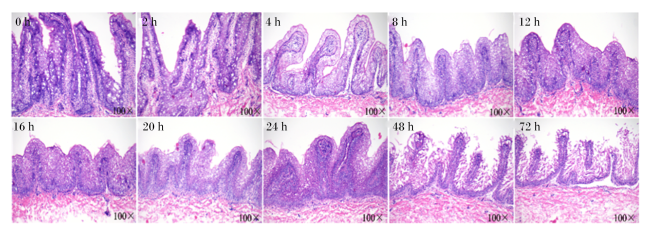
图1可见,培养时间在0~12 h瘤胃外植体的组织结构没有明显变化,且结构完整,组织紧凑,纹理清晰。当培养时间继续增加时,组织结构开始发生溃散,而且随着培养时间的延长,瘤胃组织的溃散程度不断加深,到24 h时仅大致可见组织的部分结构,而到72 h时,基本组织结构已完全溃散,初步认为24 h以内为本试验最佳培养时间。
图1 瘤胃外植体HE染色结果

Fig.1 HE staining results of rumen explants

2.1.2 不同培养时间对瘤胃外植体活性的影响

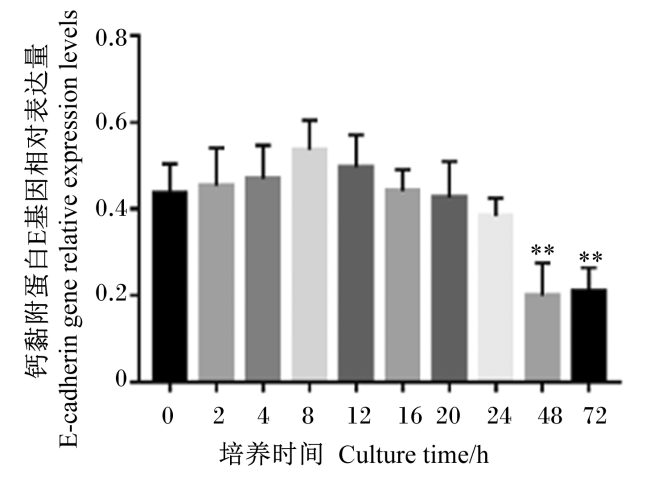
图2可见,E-cadherin在瘤胃组织中的表达水平在0~72 h呈现出先升高后降低的总体趋势。与0 h相比,孵育2~24 h时,E-cadherin在瘤胃外植体中的基因相对表达量无显著差异(P>0.05)。当培养时间大于24 h时,E-cadherin的基因相对表达量极显著下降(P<0.01),表明该阶段组织活力明显下降,这与瘤胃液外植体HE染色结果基本一致。
图2 不同培养时间钙黏附蛋白E 基因相对表达量

“**” 表示与0 h相比差异极显著(P<0.01)。

Fig.2 E-cadherin gene relative expression levels at different culture time

“**”indicates that the difference was very significant compared with 0 h (P<0.01).

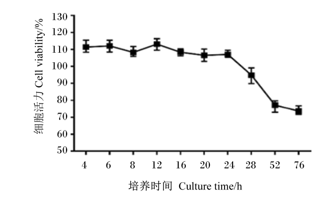
图3可知,4~24 h时瘤胃外植体细胞的活力维持在相对稳定的水平。24 h时细胞活力开始降低。当培养时间大于28 h时,细胞活力下降明显,并维持在相对较低的水平。结合HE染色和E-cadherin基因相对表达量检测结果可知,本试验以24 h作为后续试验的最佳培养时间。
图3 不同培养时间瘤胃外植体细胞活力变化

Fig.3 Cell viability changes of rumen explants at different culture time

2.2 试验2:丁酸梭菌刺激瘤胃外植体

2.2.1 丁酸梭菌活菌和灭活菌对PI3K/AKT/mTORC1信号通路中相关蛋白表达的影响
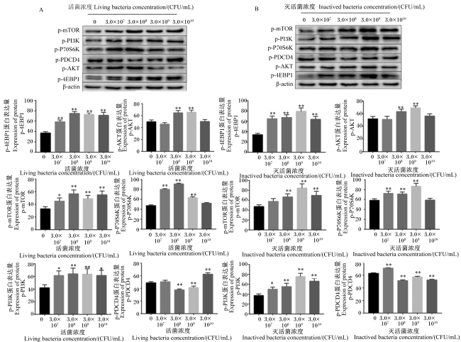
图4-A可知,添加不同浓度的丁酸梭菌活菌液对通路中关键蛋白的表达均有影响,除p-PDCD4外,该通路蛋白表达量总体上呈现出随丁酸梭菌活菌液浓度的增加先升高后降低的趋势。p-P70S6K、p-4EBP1和p-mTOR在丁酸梭菌活菌液浓度为3.0×10⁸ CFU/mL时表达量最高,PI3K和p-AKT的蛋白表达量在丁酸梭菌活菌液浓度为3.0×108和3.0×109 CFU/mL时极显著高于对照组(P<0.01),p-PDCD4主要抑制蛋白合成,丁酸梭菌活菌液浓度为3.0×108和3.0×109 CFU/mL时的蛋白表达量极显著低于对照组(P<0.01)。由于p-4EBP1蛋白表达量的提高代表了蛋白质合成效率的提高,而p-P70S6K是mTOR下游途径的关键靶蛋白,与p-4EBP1一起在蛋白质合成过程中起着非常重要的作用。因此,添加丁酸梭菌活菌积极促进了调节瘤胃外植体中蛋白质合成的PI3K/AKT/mTORC1信号传导途径,丁酸梭菌活菌液最佳浓度为3.0×109 CFU/mL。
图4 丁酸梭菌对瘤胃外植体中蛋白质合成的影响

“*”表示与对照组相比显著差异(P<0.05);“**”表示与对照组相比差异极显著(P<0.01)。图5图6同。

p-mTOR:磷酸化哺乳动物雷帕霉素靶蛋白 phosphorylated mammalian target of rapamycin;p-PI3K:磷酸化磷脂酰肌醇-3-激酶 phosphorylated phosphatidylinositol 3-kinase;p-P70S6K:磷酸化核糖体70S小亚基S6蛋白激酶 phosphorylated ribosomal 70S small subunit S6 protein kinase;p-PDCD4:磷酸化程序性细胞死亡因子4 phosphorylated programmed cell death factor 4;p-AKT:磷酸化蛋白激酶B phosphorylated protein kinase B;p-4EBP1:磷酸化真核细胞起始因子4E结合蛋白1 phosphorylated eIF4E-binding protein 1;β-actin:β-肌动蛋白。图5图6同 the same as Fig. 5 and Fig. 6

Fig.4 Effects of Clostridium butyricum on protein synthesis in rumen explants

“*” indicates significant difference compared with control group (P<0.05); “**” indicates highly significant difference compared with control group (P<0.01). The same as Fig.5 and Fig.6.

图4-B可知,丁酸梭菌灭活菌对通路相关蛋白表达也有十分明显的作用,p-PI3K、p-AKT、p-mTOR、p-P70S6K1和p-4EBP1的蛋白表达量在丁酸梭菌灭活菌液浓度为3.0×10⁹ CFU/mL时达到最高,均极显著高于对照组(P<0.01),p-PDCD4的蛋白表达量极显著低于对照组(P<0.01)。即添加灭活菌液能够影响PI3K/AKT/mTORC1通路中关键蛋白的表达,丁酸梭菌灭活菌液最佳浓度为3.0×109 CFU/mL,与添加活菌液的试验结果基本一致。
综上所述,在丁酸梭菌液浓度为3.0×10⁹ CFU/mL时,灭活菌和活菌均能够影响PI3K/AKT/mTORC1通路中关键蛋白的表达。

2.2.2 丁酸梭菌灭活菌培养上清液对PI3K/AKT/mTORC1通路中的蛋白质和基因表达的影响
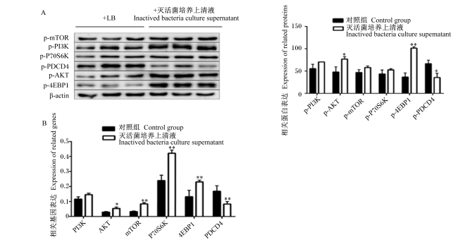
图5-A中Western Blot结果表明,丁酸梭菌灭活菌培养上清液中的p-AKT蛋白表达量显著高于对照组(P<0.05),p-4EBP1蛋白表达量极显著高于对照组(P<0.01),p-PDCD4蛋白表达量显著低于对照组(P<0.05),而PI3K、p-mTOR、p-P70S6K蛋白表达量与对照组无显著差异(P>0.05)。图5-B中RT-qPCR结果表明,AKT的基因相对表达量显著高于对照组(P<0.05),mTORP70S6K和4EBP1的基因相对表达量极显著高于对照组(P<0.01),PDCD4的基因相对表达量极显著低于对照组(P<0.01)。而P70S6K和4EBP1是通路中蛋白质合成的主要调控蛋白。综上所述,丁酸梭菌灭活菌培养上清液能够影响PI3K/AKT/mTORC1通路中关键蛋白和基因的表达,对瘤胃外植体的蛋白质合成起到促进作用。
图5 丁酸梭菌灭活菌培养上清液对PI3K/AKT/mTORC1通路关键蛋白和基因表达的影响

Fig.5 Effects of inactived bacteria culture supernatant of Clostridium butyricum on expression of key proteins and genes in PI3K/AKT/mTORC1 pathway

2.2.3 丁酸梭菌活菌和灭活菌对PI3K/AKT/mTORC1信号通路中关键蛋白和基因表达的影响
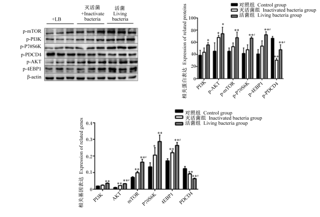
图6中Western blot结果可知,丁酸梭菌活菌和灭活菌的加入均能促进p-AKT、p-mTOR、p-PI3K、p-P70S6K和p-4EBP1的蛋白表达,而p-PDCD4的蛋白表达受到不同程度的抑制。其中,活菌组p-mTOR、p-P70S6K、p-4EBP1、p-PDCD4蛋白表达量与对照组相比有极显著差异(P<0.01),灭活菌组p-AKT蛋白表达量与对照组相比有显著差异(P<0.05),灭活菌组p-PDCD4蛋白表达量与对照组相比有极显著差异(P<0.01),灭活菌组p-PI3K、p-mTOR、p-4EBP1、p-P70S6K蛋白表达与对照组相比无显著差异(P>0.05)。活菌组p-P70S6K、p-4EBP1、p-PDCD4的蛋白表达量与灭活菌组相比有显著差异(P<0.05)。
图6 丁酸梭菌活菌和灭活菌处理的瘤胃外植体的蛋白和基因的表达

“#”表示活菌组与灭活菌组间差异显著(P<0.05)。

Fig.6 Expression of proteins and genes in rumen explants with living and inactivated bacteria of Clostridium butyricum (n=9)

“#” indicates significant difference between living bacteria group and inactivated bacteria group (P<0.05).

图6中RT-qPCR结果可知,通路中关键基因的表达变化与Western blot的结果相似,活菌和灭活菌的加入均能促进AKTmTORPI3KP70S6K和4EBP1基因相对表达量,而PDCD4的基因相对表达量受到不同程度的抑制,其中活菌组AKTmTORPI3KP70S6K、4EBP1和PDCD4基因相对表达量与对照组相比有极显著差异(P<0.01),灭活菌组P70S6K的基因相对表达量与对照组相比有显著差异(P<0.05),AKTmTOR、4EBP1和PDCD4基因相对表达量与对照组相比有极显著差异(P<0.01),灭活菌组PI3K基因相对表达量与对照组无显著差异(P>0.05)。活菌组AKTmTOR、4EBP1和PDCD4基因相对表达量与灭活菌组相比有显著差异(P<0.05)。
综上所示,添加丁酸梭菌活菌和灭活菌均能影响瘤胃外植体中蛋白质合成相关因子的表达水平,活菌对瘤胃外植体中蛋白质合成相关蛋白的影响作用大于灭活菌。

2.2.4 外植体丁酸梭菌培养上清液中游离氨基酸含量的分析

表3可以看出,对照组中谷氨酸(Glu)、甘氨酸(Gly)、苯丙氨酸(Phe)、组氨酸(His)、酪氨酸(Tyr)、精氨酸(Arg)和脯氨酸(Pro)的含量显著高于活菌培养上清液和灭活菌培养上清液组(P<0.05)。活菌培养上清液组半胱氨酸(Cys)含量最高,与其他2组存在显著差异(P<0.05),而且除了Cys以外,天冬氨酸(Asp)、苏氨酸(Thr)、丙氨酸(Ala)、缬氨酸(Val)、蛋氨酸(Met)、异亮氨酸(Ile)和亮氨酸(Leu)的含量均显著高于对照组(P<0.05)。而灭活菌培养上清液组Asp、Thr、Ser、Ala、Val、Met、Ile、Leu和Lys的含量显著高于另外2组(P<0.05)。其中,活菌培养上清液和灭活菌培养上清液组Met,Ile和Leu含量均显著高于对照组(P<0.05),说明细菌培养上清液中Met,Ile和Leu等氨基酸含量的增加可能是引起PI3K/AKT/mTORC1通路中关键蛋白和基因的表达变化的原因之一。
表3 丁酸梭菌培养24 h上清液中17种游离氨基酸含量的测定

Table 3 Determination of 17 kinds of free amino acids contents in supernatant of Clostridium butyricum culture for 24 h (n=9) mg/mL

项目
Items
对照组
Control group
活菌培养上清液组
Living bacteria culture
supernatant group
灭活菌培养上清液组
Inactivated bacteria culture
supernatant group
天冬氨酸Aspartic acid 0.044 0±0.004 6c 0.131 2±0.001 3a 0.110 9±0.000 9b
苏氨酸Threonine 0.037 1±0.000 2c 0.183 2±0.000 9a 0.102 8±0.001 1b
丝氨酸Serine 0.234 3±0.000 4a 0.068 1±0.000 9b 0.044 7±0.000 3c
谷氨酸Glutamic acid 0.443 3±0.000 2c 0.452 6±0.001 1b 0.573 5±0.004 6a
甘氨酸Glycine 0.055 9±0.000 8c 0.075 6±0.000 6b 0.092 9±0.000 2a
丙氨酸Alanine 0.158 0±0.000 8c 0.392 4±0.003 1a 0.353 3±0.001 2b
缬氨酸Valine 0.136 9±0.000 4b 0.291 1±0.002 2a 0.075 6±0.055 6b
蛋氨酸Methionine 0.068 0±0.000 2c 0.157 4±0.000 8b 0.267 4±0.000 9a
异亮氨酸Isoleucine 0.111 6±0.000 6c 0.232 8±0.000 9a 0.147 8±0.000 4b
N-羧基-DL-亮氨酸
N-carbobenzoxy-DL-leucine
0.300 9±0.001 4c 0.617 7±0.000 5a 0.431 9±0.000 6b
酪氨酸Tyrosine 0.787 3±0.000 6a 0.252 7±0.001 1c 0.616 4±0.002 1b
苯丙氨酸Phenylalanine 0.376 9±0.000 4a 0.318 7±0.004 5b 0.318 5±0.008 8b
组氨酸Histidine 0.200 7±0.000 4a 0.102 6±0.001 1c 0.137 9±0.000 4b
赖氨酸Lysine 0.010 9±0.000 6c 0.619 9±0.002 1a 0.570 6±0.002 2b
精氨酸Arginine 0.355 3±0.000 8a 0.004 6±0.000 5c 0.007 1±0.000 2b
DL-脯氨酸DL-proline 0.022 1±0.000 2b 0.009 5±0.000 4c 0.058 4±0.000 5a
半胱氨酸Cysteine 0.121 9±0.001 8b 0.151 2±0.001 6a 0.109 6±0.000 3c

同行数据肩标不同字母表示差异显著(P<0.05)。

In the same row, values with different letter superscripts mean significant differences (P<0.05).

3 讨论

通过切片、E-cadherin表达和MTT的试验结果表明,本研究成功构建了瘤胃外植体培养模型。此外,瘤胃外植体活性测定的结果显示,奶牛瘤胃外植体的最佳培养时间为24 h,这与其他研究结果[23-24]相似。在0~4 h培养期间瘤胃外植体活性持续增加,这是由于在瘤胃上皮组织分离和放置在培养基之间的这段时间内缺乏营养供应,导致外植体活力降低[25-26]。在培养基中,瘤胃外植体快速吸收营养,并开始逐渐恢复活力,与Girard等[27]的研究中观察到的现象相似。随着对益生菌的研究越来越多,益生菌的细胞组分或代谢物可发挥免疫反应、增强疾病保护和刺激相关基因的表达作用的报道也越来越多。例如,热灭活的枯草芽孢杆菌对赭曲霉毒素A的降解率也高于活枯草芽孢杆菌。热灭活解淀粉芽孢杆菌可降低蚜虫抗性,可用作蚜虫控制的新型杀虫剂[28-29]。此外,多黏芽孢杆菌培养上清液对病原微生物如气单胞菌和假单胞菌具有强烈的抑制作用[30-31]。本试验主要探讨灭活丁酸梭菌是否可以在瘤胃蛋白质合成中发挥与活菌同样的作用,并对活菌和灭活菌上清液中的各种氨基酸含量进行检测,探究哪些氨基酸在PI3K/AKT/mTORC1通路中发挥效力。
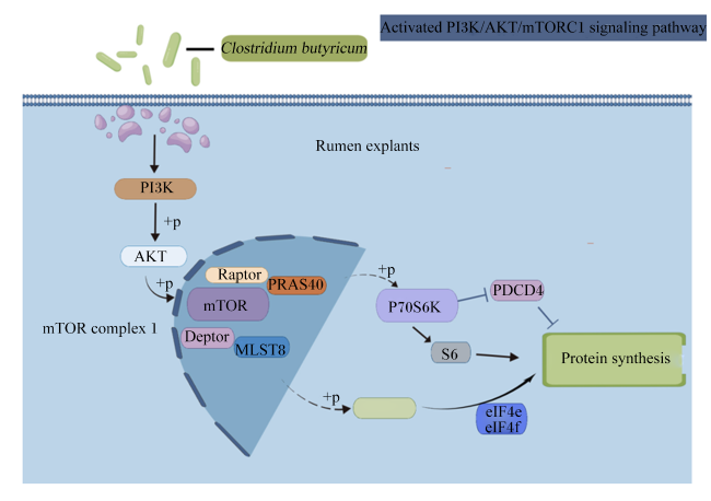
图7为PI3K/AKT/mTORC1的机制图,PI3K/AKT/mTORC1是控制组织中蛋白质合成的关键途径[32],PI3K是细胞内磷脂酰肌醇激酶类的一员[33],其下游有许多信号通路参与调控翻译起始。当PI3K被激活时,它将AKT从细胞质转移到细胞膜,激活AKT并磷酸化其Thr308位点[34]。AKT的激活导致AKT下游的mTOR等许多效应物磷酸化,mTOR是磷脂酰肌醇3-激酶相关激酶(PIKK)家族的成员[35],是蛋白质合成的重要调节因子。PIKK是一类保守的非典型丝氨酸/苏氨酸蛋白激酶,是PI3K/AKT/mTORC1途径的核心蛋白,也是雷帕霉素的靶点。mTORC1的形成受到多种营养因子调控,同时受细胞能量状态、氧、应激和氨基酸的调节[36],许多氨基酸转运载体都有在溶酶体中发现,其中一些已被证明可以调节mTORC1活性。mTORC1的激活需要营养物质和氨基酸,这导致Raptor介导的mTORC1被招募到溶酶体和细胞核中[37],并与其激活剂GTPase-RHEB1共定位。除了控制翻译和自噬外,mTORC1已被证明可靶向影响生理过程,如脂质合成、胰岛素作用和核糖体生物合成[38-39]。因此本试验利用PI3K/AKT/mTOR1通路来验证丁酸梭菌灭活菌是否具有和活菌的相同作用。
图7 PI3K/AKT/mTORC1信号通路图(由Figdraw绘制)

Rumen explants:瘤胃外植体;Clostridium butyricum:丁酸梭菌;mTOR complex 1:哺乳动物雷帕霉素靶蛋白复合体1;Protein synthesis:蛋白质合成;mTOR:哺乳动物雷帕霉素靶蛋白 mammalian target of rapamycin;PI3K:磷脂酰肌醇-3-激酶 phosphatidylinositol 3-kinase;P70S6K:核糖体70S小亚基S6蛋白激酶 ribosomal 70S small subunit S6 protein kinase;PDCD4:程序性细胞死亡因子4 programmed cell death factor 4;AKT:蛋白激酶B protein kinase B;4EBP1:真核细胞起始因子4E结合蛋白1 eIF4E-binding protein 1;eIF4e:真核起始因子4E eukaryotic initiation factor 4E;eIF4f:真核起始因子4E eukaryotic initiation factor 4F; Protein synthesis: 蛋白合成 ; mTORC1: 哺乳动物雷帕霉素靶蛋白复合物1 mammalian target protein complex of rapamycin 1。

Fig.7 PI3K/AKT/mTORC1 signaling pathway diagram (produced by Figdraw)

本试验结果表明,丁酸可以对与蛋白质合成的相关瘤胃组织中的分子反应发挥积极作用。特定靶标的mRNA和蛋白质表达以相同的方式对丁酸梭菌的活菌和灭活菌作出反应,表明这种生物体或其代谢物可以通过PI3K/AKT/mTORC1通路促进蛋白质合成。在这种情况下,灭活菌和活菌在促进瘤胃组织中蛋白质合成方面具有相似的效力。由于PI3K/AKT/mTORC1通路中关键因子的表达可受丁酸梭菌活菌和灭活菌以及灭活菌培养上清液的影响,因此除了探索丁酸梭菌生物体本身的作用之外,本试验还探讨了丁酸梭菌培养上清液中可能参与该过程的一些代谢衍生物。结果证实了这一观点,3个试验组的上清液中某些氨基酸的含量存在差异。其中,Ile和Leu在3组活菌培养上清液中含量最高。Ile和Leu可以独立地调节mTOR信号传导途径,并且Ile可以对mTOR的磷酸化发挥积极作用。这提示Ile和Leu可能是影响该通路中关键因子表达的因素之一。试验结果还表明,加入灭活菌培养上清液可影响PI3K/AKT/mTORC1途径中蛋白质和基因的表达,这与前期的研究结论一致,即Met能够通过影响mTORC1复合物来促进蛋白质合成[40-41]。Castro等[42]的研究也证实了这一想法,表明mTOR对Met、Ile、Thr和Leu更敏感,并且可以将合成信号转导至更大的蛋白质合成过程。

4 结论

综上所述,对于丁酸梭菌,活菌和灭活菌均通过改变与PI3K/AKT/mTORC1通路相关因子的表达量,促进瘤胃外植体的蛋白质合成,灭活丁酸梭菌可代替活菌调控瘤胃外植体的蛋白质合成。
[1]
JIAO P X, WANG Z W, WANG X, et al. Effect of Clostridium butyricum supplementation on in vitro rumen fermentation and microbiota with high grain substrate varying with media pH levels[J]. Frontiers in Microbiology, 2022, 13:912042.

[2]
SHEN H K, ZHAO Z T, ZHAO Z J, et al. Native and engineered probiotics:promising agents against related systemic and intestinal diseases[J]. International Journal of Molecular Sciences, 2022, 23(2):594.

[3]
TAO R Z, ZONG G F, PAN Y H, et al. Clostridium butyricum and Clostridium tyrobutyricum:angel or devil for necrotizing enterocolitis?[J]. Msystems, 2023, 8(6):e0073223.

[4]
GUPTA P, KUMAR M, GUPTA R P, et al. Fermentative reforming of crude glycerol to 1,3-propanediol using Clostridium butyricum strain L4[J]. Chemosphere, 2022, 292:133426.

[5]
DOU L, LIU C, CHEN X Y, et al. Supplemental Clostridium butyricum modulates skeletal muscle development and meat quality by shaping the gut microbiota of lambs[J]. Meat Science, 2023, 204:109235.

[6]
梁凌云. 丁酸梭菌转化甘油为1,3-丙二醇振荡机制的研究[D]. 硕士学位论文. 大连: 大连理工大学, 2022.

LIANG L Y. CDATA oscillation mechanism research of glycerol conversion to 1,3-propanediol by Clostridium butyricum[D]. Master’s Thesis. Dalian: Dalian University of Technology, 2022. (in Chinese)

[7]
LIU M, UYANGA V A, CAO X K, et al. Regulatory effects of the probiotic Clostridium butyricum on gut microbes,intestinal health,and growth performance of chickens[J]. The Journal of Poultry Science, 2023, 60(2):2023011.

[8]
YU J, DONG B, ZHAO M M, et al. Dietary Clostridium butyricum and Bacillus subtilis promote goose growth by improving intestinal structure and function,antioxidative capacity and microbial composition[J]. Animals, 2021, 11(11):3174.

[9]
易中华. 饲用微生态制剂丁酸梭菌的研究与应用进展[J]. 饲料研究, 2012(2):14-17.

YI Z H. Feed microecological preparation Clostridium butyricum[J]. Feed Research, 2012(2):14-17. (in Chinese)

[10]
冯杰, 杨旸, 周莉, 等. 丁酸梭菌的生物学功能及其在家禽生产中应用的研究进展[J]. 现代畜牧兽医, 2023(6):79-83.

FENG J, YANG Y, ZHOU L, et al. Research progress on biological function of Clostridium butyricum and its application in poultry production[J]. Modern Journal of Animal Husbandry and Veterinary Medicine, 2023(6):79-83. (in Chinese)

[11]
陈俊超, 附俊杰, 刘军, 等. 丁酸梭菌的益生特性及其活性代谢产物[J]. 食品研究与开发, 2023, 44(23):145-152.

CHEN J C, FU J J, LIU J, et al. Probiotic characteristics and active metabolites of Clostridium butyricum[J]. Food Research and Development, 2023, 44(23):145-152. (in Chinese)

[12]
ADEBAYO AROWOLO M, ZHANG X M, WANG M, et al. Proper motility enhances rumen fermentation and microbial protein synthesis with decreased saturation of dissolved gases in rumen simulation technique[J]. Journal of Dairy Science, 2022, 105(1):231-241.

[13]
LI Y J, ZANG Y T, ZHAO X H, et al. Dietary supplementation with creatine pyruvate alters rumen microbiota protein function in heat-stressed beef cattle[J]. Frontiers in Microbiology, 2021, 12:715088.

[14]
LI T, WENG J, ZHANG Y, et al. mTOR direct crosstalk with STAT5 promotes de novo lipid synthesis and induces hepatocellular carcinoma[J]. Cell Death & Disease, 2019, 10(8):619.

[15]
DE SANTIS M C, GOZZELINO L, MARGARIA J P, et al. Lysosomal lipid switch sensitises to nutrient deprivation and mTOR targeting in pancreatic cancer[J]. Gut, 2023, 72(2):360-371.

[16]
PANWAR V, SINGH A, BHATT M, et al. Multifaceted role of mTOR (mammalian target of rapamycin) signaling pathway in human health and disease[J]. Signal Transduction and Targeted Therapy, 2023, 8(1):375.

DOI PMID

[17]
YI J M, ZHU J J, WU J, et al. Oncogenic activation of PI3K-AKT-mTOR signaling suppresses ferroptosis via SREBP-mediated lipogenesis[J]. Proceedings of the National Academy of Sciences of the United States of America, 2020, 117(49):31189-31197.

DOI PMID

[18]
LEE S B, HAN S H, PARK S. Long-term culture of intestinal organoids[J]. Methods in Molecular Biology, 2018, 1817:123-135.

DOI PMID

[19]
MO Y J, KIM Y S, KIM M S, et al. Advantages of adult mouse dorsal root ganglia explant culture in investigating myelination in an inherited neuropathic mice model[J]. Methods and Protocols, 2022, 5(4):66.

[20]
王秋菊, 任雨龙, 魏笑, 等. 反刍动物外植体的研究进展[J]. 中国兽医学报, 2022, 42(5):1097-1102.

WANG Q J, REN Y L, WEI X, et al. Research progress on ruminant explants[J]. Chinese Journal of Veterinary Science, 2022, 42(5):1097-1102. (in Chinese)

[21]
OLSON A, LE V, ALDAHL J, et al. The comprehensive role of E-cadherin in maintaining prostatic epithelial integrity during oncogenic transformation and tumor progression[J]. PLoS Genetics, 2019, 15(10):e1008451.

[22]
VAN V T H, LIU Z S, HSIEH Y J, et al. Therapeutic effects of orally administration of viable and inactivated probiotic strains against murine urinary tract infection[J]. Journal of Food and Drug Analysis, 2023, 31(4):583.

DOI PMID

[23]
LI H F, JIA Y X, WENG D, et al. Clostridium butyricum inhibits fat deposition via increasing the frequency of adipose tissue-resident regulatory T cells[J]. Molecular Nutrition & Food Research, 2022, 66(12):e2100884.

[24]
ZHANG M, JIN X, YANG Y F, et al. β-glucan from Saccharomyces cerevisiae is involved in immunostimulation of ovine ruminal explants[J]. Canadian Journal of Veterinary Research, 2020, 84(4):283-293.

[25]
LI H H, SHANG Z Y, LIU X J, et al. Clostridium butyricum Alleviates enterotoxigenic Escherichia coli K88-induced oxidative damage through regulating the p62-Keap1-Nrf2 signaling pathway and remodeling the cecal microbial community[J]. Frontiers in Immunology, 2021, 12:771826.

[26]
刘琳. 丙酮酸肌酸在缓解热应激肉牛瘤胃功能上的作用及机制研究[D]. 博士学位论文. 南昌: 江西农业大学, 2020.

LIU L. Effect and mechanism of creatine pyruvate on rumen function heat-stress in beef cattle[D]. Ph.D.Thesis. Nanchang: Jiangxi Agricultural University, 2020. (in Chinese)

[27]
GIRARD F, DZIVA F, VAN DIEMEN P, et al. Adherence of enterohemorrhagic Escherichia coli O157,O26,and O111 strains to bovine intestinal explants ex vivo[J]. Applied and Environmental Microbiology, 2007, 73(9):3084-3090.

[28]
吴建民, 王雍, 周协琛, 等. 基于宏基因组学解析瘤胃微生物调节荷斯坦奶牛乳蛋白含量的研究[J]. 动物营养学报, 2020, 32(8):3843-3855.

DOI

WU J M, WANG Y, ZHOU X C, et al. Rumen microorganism regulating milk protein content in Holstein dairy cows based on metagenomic analysis[J]. Chinese Journal of Animal Nutrition, 2020, 32(8):3843-3855. (in Chinese)

[29]
TOMITA Y, GOTO Y, SAKATA S, et al. Clostridium butyricum therapy restores the decreased efficacy of immune checkpoint blockade in lung cancer patients receiving proton pump inhibitors[J]. Oncoimmunology, 2022, 11(1):2081010.

[30]
邹华威. 内侧前额叶皮质GABA(B)受体及盲肠微生物在应激“易感/非易感”中的调控作用研究[D]. 硕士学位论文. 南阳: 南阳师范学院, 2022.

ZOU H W. The functional role of mPFC GABA(B) receptors and the cecal microbiota in stress resilience and susceptibility[D]. Master’s Thesis. Nanyang: Nanyang Normal University, 2022. (in Chinese)

[31]
WU H, LI Y, MENG Q X, et al. Effect of high sulfur diet on rumen fermentation,microflora,and epithelial barrier function in steers[J]. Animals, 2021, 11(9):2545.

[32]
KIM T Y, PARK K T, CHOUNG S Y. Codonopsis lanceolata and its active component Tangshenoside I ameliorate skeletal muscle atrophy via regulating the PI3K/Akt and SIRT1/PGC-1α pathways[J]. Phytomedicine, 2022, 100:154058.

[33]
NOOROLYAI S, SHAJARI N, BAGHBANI E, et al. The relation between PI3K/AKT signalling pathway and cancer[J]. Gene, 2019, 698:120-128.

DOI PMID

[34]
LIANG X, WANG R, GUO Y, et al. Phosphorylation of Akt at Thr308 regulates p-eNOS Ser1177 during physiological conditions[J]. FEBS Open Bio, 2021, 11(7):1953-1964.

[35]
CHEN Y F, ZHOU X P. Research progress of mTOR inhibitors[J]. European Journal of Medicinal Chemistry, 2020, 208:112820.

[36]
WANG X M, CHU Y J, WANG W L, et al. mTORC signaling in hematopoiesis[J]. International Journal of Hematology, 2016, 103(5):510-518.

DOI PMID

[37]
XIAO B, ZUO D M, HIRUKAWA A, et al. Rheb1-independent activation of mTORC1 in mammary tumors occurs through activating mutations in mTOR[J]. Cell Reports, 2020, 31(4):107571.

[38]
HOSIOS A M, WILKINSON M E, MCNAMARA M C, et al. mTORC1 regulates a lysosome-dependent adaptive shift in intracellular lipid species[J]. Nature Metabolism, 2022, 4(12):1792-1811.

DOI PMID

[39]
JIAO L J, LIU Y Z, YU X Y, et al. Ribosome biogenesis in disease:new players and therapeutic targets[J]. Signal Transduction and Targeted Therapy, 2023, 8(1):15.

[40]
张曼. 酿酒酵母β-葡聚糖对绵羊瘤胃上皮细胞和外植体内β-防御素-1表达影响的研究[D]. 博士学位论文. 呼和浩特: 内蒙古农业大学, 2019.

ZHANG M. Study of effect of β-glucan on the expression of β-defensin-1 in rumen epit helial cells and explants of sheep[D]. Ph.D.Thesis. Hohhot: Inner Mongolia Agricultural University, 2019. (in Chinese)

[41]
MA Y F, BATISTEL F, XU T L, et al. Phosphorylation of AKT serine/threonine kinase and abundance of milk protein synthesis gene networks in mammary tissue in response to supply of methionine in periparturient Holstein cows[J]. Journal of Dairy Science, 2019, 102(5):4264-4274.

DOI PMID

[42]
CASTRO J J, ARRIOLA APELO S I, APPUHAMY J A D R N, et al. Development of a model describing regulation of casein synthesis by the mammalian target of rapamycin (mTOR) signaling pathway in response to insulin,amino acids,and acetate[J]. Journal of Dairy Science, 2016, 99(8):6714-6736.

Outlines

/