RESEARCH PAPER

Mechanisms of Regulatory Role of Porcine Limosilactobacillus mucosae and Tryptophan on Gastrointestinal Immune Homeostasis and Function Development of Young Mice

  • SUN Zhiyuan , 1 ,
  • HUANG Siqi 2 ,
  • DONG Wenjian 1 ,
  • HAN Dandan 1 ,
  • JIANG Lili 1 ,
  • WU Zhenlong 1 ,
  • DAI Zhaolai , 1, *
Expand
  • 1 State Key Laboratory of Animal Nutrition and Feeding, College of Animal Science and Technology, China Agricultural University, Beijing 100193, China
  • 2 College of Animal Science and Technology, China Agricultural University Beijing 100193, China
* associate professor, E-mail:

Received date: 2024-02-21

  Online published: 2024-09-08

Abstract

The aim of this experiment was to investigate the regulation and mechanism of the gastrointestinal 5-hydroxytryptamine receptor (HTR) and immune function development of young mice treated with Limosilactobacillus mucosae and tryptophan alone or in combination. Thirty-two 3-week-old male C57BL/6 mice were randomly divided into control group, tryptophan group, Limosilactobacillus mucosae group and Limosilactobacillus mucosae+tryptophan group with 8 replicates per group and 1 mouse per replicate. Mice in Limosilactobacillus mucosae group and Limosilactobacillus mucosae+tryptophan group were given 100 μL/(mouse·d) Limosilactobacillus mucosae live bacterial suspension (1×10⁸ CFU/mL) every other day, and those in the other groups were given the same amount of phosphate buffer solution; while mice in the tryptophan group and Limosilactobacillus mucosae+tryptophan group were added 0.15 mg/(g BW·d) tryptophan in drinking water (0.5 mg/mL tryptophan in drinking water), and 0.44 mg/mL alanine was added in drinking water of the other groups as iso-nitrogen treatment. The experiment lasted for 14 days. The results showed as follows: 1) after 2 weeks of treatment with Limosilactobacillus mucosae and tryptophan, the body weight and small intestine length of mice were significantly increased (P<0.05); 2) Limosilactobacillus mucosae or combined with tryptophan could regulate the metabolic homeostasis of 5-hydroxytryptamine (5-HT) in gastrointestinal tract, differentiated the expression of HTR in gastrointestinal tract, and significantly up-regulated the expression of HTR4 and HTR7 in the stomach (P<0.05); 3) Limosilactobacillus mucosae or combined with tryptophan could promote the polarization of M2-type macrophages in stomach and jejunum, up-regulate the expression of Wnt3a, and differentially regulate the expression of transforming growth factor-β (TGF-β) subtypes in gastrointestinal tract; 4) Limosilactobacillus mucosae or combined with tryptophan significantly increased the indole concentration in colon (P<0.05), but significantly decreased the 5-HT concentration in small intestine and colon (P<0.05). These results indicate that the combination of porcine Limosilactobacillus mucosae and tryptophan can promote gastrointestinal development and growth of young mice by regulating gastrointestinal 5-HT metabolism and HTR expression, up-regulating M2-type macrophage polarization and gastrointestinal TGF-β subtype expression, and activating Wnt signaling.

Cite this article

SUN Zhiyuan , HUANG Siqi , DONG Wenjian , HAN Dandan , JIANG Lili , WU Zhenlong , DAI Zhaolai . Mechanisms of Regulatory Role of Porcine Limosilactobacillus mucosae and Tryptophan on Gastrointestinal Immune Homeostasis and Function Development of Young Mice[J]. Chinese Journal of Animal Nutrition, 2024 , 36(9) : 6015 -6026 . DOI: 10.12418/CJAN2024.510

仔猪出生后的肠道功能发育与稳恒对仔猪的健康和生长至关重要。研究表明,肠道结构的成熟和功能的完善,有助于幼龄动物对营养物质的消化吸收利用,同时抵御肠道中的有害微生物[1]。哺乳仔猪出生后4周内,由于上皮更新能力强、细胞凋亡比例低,导致肠道黏膜重量、绒毛高度和隐窝深度大幅增加,有利于改善仔猪的生长[2]。而对于断奶仔猪而言,良好的肠道消化机能和屏障功能可提高饲料利用率、维持肠道微生态稳恒,从而降低断奶仔猪的腹泻率与死亡率,提高猪生产的经济效益[3]
肠道结构功能发育受多种因素影响,其中免疫及其相关信号在肠细胞增殖分化中发挥重要调节作用。二型固有淋巴细胞(group 2 ILCs)和三型固有淋巴细胞(group 3 ILCs)分别通过分泌白细胞介素(interleukin,IL)-13和IL-22等细胞因子促进肠道干细胞(intestinal stem cells,ISCs)的增殖分化并介导肠道的损伤修复与更新[4-5]。此外,巨噬细胞可通过分泌转化生长因子-β(transforming growth factor-β,TGF-β)、IL-10等细胞因子维持肠道稳态。研究表明,肠道巨噬细胞CD206+亚群分泌Wnt信号通路配体,维持肠道类器官的潘氏细胞和间充质生态位细胞的分化,降低肠道炎症的发生[6]。肠上皮细胞增殖的关键标志物为核增殖抗原(Ki67)[7],而TGF-β在肠黏膜细胞的增殖分化、损伤修复以及免疫调节中发挥重要作用。TGF-β与其特异性受体结合,激活下游Smad2/3信号通路,从而促进肠隐窝再生和ISCs增殖分化[8]
肠道中5-羟色胺(5-hydroxytryptamine,5-HT)的代谢与信号可调节肠道免疫功能,可能在肠道发育中发挥重要调节作用。体内90%~95%的5-HT在胃肠中的色氨酸羟化酶(tryptophan hydroxylase,Tph)催化下合成,发挥作用后通过肠道血清素再摄取转运载体(serotonin reuptake transporter,Sert)转运至胞内,在单胺氧化酶A(monoamine oxidase A,Maoa)作用下降解,是肠道免疫系统的重要组成部分[9]。肠道内5-HT通过激活5-HT受体(HTR)亚型,调节肠道免疫细胞行为[10]。研究表明,T细胞、B细胞、巨噬细胞和嗜中性粒细胞等多种免疫细胞表面均表达不同亚型的HTR,调节肠道免疫和神经元发育等功能[9]。其中,HTR2A和HTR7参与促炎作用,而HTR1A、HRT2B和HTR4参与抗炎作用;前期研究发现,色氨酸的肠道免疫调节作用存在HTR亚型依赖性[11-13],而其在ISCs增殖分化中的作用有待进一步研究探明。
肠道菌群和肠道先天免疫存在互作,可调节胃肠发育和适应性免疫的成熟[14]。研究表明,鼠李糖乳杆菌GG(Lactobacillus rhamnosus GG)[15]和罗伊氏乳杆菌(Lactobacillus reuteri)[16]可增强小鼠的免疫防御。此外,肠道菌群可通过促进肠神经元的Tph2的表达增加5-HT的合成,继而通过激活巨噬细胞表面的HTR2A和HTR3A激活下游的Wnt/β-连环蛋白(β-catenin)的信号,调节细胞更新[17]。已有研究表明,黏膜乳杆菌(Limosilactobacillus mucosae)具有一定的耐酸耐胆盐能力,并可通过肠上皮黏附和抑制病原菌定殖发挥其益生作用[18-19]。前期研究发现,黏膜乳杆菌是断奶仔猪胃肠中的优势乳杆菌之一,其氨基酸代谢特性有别于其他乳杆菌[20]。黏膜乳杆菌可通过调节肠道HTR4和TGF-β亚型缓解肠道免疫反应[21]。然而,黏膜乳杆菌本身及其与饲粮营养素互作在调节胃肠免疫稳恒和功能发育中的作用方式与机制有待进一步研究探明。
因此,本研究假设黏膜乳杆菌和色氨酸可通过调节肠道5-HT代谢和受体稳态改善幼龄动物的生长和肠道功能发育,并采用小鼠模型,结合肠道基因转录水平测定、免疫细胞分型以及色氨酸代谢物浓度测定,探究猪源黏膜乳杆菌和色氨酸调节幼龄小鼠肠道免疫与发育的作用方式与机制,为发掘肠道来源乳杆菌的功能以及开发新型饲用益生菌相关添加剂改善动物生长发育提供基础。

1 材料与方法

1.1 菌株和培养条件

本研究中使用的黏膜乳酸杆菌分离自仔猪小肠[20,22]。黏膜乳杆菌用MRS培养基于37 ℃厌氧培养复苏后,用继代培养18 h的培养物用于试验。采用平板计数法确定18 h培养物活菌数,按照相同方法培养离心后,用磷酸盐缓冲液(PBS,pH=7.4)重悬至1×10⁸ CFU/mL用于小鼠灌胃。

1.2 小鼠试验

选取32只3周龄、平均体重为11.0 g的雄性C57BL/6小鼠(北京维通利华实验动物技术有限公司)用于试验。小鼠饲养于温度为(22±2) ℃、光照周期为12 h的环境中,并饲喂小鼠用维持饲粮(1022号饲粮,北京华阜康生物科技股份有限公司),小鼠自由采食和饮水。小鼠维持饲粮营养成分如下:18%粗蛋白质,4%粗脂肪,5%粗纤维,8%粗灰分,1.0%~1.8%钙,0.6%~1.2%磷;饲粮中不添加抗生素及其他抗菌物质。
试验将小鼠随机分为4组,分别为对照组、色氨酸组、黏膜乳杆菌组和黏膜乳杆菌+色氨酸组,每组8个重复,每个重复1只。适应3 d后,黏膜乳杆菌组和黏膜乳杆菌+色氨酸组小鼠每隔1 d灌胃100 μL/(只·d)的黏膜乳杆菌活菌悬液(1×108 CFU/mL),其余组灌胃等量PBS。色氨酸组和黏膜乳杆菌+色氨酸组小鼠通过饮水添加0.15 mg/(g BW·d)的色氨酸(0.5 mg/mL色氨酸饮水),添加量参照本实验室前期哺乳仔猪氨基酸试验中关于等氮对照的试验设计原理和计算方法[23];其余组饮水中添加0.44 mg/mL的丙氨酸作为等氮处理。正试期持续2周(14 d),每天记录幼鼠体重、采食量和饮水量。试验期间,各组小鼠饮水量无显著差异(P>0.05)。试验结束后,小鼠眼球采血并颈椎脱臼处死,分离肠道并测量小肠、结肠长度;采样胃、空肠和结肠组织于1.5 mL离心管中,液氮速冻后保存至-80 ℃待测。以上动物试验经中国农业大学实验动物福利与动物实验伦理审查委员会批准(批准号:AW11013202-1-5)。

1.3 测定指标与方法

1.3.1 实时荧光定量PCR

肠道组织样品采用TRIpure试剂盒(北京艾德莱生物科技有限公司)提取mRNA,反转录为cDNA后,采用荧光定量PCR试剂盒[天根生化科技(北京)有限公司]进行扩增(7500 Real-Time PCR System,Applied Biosystems,美国),除精氨酸酶1(Arg1,F-GAATCTGCATGGGCAACC,R-GAATCCTGGTACATCTGGGAAC)、Axin2(F-GAGATGACGCCTGTGGAACC,R-CCTGCTCAGACCCCTCCTTT)、Ki67(F-GCATCGAGTGTGAGCAAGTT,R-TGTCCTGCACCTGTTGATCT)和Wnt3a(F-GGCGGCTGTAGTGAGGACAT,R-GTGCATGTGACTGGCGATGG)基因外,其他目的基因和内参基因的引物序列和表达量计算参考过往研究[13,21]

1.3.2 流式细胞术分离和脾脏免疫细胞分析

参考Sun等[24]的方法,用2种不同孔径(70和50 μm)的细胞筛和Percoll从脾脏中分离免疫细胞,孵育抗体后用流式细胞仪(Beckman Coulter,美国)进行流式细胞术分析。不同类型免疫细胞丰度采用占代表总免疫细胞丰度的CD45+群体的百分比(%)表示,如CD11b+Ly6G+嗜中性粒细胞、CD11b+CD64+巨噬细胞、CD4+Foxp3+调节性T细胞(regulatory T cells,Treg)、CD11c+MHCⅡ+树突状细胞和CD3-CD19+ B淋巴细胞。采用CytExpert软件(Beckman Coulter,美国)对数据进行分析。

1.3.3 高效液相色谱法测定色氨酸代谢物浓度

称取20 mg液氮研磨后的组织样品或肠道内容物样品于1.5 mL离心管中,用预冷的50%甲醇水溶液除蛋白后,取50 μL上清与50 μL样品提取液混匀后用高效液相色谱仪(e2695,Waters Corporation,美国)检测色氨酸代谢物浓度[25]

1.4 数据统计分析

试验数据采用SPSS 16.0进行统计分析,结果采用平均值和均值标准误(SEM)表示。采用双因素方差分析比较黏膜乳杆菌和色氨酸的效应及其是否有互作效应,并采用Duncan氏法进行组间多重比较检验。P<0.05表示差异显著,0.05<P<0.10表示差异有显著趋势。

2 结果与分析

2.1 猪源黏膜乳杆菌和色氨酸对小鼠体重和小肠长度的影响

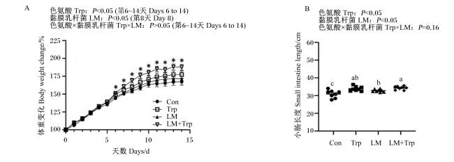
图1所示,黏膜乳杆菌和色氨酸处理后,色氨酸组和黏膜乳杆菌组小鼠体重与对照组相比无显著差异(P>0.05);黏膜乳杆菌+色氨酸组小鼠从处理后的第6天开始体重显著高于对照组(P<0.05)。对于小肠长度而言,与对照组相比,色氨酸和黏膜乳杆菌处理2周后均可显著增加小鼠小肠长度(P<0.05),且黏膜乳杆菌+色氨酸组小鼠小肠长度显著高于黏膜乳杆菌组(P<0.05)。
图1 猪源黏膜乳杆菌和色氨酸对小鼠体重和小肠长度的影响

Con:对照组;Trp:色氨酸组;LM:黏膜乳杆菌组;LM+Trp:黏膜乳杆菌+色氨酸组。*表示与对照组相比差异显著(P<0.05);数据标注不同字母表示差异显著(P<0.05)。下图同。

Fig.1 Effects of porcine Limosilactobacillus mucosae and tryptophan on body weight and small intestine length of mice

Con: control group; Trp: tryptophan group; LM: Limosilactobacillus mucosae group; LM+Trp: Limosilactobacillus mucosae+tryptophan group. * indicated significant difference compared with control group (P<0.05); values with different letters indicated significant difference (P<0.05). The same as below.

2.2 猪源黏膜乳杆菌和色氨酸对小鼠脾脏免疫细胞相对丰度的影响

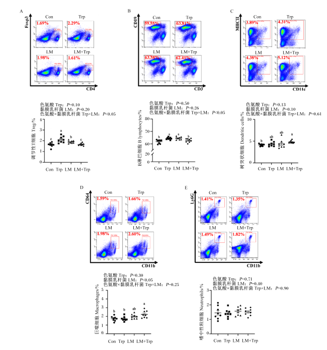
图2所示,与对照组相比,色氨酸和黏膜乳杆菌单独添加均可显著提高小鼠脾脏中Treg和B淋巴细胞相对丰度(P<0.05),而联合添加并未表现出显著影响(P>0.05)。与对照组相比,黏膜乳杆菌单独添加有提高小鼠脾脏中树突状细胞和巨噬细胞相对丰度的趋势(P<0.10),黏膜乳杆菌和色氨酸联合添加可显著提高这2种免疫细胞的相对丰度(P<0.05)。试验各处理对小鼠脾脏中嗜中性粒细胞相对丰度无显著影响(P>0.05)。
图2 猪源黏膜乳杆菌和色氨酸对小鼠脾脏免疫细胞相对丰度的影响

Fig.2 Effects of porcine Limosilactobacillus mucosae and tryptophan on relative abundance of immune cells in spleen of mice

2.3 猪源黏膜乳杆菌和色氨酸对小鼠胃肠M2型巨噬细胞标志物和Wnt信号通路表达的影响
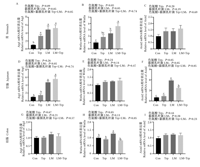
图3所示,与对照组相比,色氨酸组和黏膜乳杆菌+色氨酸组小鼠胃中Arg1的相对表达量显著提高(P<0.05),黏膜乳杆菌和黏膜乳杆菌+色氨酸组胃和空肠中Arg1的相对表达量显著提高(P<0.05),各处理对结肠中Arg1的相对表达量均无显著影响(P>0.05)。此外,与对照组相比,黏膜乳杆菌和黏膜乳杆菌+色氨酸组小鼠胃中Wnt3a的相对表达量显著提高(P<0.05),同时空肠中Axin2的相对表达量也显著提高(P<0.05)。
图3 猪源黏膜乳杆菌和色氨酸对小鼠胃肠M2型巨噬细胞标志物和Wnt信号通路表达的影响

Fig.3 Effects of porcine Limosilactobacillus mucosae and tryptophan on expression of M2 macrophage markers and Wnt signaling pathway in gastrointestinal tract of mice

2.4 猪源黏膜乳杆菌和色氨酸对小鼠胃肠增殖分化因子表达的影响

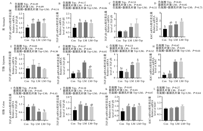
图4所示,与对照组相比,黏膜乳杆菌组小鼠胃中TGF-β1的相对表达量显著提高(P<0.05),各试验组胃中TGF-β2的相对表达量均显著提高(P<0.05);而在空肠和结肠中,黏膜乳杆菌组TGF-β2的相对表达量显著提高(P<0.05),黏膜乳杆菌组和黏膜乳杆菌+色氨酸组TGF-β3的相对表达量显著提高(P<0.05)。与对照组相比,黏膜乳杆菌组和黏膜乳杆菌+色氨酸组小鼠胃和空肠中Ki67的相对表达量显著提高(P<0.05)。
图4 猪源黏膜乳杆菌和色氨酸对小鼠胃肠增殖分化因子表达的影响

Fig.4 Effects of porcine Limosilactobacillus mucosae and tryptophan on expression of proliferative differentiation factors in gastrointestinal tract of mice

2.5 猪源黏膜乳杆菌和色氨酸对小鼠胃肠5-HT代谢和转运关键基因表达以及色氨酸代谢物浓度的影响
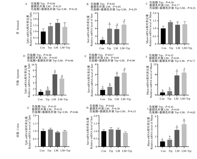
图5所示,与对照组相比,各试验组小鼠胃中Sert的相对表达量均显著提高(P<0.05);黏膜乳杆菌组和黏膜乳杆菌+色氨酸组空肠中Tph1和Sert的相对表达量显著提高(P<0.05);同时,黏膜乳杆菌组和黏膜乳杆菌+色氨酸组空肠和结肠中Maoa的相对表达量显著提高(P<0.05)。
图5 猪源黏膜乳杆菌和色氨酸对小鼠胃肠5-HT代谢和转运关键基因表达的影响

Fig.5 Effects of porcine Limosilactobacillus mucosae and tryptophan on expression of key genes in 5-HT metabolism and transport in gastrointestinal tract of mice

表1可知,与对照组相比,黏膜乳杆菌组和黏膜乳杆菌+色氨酸组小鼠胃组织中5-羟吲哚乙酸(5-HIAA)和3-吲哚乳酸(3-ILA)浓度显著提高(P<0.05),空肠组织中5-HT浓度显著降低(P<0.05);黏膜乳杆菌+色氨酸组空肠组织中3-ILA和3-吲哚乙酸(3-IAA)浓度显著降低(P<0.05);色氨酸组和黏膜乳杆菌组结肠组织中5-HT和3-IAA浓度显著降低(P<0.05),黏膜乳杆菌组和黏膜乳杆菌+色氨酸组结肠组织中吲哚浓度显著提高(P<0.05);黏膜乳杆菌组回肠内容物中5-HIAA浓度显著降低(P<0.05);黏膜乳杆菌组和黏膜乳杆菌+色氨酸组结肠内容物中3-吲哚丙酸(3-IPA)和3-甲基吲哚浓度显著降低(P<0.05),但结肠内容物中吲哚浓度显著提高(P<0.05)。
表1 猪源黏膜乳杆菌和色氨酸对小鼠肠道内容物和组织中色氨酸代谢物浓度的影响

Table 1 Effects of porcine Limosilactobacillus mucosae on concentrations of tryptophan metabolites in intestinal contents and tissues of micenmol/g

项目
Items
对照组
Control
group
色氨酸组
Trp
group
黏膜乳
杆菌组
LM group
黏膜乳杆菌+
色氨酸组
LM+Trp
group
均值
标准误
SEM
PP-value
色氨酸
Trp
黏膜
乳杆菌
LM
色氨酸×
黏膜乳杆菌
Trp×LM
胃组织Stomach tissue
5-羟色胺5-HT 2.13 2.70 3.48 2.42 0.21 0.54 0.19 0.06
5-羟吲哚乙酸5-HIAA 1.27c 1.31c 1.88b 2.37a 0.11 0.12 <0.05 0.18
3-吲哚乳酸3-ILA 1.18b 1.15b 1.67a 1.80a 0.11 0.81 <0.05 0.68
3-吲哚乙酸3-IAA 0.92 1.21 1.02 0.60 0.10 0.74 0.20 0.07
空肠组织Jejunum tissue
5-羟色胺5-HT 9.34a 8.92a 6.54b 6.61b 0.51 0.86 <0.05 0.80
5-羟吲哚乙酸5-HIAA 9.43 9.34 11.10 11.70 0.56 0.82 0.08 0.76
3-吲哚乳酸3-ILA 1.97a 1.84a 1.96a 1.42b 0.08 <0.05 0.12 0.14
3-吲哚乙酸3-IAA 4.13a 3.70a 1.61c 2.66b 0.20 0.12 <0.05 <0.05
结肠组织Colon tissue
5-羟色胺5-HT 24.50a 15.60bc 11.90c 20.80ab 1.43 1.00 0.13 <0.05
5-羟吲哚乙酸5-HIAA 5.36 8.10 9.11 7.03 0.51 0.74 0.17 <0.05
3-吲哚乳酸3-ILA 3.42 3.34 2.81 2.91 0.14 0.97 0.07 0.74
3-吲哚乙酸3-IAA 10.80a 7.31b 5.51b 10.70a 0.64 0.43 0.37 <0.05
3-吲哚丙酸3-IPA 0.71 0.62 0.38 0.60 0.05 0.48 0.07 0.12
吲哚Indole 9.49b 10.20b 12.10a 14.10a 0.80 0.40 <0.05 0.68
回肠内容物Ileal content
5-羟吲哚乙酸5-HIAA 21.40a 17.40ab 12.00b 22.50a 1.26 0.15 0.33 <0.05
吲哚Indole 8.65 8.51 7.28 8.24 0.43 0.64 0.36 0.54
结肠内容物Colonic content
5-羟吲哚乙酸5-HIAA 6.73ab 7.07ab 8.39a 5.61b 0.39 0.10 0.89 <0.05
3-吲哚乳酸3-ILA 9.15 7.39 7.68 6.46 0.43 0.09 0.16 0.75
3-吲哚乙酸3-IAA 2.00 1.74 1.73 1.77 0.09 0.58 0.54 0.43
3-吲哚丙酸3-IPA 1.97a 1.30b 1.11b 1.34b 0.11 0.29 0.06 <0.05
吲哚Indole 19.80b 17.40b 25.90a 31.00a 2.30 0.77 <0.05 0.40
3-甲基吲哚3-methylindole 0.10a 0.06b 0.05b 0.05b 0.01 0.12 <0.05 0.11

Trp:色氨酸;LM:黏膜乳杆菌。同行数据肩标不同字母表示差异显著(P<0.05)。表2同。

Trp: tryptophan; LM: Limosilactobacillus mucosae. In the same row, values with different letter superscripts mean significant difference (P<0.05). The same as Table 2.

2.6 猪源黏膜乳杆菌和色氨酸对小鼠胃肠HTR亚型表达的影响

表2可知,与对照组相比,黏膜乳杆菌+色氨酸组小鼠胃中HTR2AHTR7的相对表达量显著提高(P<0.05);黏膜乳杆菌组空肠和结肠中HTR1A的相对表达量显著提高(P<0.05),结肠中HTR2A的相对表达量显著提高(P<0.05);黏膜乳杆菌组和黏膜乳杆菌+色氨酸组胃中的HTR4的相对表达量以及空肠和结肠中HTR2B的相对表达量显著提高(P<0.05)。
表2 猪源黏膜乳杆菌和色氨酸对小鼠胃肠HTR亚型表达的影响

Table 2 Effects of porcine Limosilactobacillus mucosae and tryptophan on expression of HTR subtypes in gastrointestinal tract of mice

项目
Items
对照组
Control
group
色氨酸组
Trp
group
黏膜
乳杆菌组
LM group
黏膜乳杆菌+
色氨酸组
LM+Trp
group
均值
标准误
SEM
PP-value
色氨酸
Trp
黏膜
乳杆菌
LM
色氨酸×
黏膜乳杆菌
Trp×LM
胃Stomach
5-羟色胺受体1A HTR1A 1.00a 0.22b 0.83a 0.57ab 0.09 <0.05 0.54 0.09
5-羟色胺受体2A HTR2A 1.00b 1.63b 1.52b 3.06a 0.26 <0.05 <0.05 0.31
5-羟色胺受体2B HTR2B 1.00 0.69 1.05 1.16 0.08 0.53 0.11 0.19
5-羟色胺受体4 HTR4 1.00c 1.98c 3.29b 5.52a 0.40 <0.05 <0.05 0.11
5-羟色胺受体7 HTR7 1.00b 0.87b 0.91b 5.50a 0.46 <0.05 <0.05 <0.05
空肠Jejunum
5-羟色胺受体1A HTR1A 1.00bc 0.85c 2.71a 2.28ab 0.26 0.54 <0.05 0.76
5-羟色胺受体2A HTR2A 1.00 1.19 1.17 1.32 0.12 0.50 0.54 0.94
5-羟色胺受体2B HTR2B 1.00b 1.34ab 2.01a 2.00a 0.16 0.57 <0.05 0.54
5-羟色胺受体4 HTR4 1.00 0.99 1.08 1.13 0.07 0.92 0.49 0.85
5-羟色胺受体7 HTR7 1.00 0.85 1.34 1.07 0.11 0.36 0.22 0.79
结肠Colon
5-羟色胺受体1A HTR1A 1.00b 1.03b 4.94a 0.23c 0.41 <0.05 <0.05 <0.05
5-羟色胺受体2A HTR2A 1.00b 1.20b 2.81a 0.71b 0.18 <0.05 <0.05 <0.05
5-羟色胺受体2B HTR2B 1.00b 0.87b 1.31a 1.45a 0.07 0.96 <0.05 0.27
5-羟色胺受体4 HTR4 1.00 0.90 1.01 0.83 0.04 0.07 0.67 0.55
5-羟色胺受体7 HTR7 1.00b 1.10b 1.50a 0.79b 0.07 <0.05 0.40 <0.05

3 讨论

新生动物出生后的胃肠功能发育与成熟对畜禽的生长和健康以及生产性能的发挥至关重要。大量研究证实,功能性氨基酸以及以乳杆菌为代表的益生菌可改善幼龄畜禽的生长和健康。前期研究发现,色氨酸可改善断奶仔猪生长并提高了肠道中乳杆菌的相对丰度[26],而色氨酸可在体外培养条件下调节猪源黏膜乳杆菌的代谢[22]。肠道5-HT代谢以及HTR信号介导了色氨酸和黏膜乳杆菌缓解仔猪和小鼠肠道免疫反应与肠道炎症[21,27]。因此,黏膜乳杆菌与色氨酸可能通过调节仔猪胃肠发育和功能发挥作用,两者联合使用的作用效果和机制有待进一步研究明确。本试验通过幼龄小鼠模型,结合猪源黏膜乳杆菌灌胃和色氨酸饮水处理,主要发现如下:1)黏膜乳杆菌+色氨酸可促进幼龄小鼠体重增长和小肠长度增加;2)黏膜乳杆菌+色氨酸可调节胃肠5-HT代谢,并差异化调节HTR亚型在胃、空肠和结肠中的表达;3)黏膜乳杆菌可增加肠道M2型巨噬细胞极化,并差异化调节TGF-β亚型和Wnt信号通路在胃肠表达;4)黏膜乳杆菌+色氨酸可能通过提高后肠肠腔中吲哚浓度来发挥作用。因此,猪源黏膜乳杆菌与色氨酸联合使用可通过调节胃肠5-HT信号以及免疫稳态促进幼龄小鼠胃肠发育,其在仔猪生产中的作用效果与机制有待进一步验证。
幼龄动物肠道先天免疫的发育对适应性免疫的成熟以及抵抗病原菌的感染至关重要[28]。本研究结果表明,黏膜乳杆菌或色氨酸可提高幼龄小鼠脾脏中巨噬细胞和B淋巴细胞的相对丰度,并促进抗原递呈细胞树突状细胞的分化,黏膜乳杆菌和色氨酸联合处理显著提高巨噬细胞相对丰度并促进肠道M2型巨噬细胞极化。研究表明,将妊娠母猪粪便菌群与丁酸梭菌和布拉氏链球菌移植给新生仔猪后,仔猪血浆中IL-22和IL-17的浓度升高[29]。丁酸梭菌的早期干预提高大黄鱼内脏中酸性磷酸酶、过氧化氢酶和溶菌酶等免疫相关酶的活性[30]。免疫细胞相对丰度提高伴随一系列调节肠道增殖分化相关的细胞因子的产生,具体调控规律有待进一步动物试验探明。
本研究结果初步证明,HTR和TGF-β可能在巨噬细胞调节胃肠细胞增殖分化和损伤修复中发挥重要作用。研究表明,M2型巨噬细胞可特异性表达Arg1[31]。而Arg1催化底物产生的L-脯氨酸和多胺参与组织修复和伤口愈合[32]。HTR2B和HTR7的激活可特异性提高M2型巨噬细胞的相对丰度,并促进M2型巨噬细胞来源TGF-β浓度的升高[33]。此外,M2型巨噬细胞可通过表达Wnt配体(如Wnt1和Wnt3a)上调Wnt信号转导。本研究中,黏膜乳杆菌或黏膜乳杆菌+色氨酸处理可上调Arg1、Wnt3a以及Wnt信号通路激活标志物Axin2在胃和空肠中的表达;且黏膜乳杆菌或黏膜乳杆菌+色氨酸处理可同时差异化上调TGF-β2、TGF-β3或Ki67在胃肠的表达。这与以上2种处理可差异化上调HTR4和HTR7在胃中的表达以及HTR2B在空肠和结肠中的表达相关联。已有的研究表明,黏膜乳杆菌和色氨酸可差异化调节后肠炎症状态下TGF-β亚型的表达,TGF-β的表达和HTR4的表达呈正相关[13,21],然而HTR调节巨噬细胞功能调控肠细胞增殖分化的机制有待进一步试验探明。
本研究结果提示,肠道5-HT代谢以及信号稳态可能在幼龄动物肠道功能发育中发挥重要作用。肠道内的5-HT主要由色氨酸经Tph的催化合成,5-HT与受体结合发挥作用后在胞内经单胺氧化酶作用降解为5-HIAA失活[9]。关于生命早期肠道5-HT代谢和信号的研究大多集中在神经系统功能的调节作用,对幼龄动物肠道免疫的调节作用研究较少[34]。有研究表明,发育早期的应激可导致肠道5-HT产生增加,引起肠功能失调和免疫激活[35]。饲粮中额外添加色氨酸可降低仔猪结肠组织中5-HT浓度以及下调结肠中HTR2A的表达并上调HTR4的表达,缓解乙酸诱导的仔猪结肠免疫反应[27]。本研究表明,黏膜乳杆菌处理可降低小鼠空肠和结肠组织中5-HT浓度,这与黏膜乳杆菌上调了SertMaoa的表达并提高了空肠和结肠组织中的5-HIAA浓度相关。有趣的是,黏膜乳杆菌可上调HTR2B在空肠和结肠中的表达,HTR2B的激活已被证明可抑制结肠炎的发生[11]HTR2B敲除会促进小鼠肠上皮细胞的凋亡,并降低小鼠抵抗结肠炎症的能力[11]。此外,黏膜乳杆菌上调了胃中HTR4的表达。肠道HTR4可以通过增加肠上皮细胞增殖、促进细胞迁移以及抑制氧化应激诱导的细胞凋亡发挥肠道保护作用[36]。由于小肠和大肠的免疫功能存在差异[37-38],肠道HTR介导的胃肠免疫稳态及其调节肠道功能的机制有待进一步研究探明。

4 结论

① 黏膜乳杆菌和色氨酸联合处理2周后,可提高幼龄小鼠体重,黏膜乳杆菌和色氨酸单独或联合处理均可增加小鼠小肠长度。
② 黏膜乳杆菌单独处理或与色氨酸联合处理可差异化调节胃肠5-HT代谢以及HTR2BHTR4、HTR7的表达。
③ 黏膜乳杆菌单独处理或与色氨酸联合处理可上调胃肠Wnt配体及信号、增加胃肠M2型巨噬细胞极化以及促进TGF-β亚型的表达。
[1]
FRAZER L C, GOOD M. Intestinal epithelium in early life[J]. Mucosal Immunology, 2022, 15(6):1181-1187.

DOI PMID

[2]
WANG Q Y, XIONG X, LI J Z, et al. Energy metabolism in the intestinal crypt epithelial cells of piglets during the suckling period[J]. Scientific Reports, 2018, 8(1):12948.

DOI PMID

[3]
HUTING A M S, MIDDELKOOP A, GUAN X N, et al. Using nutritional strategies to shape the gastro-intestinal tracts of suckling and weaned piglets[J]. Animals:an open access journal from MDPI, 2021, 11(2):402.

[4]
LINDEMANS C A, CALAFIORE M, MERTELSMANN A M, et al. Interleukin-22 promotes intestinal-stem-cell-mediated epithelial regeneration[J]. Nature, 2015, 528(7583):560-564.

[5]
ZHU P P, ZHU X X, WU J Y, et al. IL-13 secreted by ILC2s promotes the self-renewal of intestinal stem cells through circular RNA circPan3[J]. Nature Immunology, 2019, 20(2):183-194.

DOI PMID

[6]
KIM J E, LI B, FEI L J, et al. Gut microbiota promotes stem cell differentiation through macrophage and mesenchymal niches in early postnatal development[J]. Immunity, 2022, 55(12):2300-2317.e6.

[7]
SONG S, BAI M J, LING Z Q, et al. Intermittent administration of a fasting-mimicking diet reduces intestinal inflammation and promotes repair to ameliorate inflammatory bowel disease in mice[J]. The Journal of Nutritional Biochemistry, 2021, 96:108785.

[8]
FISCHER J M, CALABRESE P P, MILLER A J, et al. Single cell lineage tracing reveals a role for TgfβR2 in intestinal stem cell dynamics and differentiation[J]. Proceedings of the National Academy of Sciences of the United States of America, 2016, 113(43):12192-12197.

PMID

[9]
JIANG L L, HAN D D, HAO Y L, et al. Linking serotonin homeostasis to gut function: nutrition, gut microbiota and beyond[J/OL]. Critical Reviews in Food Science and Nutrition, 2023:1-20(2023-03-01)[2024-01-10].https://doi.org/10.1080/10408398.2023.2183935.

[10]
AGUS A, PLANCHAIS J, SOKOL H. Gut microbiota regulation of tryptophan metabolism in health and disease[J]. Cell Host & Microbe, 2018, 23(6):716-724.

[11]
MAO L Y, XIN F, REN J, et al. 5-HT2B-mediated serotonin activation in enterocytes suppresses colitis-associated cancer initiation and promotes cancer progression[J]. Theranostics, 2022, 12(8):3928-3945.

DOI PMID

[12]
RAPALLI A, BERTONI S, ARCARO V, et al. Dual role of endogenous serotonin in 2,4,6-trinitrobenzene sulfonic acid-induced colitis[J]. Frontiers in Pharmacology, 2016, 7:68.

DOI PMID

[13]
WANG B, SUN S Q, LIU M Y, et al. Dietary L-tryptophan regulates colonic serotonin homeostasis in mice with dextran sodium sulfate-induced colitis[J]. The Journal of Nutrition, 2020, 150(7):1966-1976.

[14]
MARSHALL J S, WARRINGTON R, WATSON W, et al. An introduction to immunology and immunopathology[J]. Allergy,Asthma,and Clinical Immunology:Official Journal of the Canadian Society of Allergy and Clinical Immunology, 2018, 14(Suppl 2):49.

[15]
PATEL R M, MYERS L S, KURUNDKAR A R, et al. Probiotic bacteria induce maturation of intestinal claudin 3 expression and barrier function[J]. The American Journal of Pathology, 2012, 180(2):626-635.

[16]
PREIDIS G A, SAULNIER D M, BLUTT S E, et al. Probiotics stimulate enterocyte migration and microbial diversity in the neonatal mouse intestine[J]. The FASEB Journal, 2012, 26(5):1960-1969.

[17]
ZHU P P, LU T K, WU J Y, et al. Gut microbiota drives macrophage-dependent self-renewal of intestinal stem cells via niche enteric serotonergic neurons[J]. Cell Research, 2022, 32(6):555-569.

DOI PMID

[18]
ROOS S, KARNER F, AXELSSON L, et al. Lactobacillus mucosae sp.nov.,a new species with in vitro mucus-binding activity isolated from pig intestine[J]. International Journal of Systematic and Evolutionary Microbiology, 2000, 50(1):251-258.

[19]
DE MORAES G M D, DE ABREU L R, DO EGITO A S, et al. Functional properties of Lactobacillus mucosae strains isolated from Brazilian goat milk[J]. Probiotics and Antimicrobial Proteins, 2017, 9(3):235-245.

[20]
刘沫言, 郝友玲, 宋庆庆, 等. 仔猪小肠优势乳酸杆菌断奶前后全肠道演替及其比较代谢组学研究[J]. 动物营养学报, 2023, 35(3):1501-1512.

DOI

LIU M Y, HAO Y L, SONG Q Q, et al. Whole intestinal succession of dominant lactobacilli in small intestine of piglets before and after weaning and their comparative metabolomics[J]. Chinese Journal of Animal Nutrition, 2023, 35(3):1501-1512. (in Chinese)

[21]
HAO Y L, JIANG L L, HAN D D, et al. Limosilactobacillus mucosae and Lactobacillus amylovorus protect against experimental colitis via upregulation of colonic 5-hydroxytryptamine receptor 4 and transforming growth factor-β2[J]. The Journal of Nutrition, 2023, 153(8):2512-2522.

[22]
WU S Z, LIU M Y, CHEN H, et al. Tryptophan regulates bile and nitrogen metabolism in two pig gut lactobacilli species in vitro based on metabolomics study[J]. Amino Acids, 2022, 54(10):1421-1435.

[23]
SUN Y L, WU Z L, LI W, et al. Dietary L-leucine supplementation enhances intestinal development in suckling piglets[J]. Amino Acids, 2015, 47(8):1517-1525.

[24]
SUN Z Y, HUANG S Q, YAN X, et al. Living,heat-killed Limosilactobacillus mucosae and its cell-free supernatant differentially regulate colonic serotonin receptors and immune response in experimental colitis[J]. Nutrients, 2024, 16(4):468.

[25]
DAI Z L, SUN S Q, CHEN H, et al. Analysis of tryptophan and its metabolites by high-performance liquid chromatography[M]// ALTERMAN M A.Amino acid analysis:methods and protocols. New Jersey: Humana, 2019:131-142.

[26]
LIANG H W, DAI Z L, KOU J, et al. Dietary L-tryptophan supplementation enhances the intestinal mucosal barrier function in weaned piglets:implication of tryptophan-metabolizing microbiota[J]. International Journal of Molecular Sciences, 2018, 20(1):20.

[27]
WANG B, CUI L, SONG Q Q, et al. Excessive dietary L-tryptophan regulated amino acids metabolism and serotonin signaling in the colon of weaning piglets with acetate-induced gut inflammation[J]. Amino Acids, 2023, 55(3):403-412.

[28]
WYNN J L, LEVY O. Role of innate host defenses in susceptibility to early-onset neonatal sepsis[J]. Clinics in Perinatology, 2010, 37(2):307-337.

DOI PMID

[29]
XIANG Q H, WU X Y, PAN Y, et al. Early-life intervention using fecal microbiota combined with probiotics promotes gut microbiota maturation,regulates immune system development,and alleviates weaning stress in piglets[J]. International Journal of Molecular Sciences, 2020, 21(2):503.

[30]
YIN Z Y, LIU Q D, LIU Y T, et al. Early life intervention using probiotic Clostridium butyricum improves intestinal development,immune response,and gut microbiota in large yellow croaker (Larimichthys crocea) larvae[J]. Frontiers in Immunology, 2021, 12:640767.

[31]
WYNN T A, CHAWLA A, POLLARD J W. Macrophage biology in development,homeostasis and disease[J]. Nature, 2013, 496(7446):445-455.

[32]
PESCE J T, RAMALINGAM T R, MENTINK-KANE M M, et al. Arginase-1-expressing macrophages suppress Th2 cytokine-driven inflammation and fibrosis[J]. PLoS Pathogens, 2009, 5(4):e1000371.

[33]
DE LAS CASAS-ENGEL M, DOMÍNGUEZ-SOTO A, SIERRA-FILARDI E, et al. Serotonin skews human macrophage polarization through HTR2B and HTR7[J]. Journal of Immunology(Baltimore,Md.: 1950),2013, 190(5):2301-2310.

[34]
MALAVE L, VAN DIJK M T, ANACKER C. Early life adversity shapes neural circuit function during sensitive postnatal developmental periods[J]. Translational Psychiatry, 2022, 12(1):306.

DOI PMID

[35]
CHOW C F W, CHE S J, QIN H Y, et al. From psychology to physicality:how nerve growth factor transduces early life stress into gastrointestinal motility disorders later in life[J]. Cell Cycle, 2019, 18(16):1824-1829.

[36]
SPOHN S N, BIANCO F, SCOTT R B, et al. Protective actions of epithelial 5-hydroxytryptamine 4 receptors in normal and inflamed colon[J]. Gastroenterology, 2016, 151(5):933-944.e3.

DOI PMID

[37]
MOWAT A M, AGACE W W. Regional specialization within the intestinal immune system[J]. Nature Reviews Immunology, 2014, 14(10):667-685.

DOI PMID

[38]
DENNING T L, NORRIS B A, MEDINA-CONTRERAS O, et al. Functional specializations of intestinal dendritic cell and macrophage subsets that control Th17 and regulatory T cell responses are dependent on the T cell/APC ratio,source of mouse strain,and regional localization[J]. Journal of Immunology(Baltimore,Md.: 1950),2011, 187(2):733-747.

Outlines

/