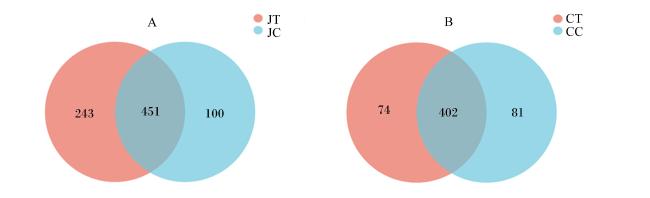
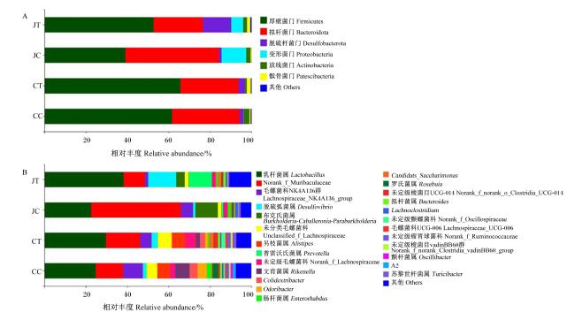
1 材料与方法
1.1 菌株的分离和鉴定
1.2 温度耐受性试验
1.3 人工胃液和人工肠液耐受性试验
1.4 耐药性试验
1.5 动物试验
1.5.1 肠道组织形态分析
1.5.2 肠道菌群分析
1.6 数据统计分析
2 结果与分析
2.1 菌株的分离鉴定结果
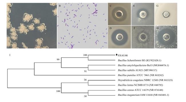
图1 分离菌株鉴定结果A为菌落形态,B为革兰氏染色结果,C~H分别表示对金黄色葡萄球菌、表皮葡萄球菌、大肠杆菌、单增李斯特菌、短小芽孢杆菌和藤黄微球菌的抑菌效果,I为系统发育进化树。 Bacillus licheniformis:地衣芽孢杆菌;Bacillus amyloliquefaciens:解淀粉芽孢杆菌;Bacillus subtilis:枯草芽孢杆菌;Bacillus pumilus:短小芽孢杆菌;Heyndrickxia coagulans:凝结芽孢杆菌;Bacillus lentus:迟缓芽孢杆菌;Bacillus cereus:蜡样芽孢杆菌;Bacillus megaterium:巨大芽孢杆菌。 Fig.1 Identification results of isolated strain A was colony morphology, B was Gram staining result, C to H represented the antibacterial effects on Staphylococcus aureus, Staphylococcus epidermidis, Escherichia coli, Listeria monocytogenes, Bacillus pumilus and Micrococcus luteus, respectively, and I was phylogenetic evolutionary tree. |
2.2 地衣芽孢杆菌SXAU08对温度的耐受性
表1 温度对地衣芽孢杆菌SXAU08生长的影响Table 1 Effects of temperature on growth of Bacillus licheniformis SXAU08 |
| 时间 Time/h | 温度 Temperature/℃ | ||||
|---|---|---|---|---|---|
| 37 | 40 | 60 | 80 | 100 | |
| 6 | 0.30±0.01a | 0.30±0.00a | 0.25±0.00b | 0.10±0.00c | 0.05±0.00d |
| 12 | 0.43±0.10a | 0.42±0.00a | 0.37±0.00b | 0.31±0.00c | 0.33±0.00c |
| 18 | 0.51±0.00c | 0.49±0.00c | 0.56±0.02ab | 0.59±0.01a | 0.53±0.01bc |
| 24 | 0.53±0.01b | 0.46±0.00c | 0.56±0.02b | 0.64±0.01a | 0.43±0.01c |
| 30 | 0.55±0.01b | 0.52±0.01c | 0.58±0.00b | 0.63±0.01a | 0.51±0.01c |
| 36 | 0.55±0.01b | 0.51±0.01c | 0.52±0.01c | 0.65±0.01a | 0.46±0.00d |
| 42 | 0.57±0.00b | 0.54±0.01bc | 0.55±0.01b | 0.62±0.01a | 0.50±0.00c |
| 48 | 0.55±0.02b | 0.55±0.03b | 0.56±0.01b | 0.65±0.01a | 0.41±0.01c |
2.3 地衣芽孢杆菌SXAU08对人工胃液和人工肠液的耐受性
表2 人工胃液和人工肠液对地衣芽孢杆菌SXAU08生长的影响Table 2 Effects of simulated gastric fluid and simulated intestinal fluid on growth of Bacillus licheniformis SXAU08 |
| 项目 Items | 时间 Time/h | ||||||
|---|---|---|---|---|---|---|---|
| 1 | 2 | 3 | 4 | 5 | 6 | ||
| 人工胃液 Simulated gastric fluid | |||||||
| pH | 2.0 | 1.54±0.01 | 1.58±0.01 | 1.54±0.02 | 1.56±0.04 | 1.59±0.01 | 1.61±0.01 |
| 3.0 | 1.74±0.01c | 1.86±0.01bc | 1.85±0.03c | 1.81±0.03c | 1.97±0.00ab | 1.98±0.02a | |
| 4.0 | 1.78±0.01c | 1.85±0.02c | 1.87±0.01c | 1.87±0.01c | 2.14±0.04a | 1.97±0.04b | |
| 人工肠液 Simulated intestinal fluid | |||||||
| 胆盐浓度 Bile salt concentration/% | 0 | 0.42±0.01e | 0.44±0.01e | 0.62±0.01d | 0.70±0.00c | 0.75±0.01b | 0.81±0.00a |
| 0.2 | 0.34±0.00e | 0.40±0.01d | 0.41±0.01d | 0.49±0.00c | 0.51±0.00b | 0.66±0.01a | |
| 0.4 | 0.34±0.00c | 0.32±0.01c | 0.41±0.01b | 0.43±0.01b | 0.44±0.01b | 0.50±0.01a | |
| 0.6 | 0.34±0.01c | 0.36±0.00c | 0.39±0.01b | 0.37±0.01c | 0.46±0.01a | 0.45±0.01a | |
| 0.8 | 0.35±0.00d | 0.36±0.00cd | 0.40±0.02ab | 0.43±0.00a | 0.39±0.00bc | 0.42±0.00a | |
| 1.0 | 0.27±0.01b | 0.31±0.04b | 0.37±0.00a | 0.41±0.01a | 0.40±0.01a | 0.42±0.00a | |
2.4 地衣芽孢杆菌SXAU08的耐药性分析
表3 地衣芽孢杆菌SXAU08对抗生素的敏感性Table 3 Sensitivity of Bacillus licheniformis SXAU08 to antibioticsmm |
| 项目 Items | 浓度 Concentration/(mg/mL) | ||||
|---|---|---|---|---|---|
| 1 | 10-1 | 10-2 | 10-3 | 10-4 | |
| 磺胺甲恶唑 Sulfamethoxazole | 41.15±0.94a | 33.07±1.58b | 21.89±0.29c | 0.00±0.00d | 0.00±0.00d |
| 磺胺嘧啶钠 Sodium sulfadiazine | 16.65±0.50a | 0.00±0.00b | 0.00±0.00b | 0.00±0.00b | 0.00±0.00b |
| 庆大霉素 Gentamicin | 27.81±0.37a | 21.04±0.23b | 14.27±0.09c | 0.00±0.00d | 0.00±0.00d |
| 新霉素 Neomycin | 23.32±0.11a | 16.79±0.89b | 9.46±0.39c | 0.00±0.00d | 0.00±0.00d |
| 盐酸土霉素 Oxytetracycline hydrochloride | 25.34±0.53a | 16.08±0.20b | 0.00±0.00c | 0.00±0.00c | 0.00±0.00c |
| 氟氧沙星 Ofloxacin | 45.11±0.34a | 37.42±0.47b | 29.94±0.48c | 16.32±0.15d | 0.00±0.00e |
| 氟苯尼考 Florfenicol | 40.38±0.28a | 28.41±0.41b | 18.14±0.34c | 0.00±0.00d | 0.00±0.00d |
| 红霉素 Erythromycin | 24.15±0.12a | 20.14±0.27b | 15.59±0.59c | 12.63±0.38d | 0.00±0.00e |
| 甲硝唑 Metronidazole | 25.12±0.60a | 0.00±0.00b | 0.00±0.00b | 0.00±0.00b | 0.00±0.00b |
| 阿莫西林 Amoxicillin | 32.71±0.41a | 25.78±0.21b | 24.93±4.53b | 0.00±0.00c | 0.00±0.00c |
2.5 地衣芽孢杆菌SXAU08对小鼠十二指肠组织形态的影响
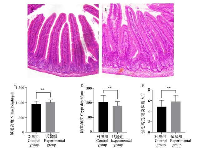
图2 地衣芽孢杆菌SXAU08对小鼠十二指肠组织形态的影响A:对照组小鼠十二指肠HE染色(100×);B:试验组小鼠十二指肠HE染色(100×);C:十二指肠绒毛高度分析;D:十二指肠隐窝深度分析;E:十二指肠V/C值分析。**表示差异极显著(P<0.01)。 Fig.2 Effects of Bacillus licheniformis SXAU08 on duodenal tissue morphology of mice A: HE staining in duodenum of mice in control group (100×); B: HE staining in duodenum of mice in experiment group (100×); C: villus height analysis in duodenum; D: crypt depth analysis in duodenum; E: V/C value analysis in duodenum. ** mean extremely significant difference (P<0.01). |