RESEARCH PAPER

Diversity Analysis of Rumen Fiber-Degrading Bacteria in Dairy Cows Enriched with Different Fiber Sources

  • CHANG Shuaifei ,
  • CHENG Yuchen ,
  • REN Wenyi ,
  • LI Haibo ,
  • HE Jintong ,
  • KANG Yan ,
  • KONG Xiaoli ,
  • ZHANG Lili ,
  • XU Xiaofeng , *
Expand
  • College of Animal Science and Technology, Ningxia University, Yinchuan 750021, China
* professor, E-mail:

Received date: 2024-04-01

  Online published: 2024-11-09

Abstract

The aim of this experiment was to investigate the changes in the diversity of rumen fiber-degrading bacteria in dairy cows enriched with different fiber sources. The experiment was conducted in a completely randomized design and divided into four groups, namely, control group without carbon source (CK group), sodium carboxymethylcellulose group (CMC group), microcrystalline cellulose group (MCC group) and Whatman No.1 filter paper group (WN group), with six replicates in each group. In vitro rumen fermentation tests were carried out to study the effects of different fiber sources on fermentation parameters and microflora structure. The results showed as follows: 1) the pH in both MCC and WN groups was significantly lower than that in CK and CMC groups (P<0.05). The concentrations of acetic acid, butyric acid and valeric acid as well as the acetic acid to propanoic acid ratio in all experimental groups were significantly higher than those in CK group (P<0.05); the isobutyric acid concentration in CMC group was significantly higher than that in CK group (P<0.05); the concentrations of propionic acid, isobutyric acid and total volatile fatty acids in MCC group were significantly higher than those in CK group (P<0.05), and the isovaleric acid concentration was significantly lower than that in CK group (P<0.05); the concentrations of propionic acid and total volatile fatty acids in WN group were significantly higher than those in CK group (P<0.05), and the isovaleric acid concentration was significantly lower than that in CK group (P<0.05). 2) In α diversity analysis, the indices of Chao1, Sobs, Simpson, Shannon, Pielou_e and PD in MCC and WN group were significantly lower than those in CK and CMC groups (P<0.05). At the phylum level, the Firmicutes relative abundance in all experimental groups was significantly higher than that in CK group (P<0.05), and the relative abundances of Bacteroidetes and Synergistetes were significantly lower than those in CK group (P<0.05); the Fibrobacteres relative abundance in MCC group was significantly higher than that in the other experimental groups and CK group (P<0.05). At the genus level, the relative abundances of Bacteroides, Pyramidobacter and Oscillospira in all experimental groups were significantly lower than that in CK group (P<0.05); the Clostridium relative abundance in CMC group was significantly higher than that in CK group (P<0.05), and the relative abundances of Prevotella and Succiniclasticum were significantly lower than those in CK group (P<0.05); the relative abundances of Lachnospira, Clostridium, Ruminococcus, Prevotella and Fibrobacter in MCC group were significantly higher than those in CK group (P<0.05), and the relative abundances of Succiniclasticum and Butyrivibrio in MCC group were significantly lower than those in CK group (P<0.05); the relative abundances of Lachnospira and Ruminococcus in WN group were significantly higher than those in CK group (P<0.05), and the Butyrivibrio relative abundance was significantly lower than that in CK group (P<0.05). 3) The Bacteroides relative abundance had a significant negative correlation with concentrations of acetic acid and valeric acid (P<0.05); the relative abundances of Lachnospira and Ruminococcus had a significant positive correlation with the concentrations of acetic acid, butyric acid and total volatile fatty acids (P<0.05), and had a significant negative correlation with the isovaleric acid concentration and pH (P<0.05); the Clostridium relative abundance had a significant positive correlation with the isobutyric acid concentration (P<0.05); the Prevotella relative abundance had a significant positive correlation with the propionic acid content (P<0.05). In conclusion, the highest degradation rate of Whatman No.1 filter paper was achieved by the rumen fiber-degrading bacteria in dairy cows; the preference of rumen fiber-degrading bacteria in dairy cows for different fiber sources varies markedly, with sodium carboxymethylcellulose being enriched for Clostridium, microcrystalline cellulose being enriched for Fibrobacter, and Whatman No.1 filter paper being enriched for Ruminococcus.

Cite this article

CHANG Shuaifei , CHENG Yuchen , REN Wenyi , LI Haibo , HE Jintong , KANG Yan , KONG Xiaoli , ZHANG Lili , XU Xiaofeng . Diversity Analysis of Rumen Fiber-Degrading Bacteria in Dairy Cows Enriched with Different Fiber Sources[J]. Chinese Journal of Animal Nutrition, 2024 , 36(11) : 7068 -7080 . DOI: 10.12418/CJAN2024.603

反刍动物的瘤胃是降解纤维素的高效的“生物反应器”。大量的瘤胃微生物将植物纤维物质转化为微生物蛋白和挥发性脂肪酸,在满足自身的营养需要的同时,还能够满足宿主的营养需要[1-3]。随着人们对瘤胃微生物的不断探索,已有大量文献报道了用于分离瘤胃微生物的培养基及分离方法。刘占英[4]使用羧甲基纤维素钠作为唯一碳源依据“透明圈”的大小来筛选到绵羊瘤胃内23株纤维降解菌。但由于羧甲基纤维素钠的羧甲基对外切纤维素酶有阻碍作用,使用“透明圈法”筛选到的纤维降解菌还需要使用Whatman No.1滤纸进行复筛。程超[5]先使用Whatman No.1滤纸作为唯一碳源配制液体培养基对瘤胃纤维降解菌初筛后,再使用纤维二糖分离纯化。刘玉承[6]使用Whatman No.1滤纸初筛,再使用纤维素粉分离纯化。国内外学者使用不同的分离方法和不同的碳源分离瘤胃纤维降解菌。然而,目前关于瘤胃纤维降解菌对不同纤维源的偏好性还鲜有报道。因此,本试验旨在通过在体外条件下利用3种代表性纤维碳源(羧甲基纤维素钠、微晶纤维素和Whatman No.1滤纸)富集奶牛瘤胃纤维降解菌,研究其菌群多样性的变化,以期为瘤胃菌群纤维降解机制的阐明和反刍动物饲粮纤维的高效利用提供理论参考。

1 材料与方法

1.1 培养基的配制

对文献[4]培养基配方适当调整后配制基础培养基,具体成分如下(每升含):碳酸氢钠(NaHCO3),5.0 g;蛋白胨,1.0 g;酵母粉,1.0 g;无细胞瘤胃液,170 mL;磷酸二氢钾(KH2PO4),0.495 g;硫酸铵[(NH4)2SO4],0.495 g;氯化钠(NaCl),0.99 g;二水合氯化钙(CaCl2·2H2O),0.066 g;七水合硫酸锰(MgSO4·7H2O),0.095 7 g;三水合磷酸氢二钾(K2HPO4·3H2O),0.653 4 g;0.1%刃天青,1 mL。
无细胞瘤胃液的制备方法:新鲜瘤胃液经高速冷冻离心机(2 743×g,4 ℃)离心15 min后,取上清液在15 710×g、4 ℃条件下离心30 min,分装上清液,于-20 ℃保存备用。
将基础培养基分别添加10 g/L的羧甲基纤维素钠、微晶纤维素和Whatman No.1滤纸(将滤纸剪切成滤纸碎片)配制羧甲基纤维素钠培养基、微晶纤维素培养基和Whatman No.1滤纸培养基。将培养基持续通二氧化碳(CO2),待培养基颜色变浅时加入1.5 g/L的半胱氨酸盐酸盐,继续通CO2至培养基淡黄色后在厌氧条件下分装到20 mL亨氏管中,每管装9 mL,121 ℃灭菌15 min。

1.2 试验设计

试验采用完全随机设计,共分为4组。以不加任何纤维素的基础培养基为对照组(CK组),羧甲基纤维素钠培养基为羧甲基纤维素钠组(CMC组),微晶纤维素培养基为微晶纤维素组(MCC组),Whatman No.1滤纸培养基为Whatman No.1滤纸组(WN组),每组设置6个重复。

1.3 瘤胃发酵液的制备

自屠宰场采集现场屠宰的新鲜瘤胃液。每次取样时,采集3头奶牛的瘤胃液,混匀后装入充有CO2的保温瓶,迅速带回实验室。奶牛饲粮精粗比为30∶70(风干基础),基础饲粮组成及营养水平见表1。接种时,将新鲜瘤胃液匀浆10 s后经4层纱布过滤去掉残渣后分别接种至基础培养基、羧甲基纤维素钠培养基、微晶纤维素培养基和Whatman No.1滤纸培养基,每管接种1 mL。将接种后的培养基39 ℃、120 r/min振荡培养。48 h后采集样品,测定pH后将所有发酵液分装于2 mL冻存管,于-80 ℃保存备用。
表1 基础饲粮组成及营养水平(干物质基础)

Table 1 Composition and nutrient levels of the basal diet (DM basis) %

项目Items 含量Content
原料Ingredients
玉米青贮Corn silage 45.06
苜蓿青贮Alfalfa silage 4.12
啤酒糟Brewer’s grains 1.53
蒸汽压片玉米Steam-flaked corn 22.50
苜蓿干草Alfalfa hay 10.32
浓缩料Concentrated feed1) 14.30
甜菜颗粒Beet granules 2.00
碳酸氢钠NaHCO3 0.17
合计Total 100.00
营养水平Nutrient levels2)
净能NE/(MJ/kg) 5.94
粗蛋白质CP 11.69
中性洗涤纤维NDF 47.32
酸性洗涤纤维ADF 30.74
粗脂肪EE 2.54
钙Ca 0.67
磷P 0.28

1)浓缩料的营养水平如下 Nutrient levels of the concentrated feed were as follows:干物质 DM 86%,粗蛋白质 CP 29.5%,粗灰分 Ash 18%,粗纤维 CF 12%,钙 Ca 1.3%,总磷 TP 0.6%,氯化钠 NaCl 1.3%,赖氨酸 Lys 0.8%。每千克浓缩料含有 One kilogram of the concentrated feed contained the following:维生素A乙酸酯 vitamin A acetate 12 000 IU,VD3 3 300 IU,DL-α-生育酚乙酸酯 DL-α-tocopheryl acetate 4 200 mg,烟酰胺 nicotinamide 6 700 mg,Mn 6 200 mg,Fe 1 800 mg,Zn 8 900 mg,Cu 1 700 mg,Se 41.7 mg,I 120 mg,Co 90 mg。

2)净能参照NRC(2001)计算所得,其余为实测值。NE was calculated by reference to NRC (2001), while the others were measured values.

1.4 测定指标及方法

1.4.1 饲粮营养成分的测定

饲粮粗蛋白质含量参照GB/T 6432—2018方法测定,粗脂肪含量参照GB/T 6433—2006方法测定,钙和磷含量分别参照GB/T 6436—2018和GB/T 6437—2018方法测定;中性洗涤纤维和酸性洗涤纤维含量参照Van Soest等[7]的方法测定。

1.4.2 发酵参数的测定

发酵结束后,立即用冰水冷却发酵液,使用便携式pH计测量pH。随后将剩余发酵液全部置于-80 ℃冰箱保存。参照李娅楠[8]所述方法测定挥发性脂肪酸浓度。

1.4.3 微生物测序

将-80 ℃保存的发酵液样品送往上海拜谱生物科技有限公司进行细菌测序。先对样品进行DNA提取,然后利用引物338F(5'-ACTCCTACGGGAGGCAGCA-3')和806R(5'-GGACTACHVGGGTWTCTAAT-3')对样品DNA进行PCR扩增。将PCR产物用1%琼脂糖凝胶电泳检测,再用Agencourt AMPure XP核酸纯化试剂盒进行分离纯化。利用Illumina HiSeq PE 2500平台对端测序,进行16S rDNA序列测定。利用DADA2软件对原始数据进行质量过滤、去噪、去重、拼接及去嵌合体。利用Greengenes数据库(gg_13)对扩增子序列变异(amplicon sequence variants,ASV)进行聚类[9]。α多样性指数通过QIIME2软件计算[10],微生物的相对丰度和多样性差异使用单因素方差分析来比较。

1.5 数据统计与分析

使用SAS 9.4版本对试验数据进行单因素方差分析,并采用Duncan氏法检验不同样品平均值之间的差异显著性。试验结果数据以“平均值±标准差”形式表示,所有分析均以P<0.05为差异显著,以0.05<P<0.10为差异有显著趋势。

2 结果与分析

2.1 不同纤维源发酵液发酵参数的变化

表2可知,针对发酵液pH,MCC组和WN组pH均显著低于CK组(P<0.05),CMC组pH与CK组相比无显著差异(P>0.05)。针对发酵液挥发性脂肪酸浓度,各试验组乙酸、丁酸、戊酸浓度以及乙丙比均显著高于CK组(P<0.05);CMC组异丁酸浓度显著高于CK组(P<0.05),而丙酸、异戊酸和总挥发性脂肪酸浓度与CK组相比均无显著差异(P>0.05);MCC组丙酸、异丁酸和总挥发性脂肪酸浓度均显著高于CK组(P<0.05),异戊酸浓度显著低于CK组(P<0.05);WN组丙酸和总挥发性脂肪酸浓度显著高于CK组(P<0.05),异戊酸浓度显著低于CK组(P<0.05),异丁酸浓度与CK组相比无显著差异(P>0.05)。
表2 不同纤维源发酵液发酵参数的变化

Table 2 Changes of fermentation parameters in fermentation broth from different fiber sources

项目
Items
组别Groups P
P-value
CK CMC MCC WN
pH 6.54±0.03a 6.56±0.02a 6.23±0.06b 6.09±0.04c <0.001
乙酸Acetic acid/(mmol/L) 12.11±0.79c 13.35±0.34b 19.74±0.35a 20.20±0.84a <0.001
丙酸Propionic acid/(mmol/L) 3.30±0.39b 2.80±0.44b 4.61±0.35a 4.70±0.31a <0.001
丁酸Butyric acid/(mmol/L) 1.62±0.17d 1.93±0.24c 3.02±0.03b 3.68±0.18a <0.001
异丁酸Isobutyric acid/(mmol/L) 0.00±0.00b 0.21±0.02a 0.15±0.09a 0.01±0.01b 0.012
戊酸Valeric acid/(mmol/L) 0.34±0.05c 0.49±0.10b 0.44±0.03b 0.58±0.04a <0.001
异戊酸Isovaleric acid/(mmol/L) 0.47±0.05a 0.42±0.06a 0.35±0.03b 0.19±0.04c <0.001
乙丙比Acetic acid/propionic acid 3.69±0.37b 4.86±0.71a 4.29±0.27a 4.30±0.22a 0.006
总挥发性脂肪酸TVFA/(mmol/L) 17.71±1.16c 19.19±0.99c 30.71±0.74b 32.96±1.31a <0.001

同行数据肩标相同小写字母或无字母表示差异不显著(P>0.05),不同小写字母表示差异显著(P<0.05)。下表同。

In the same row, values with the same small letter or no letter superscript mean no significant difference (P>0.05), while with different small letter superscripts mean significant difference (P<0.05). The same as below.

2.2 不同纤维源发酵液细菌群落的变化

2.2.1 不同纤维源发酵液细菌群落多样性分析
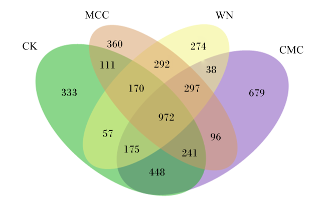
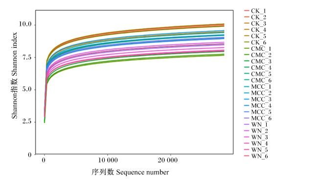
对CK组、CMC组、MCC组和WN组共计24个样品进行16S rRNA基因测序,下机序列经拼接、优化和质控后共得到1 198 849条优质序列,共获得4 543个ASV。由图1可知,CK组特有ASV数目为333个,CMC组特有ASV数目为679个,MCC组特有ASV数目为360个,WN组特有ASV数目为274个。由图2可知,随着测序深度的增加,各样品菌群稀释曲线末端均趋向平稳,表明测序数据能够反映菌群结构。
图1 不同纤维源发酵液细菌群落ASV分布韦恩图

CK:对照组;CMC:羧甲基纤维素钠组;MCC:微晶纤维素组;WN:Whatman No.1滤纸组。下图同。

Fig.1 Venn graph of ASV distribution of bacterial community in fermentation broth from different fiber sources

CK: control group, CMC: sodium carboxymethylcellulose group; MCC: microcrystalline cellulose group; WN: Whatman No.1 filter paper group. The same as below.

图2 不同纤维源发酵液菌群稀疏曲线

Fig.2 Rarefaction curves of bacteria in fermentation broth from different fiber sources

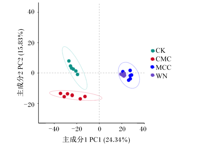
表3可知,各组间Goods_coverage指数无显著差异(P>0.05)。MCC组和WN组Chao1指数、Sobs指数、Simpson指数、Shannon指数、Pielou_e指数和PD指数均显著低于CK组和CMC组(P<0.05)。MCC组Shannon指数和Pielou_e显著低于WN组(P<0.05),CMC组Pielou_e指数显著高于CK组(P<0.05)。由图3可知,4组发酵液样品呈现各自的聚类性,在细菌群落组成上具有明显差异。
表3 不同纤维源发酵液细菌群落α多样性的变化

Table 3 Changes of α diversity of bacterial community in fermentation broth from different fiber sources

项目
Items
组别Groups P
P-value
CK CMC MCC WN
Goods_coverage指数
Goods_coverage index
0.99±0.00 0.99±0.00 0.99±0.00 0.99±0.00 0.291
Chao1指数Chao1 index 1 193.46±38.18a 1 224.80±70.78a 963.86±84.72b 940.03±75.92b <0.001
Sobs指数Sobs index 1 184.33±38.14a 1 212.83±73.83a 953.00±84.13b 926.33±75.40b <0.001
Simpson指数Simpson index 0.99±0.00a 0.99±0.00a 0.98±0.01b 0.99±0.01b 0.001
Shannon指数Shannon index 8.23±0.22a 8.30±0.07a 7.36±0.24c 7.71±0.21b <0.001
Pielou_e指数Pielou_e index 0.89±0.02b 0.90±0.01a 0.83±0.02d 0.86±0.01c <0.001
PD指数PD index 98.65±2.50a 99.62±3.55a 83.64±5.01b 80.14±3.98b <0.001
图3 不同发酵液菌群的PCA图

Fig.3 PCA graph of rumen microflora of different fermentation broth flora

2.2.2 不同纤维源发酵液细菌群落组成分析

表4可知,各试验组厚壁菌门(Firmicutes)相对丰度均显著高于CK组(P<0.05),拟杆菌门(Bacteroidetes)和互养菌门(Synergistetes)相对丰度均显著低于CK组(P<0.05)。WN组厚壁菌门相对丰度最高,显著高于其他试验组和CK组(P<0.05)。MCC组纤维杆菌门(Fibrobacteres)相对丰度最高,显著高于其他试验组和CK组(P<0.05)。
表4 不同种纤维素发酵液细菌群落在门水平上的变化

Table 4 Changes of bacterial community in fermentation broth from different fiber sources at phylum level %

项目
Items
组别Groups P
P-value
CK CMC MCC WN
厚壁菌门Firmicutes 40.18±1.75d 66.72±5.24b 58.63±3.96c 71.19±2.08a <0.001
拟杆菌门Bacteroidetes 51.85±0.94a 27.92±4.80c 36.34±2.02b 24.42±1.70d <0.001
互养菌门Synergistetes 2.70±0.39a 0.41±0.13d 0.84±0.16c 0.15±0.06b <0.001
纤维杆菌门Fibrobacteres 0.00±0.00b 0.01±0.01b 0.68±0.37a 0.00±0.00b <0.001
其他Others 5.25±0.92 4.93±0.75 3.51±1.89 4.14±0.78 0.236
表5可知,各试验组拟杆菌属(Bacteroides)、Pyramidobacter和颤螺菌属(Oscillospira)相对丰度显著低于CK组(P<0.05);CMC组梭菌属(Clostridium)相对丰度显著高于CK组(P<0.05),普雷沃氏菌属(Prevotella)和琥珀酸单胞菌属(Succiniclasticum)相对丰度显著低于CK组(P<0.05);MCC组毛螺菌属(Lachnospira)、梭菌属、瘤胃球菌属(Ruminococcus)、普雷沃氏菌属和纤维杆菌属(Fibrobacter)相对丰度显著高于CK组(P<0.05),琥珀酸单胞菌属和丁酸弧菌属(Butyrivibrio)相对丰度均显著低于CK组(P<0.05);WN组毛螺菌属和瘤胃球菌属相对丰度显著高于CK组(P<0.05),而丁酸弧菌属相对丰度显著低于CK组(P<0.05)。WN组毛螺菌属和瘤胃球菌属相对丰度最高,显著高于其他试验组和CK组(P<0.05);MCC组普雷沃氏菌属和纤维杆菌属相对丰度最高,显著高于其他试验组和CK组(P<0.05);CMC组梭菌属相对丰度最高,显著高于其他试验组和CK组(P<0.05)。
表5 不同种纤维素发酵液细菌群落在属水平上的变化

Table 5 Changes of bacterial community in fermentation broth from different fiber sources at genus level %

项目
Items
组别Groups P
P-value
CK CMC MCC WN
拟杆菌属Bacteroides 31.12±1.03a 13.30±4.24c 16.75±2.95c 10.19±0.72b <0.001
毛螺菌属Lachnospira 9.33±0.63c 9.22±2.07c 27.71±2.20b 30.52±1.14a <0.001
梭菌属Clostridium 10.18±0.78c 37.40±8.07a 18.33±1.78b 10.57±0.94c <0.001
瘤胃球菌属Ruminococcus 1.78±0.15c 0.76±0.11c 13.54±3.48b 21.67±2.47a <0.001
普雷沃氏菌属Prevotella 4.27±0.82b 0.37±0.16c 10.00±2.40a 2.61±0.28b <0.001
琥珀酸单胞菌属Succiniclasticum 2.19±0.44a 1.18±0.24b 1.13±0.55b 2.17±0.21a <0.001
Pyramidobacter 2.59±0.37a 0.38±0.13d 0.72±0.21c 1.38±0.09b <0.001
颤螺菌属Oscillospira 2.26±1.02a 1.35±0.21b 0.25±0.28c 0.29±0.05c <0.001
丁酸弧菌属Butyrivibrio 0.57±0.07a 0.51±0.19a 0.24±0.04b 0.12±0.04b <0.001
纤维杆菌属Fibrobacter 0.00±0.00b 0.00±0.00b 0.68±0.37a 0.00±0.00b <0.001
其他Others 33.25±2.05a 33.19±1.30a 17.60±2.95b 21.26±1.93b <0.001

2.3 不同纤维源发酵液发酵参数与细菌群落相对丰度关联分析

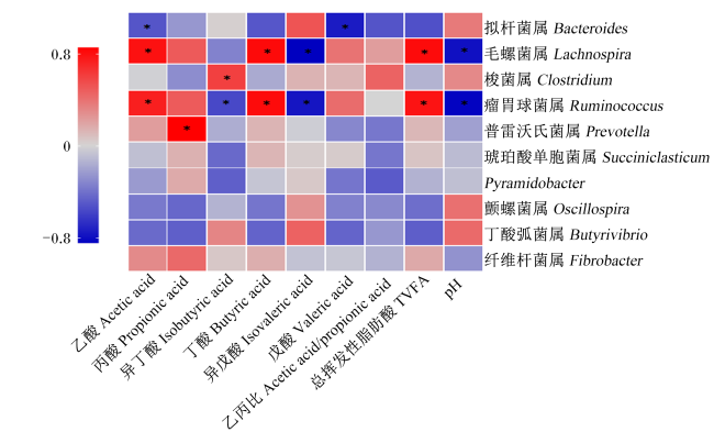
图4可知,不同纤维源发酵液拟杆菌属相对丰度与乙酸和戊酸浓度呈显著负相关(P<0.05);毛螺菌属和瘤胃球菌属相对丰度与乙酸、丁酸和总挥发性脂肪酸浓度呈显著正相关(P<0.05),与异戊酸浓度和pH呈显著负相关(P<0.05);梭菌属相对丰度与异丁酸浓度呈显著正相关(P<0.05);普雷沃氏菌属相对丰度与丙酸浓度呈显著正相关(P<0.05)。
图4 不同纤维源发酵液发酵参数与细菌群落相对丰度关联分析

*表示显著相关(P<0.05)。

Fig.4 Correlation analysis between fermentation parameters and relative abundance of bacterial community in fer-mentation broth from different fiber sources

* indicated significant correlation (P<0.05).

3 讨论

3.1 不同纤维源对发酵液发酵参数的影响

挥发性脂肪酸产量是衡量发酵品质的重要指标[8]。一般而言,正常瘤胃和体外产气的总挥发性脂肪酸浓度应至少在80 mmol/L以上[11]。本试验的总挥发性脂肪酸浓度很低,可能有两方面的原因:1)本次试验使用的培养底物是纯度达到99%的纤维素,极难降解。2)培养技术的差异。本试验借鉴了苛养厌氧菌(瘤胃球菌)的体外培养技术,培养过程在亨氏管中进行,强调了培养全程的厌氧性。在瘤胃液接种时,产气试验普遍使用瘤胃液与缓冲液1∶2混合,本试验瘤胃液与培养基1∶9混合。
羧甲基纤维素钠化学式为[C6H7O2(OH)2CH2COONa]n,是由天然纤维素经过化学改性得到的一种水溶性纤维素醚。研究表明,羧甲基纤维素钠的羧甲基对外切纤维素酶有阻碍作用,可用于内切纤维素酶活的测定[1]。微晶纤维素是一种以β-1,4-糖苷键结合的直链式多糖,可用于外切纤维素酶活的测定。而Whatman No.1滤纸是一种经过特殊处理的高纯度不溶性纤维素,其α-纤维素含量在98%以上。Whatman No.1滤纸广泛用于总纤维素酶活的测定。在本试验条件下,Whatman No.1滤纸组(WN组)发酵液总挥发性脂肪酸浓度最高,其次是MCC组,最后是CMC组,且pH排序正好相反。产酸效率和pH可以间接反映瘤胃微生物对纤维素的降解效率[12]。由于羧甲基对外切纤维素酶的阻碍作用,瘤胃微生物对羧甲基纤维素钠的降解效率较低。而外切葡聚糖酶在纤维降解过程中起中心作用[4],瘤胃微生物对Whatman No.1滤纸和微晶纤维素均有较高的分解效率。此外,瘤胃微生物对不同纤维源降解速率的差异可能与纤维降解机制有关。瘤胃纤维降解菌往往需要附着于纤维素表面,将分泌的纤维素酶与纤维素紧紧黏结在一起来发挥纤维降解功能[13]。相比于另2种纤维素,Whatman No.1滤纸具有疏松多孔的特性,易于附着。因此,瘤胃微生物对Whatman No.1滤纸的降解速率最高。
大多数纤维降解菌(黄色瘤胃球菌和白色瘤胃球菌)的发酵终产物是乙酸而不是丙酸,当饲料纤维含量增加时,瘤胃液的乙丙比往往增加[14]。这与本研究结果一致,添加不同纤维源的发酵液乙丙比均显著提高,并在CMC组达到最高。支链脂肪酸(异丁酸和异戊酸)主要来源于蛋白质降解后的氨基酸(缬氨酸、亮氨酸和异亮氨酸)经氧化脱氨基和脱羧基产物[15]。大量研究表明,反刍动物饲粮中添加异丁酸[16]和异戊酸[17]能显著提高纤维分解菌数量和纤维素酶活性。本试验中,发酵液的异丁酸和异戊酸含量分别于CMC组、MCC组和WN组依次降低,与纤维降解效率成反比。这可能与纤维降解菌的利用有关,瘤胃内的主要纤维降解菌(白色瘤胃球菌、黄色瘤胃球菌和琥珀酸丝状杆菌)往往需要在低浓度的支链脂肪酸条件下生长[18]。在瘤胃球菌的分离培养基中需要加入3%的支链脂肪酸混合液,且当培养基中添加100~300 mmol/L的挥发性脂肪酸混合物时,白色瘤胃球菌对羧甲基纤维素的降解能力会显著提高[4]。戊酸的生物合成与丙酸有关,当丙酸与电子供体之间发生链延伸反应过程时,戊酸的偶链和奇链可以作为中间产物产生[19]。在纯培养条件下,克氏梭状芽孢杆菌(Clostridium kluyveri)可以使用乙醇作为电子供体将丙酸转化为戊酸[20]。本次的试验结果验证了戊酸和丙酸之间的相关性,发酵液中戊酸的变化趋势与丙酸相同。

3.2 不同纤维源对发酵液细菌群落的影响

pH对微生物的生长繁殖有较大影响[21]。研究表明,当瘤胃内pH由6.5下降到5.8时,瘤胃菌群密度显著下降[22]。同时,瘤胃内的纤维素降解与pH有较大的依赖性,瘤胃内主要的纤维降解菌(白色瘤胃球菌、黄色瘤胃球菌和产琥珀酸丝状杆菌)不能在pH<6的酸性条件下正常生长[23]。本试验中,CMC组和CK组发酵液的菌群α多样性显著高于MCC组和WN组,这可能与发酵液pH变化有关,pH的变化影响了菌群的丰富度和均匀度。
瘤胃微生物是具有高适应性的生态群,菌群结构会根据底物类型发生变化。富纤维饲粮使瘤胃内纤维降解菌增加,富淀粉饲粮使淀粉降解菌增加[24]。厚壁菌门和拟杆菌门是本试验条件下的优势菌门,当Whatman No.1滤纸为唯一碳源时,厚壁菌门相对丰度最高(68.8%~74.4%)。厚壁菌门和拟杆菌门是反刍动物瘤胃内的主要菌门[25]。厚壁菌门主要参与纤维物质的分解代谢[26],目前对牛瘤胃中纤维素降解的研究主要集中在厚壁菌门中分离的少数菌株,即白色瘤胃球菌和黄色瘤胃球菌。拟杆菌门主要参与非纤维碳水化合物的降解。研究表明,拟杆菌门的碳水化合物降解能力与一种被称为多糖利用位点(PULs)的基因簇有关[27]。PULs主要作用于淀粉、半纤维素和果胶,可能有助于从半纤维素基质中释放纤维素[28]。纤维杆菌门是另一个参与木质纤维素降解的重要菌门。关鹏等[29]研究表明,纤维杆菌门和纤维杆菌属相对丰度变化与饲粮中性洗涤纤维含量有关。
瘤胃球菌属是奶牛瘤胃中最重要的纤维降解类群,瘤胃球菌属的成员(白色瘤胃球菌和黄色瘤胃球菌)是瘤胃中主要的纤维降解菌。黄色瘤胃球菌和白色瘤胃球菌会利用纤维小体来促进植物纤维在瘤胃内的降解。纤维小体是一种能够使微生物黏附到纤维素上,从而在超微结构上降解纤维素的多模块酶复合物[1]。研究证实,黄色瘤胃球菌能够编码大量的锚定蛋白和新型碳水化合物结合模块(CBMs)[30]。基因组分析表明,纤维小体蛋白在黄色瘤胃球菌的不同菌株间存在差异,其锚定蛋白数量在53~223[31]。黄色瘤胃球菌FD-1的纤维小体多达14酶亚基,这些酶亚基被组装在4个不同的支架蛋白上[32]。但与黄色瘤胃球菌相比,白色瘤胃球菌锚定蛋白编码基因较少,在3株已测序的菌株中,2株仅含有1个锚定蛋白编码基因[33]。除此之外,瘤胃球菌属的很多菌株都可以降解十分牢固的纤维素种类,如棉花纤维[34]。反刍动物瘤胃中有大量瘤胃球菌属来源糖苷水解酶第48家族(GH48)基因序列,并且这些基因有丰富的多样性[35]。而GH48家族成员是外切纤维素酶的重要来源之一,可以通过和其它糖苷水解酶形成纤维小体或者自由酶体系2种方式来降解纤维素[36]。本试验中,瘤胃球菌属在固态纤维素(滤纸和微晶纤维素)发酵液的相对丰度显著高于液态纤维素(羧甲基纤维素钠)发酵液。这可能与瘤胃球菌的纤维小体酶解机制有关。通过纤维小体降解机制,使瘤胃球菌黏附于纤维素表面[28],可以使纤维降解菌分泌的纤维素酶与底物直接接触,最大限度减少纤维素酶进入瘤胃液中造成的浪费。黏附可以使瘤胃球菌优先获得纤维素水解产物(如纤维二糖和纤维三糖),避免这些纤维降解产物被非纤维降解菌利用[37]。并有研究表明,当在可溶性的甲基纤维素上培养黄色瘤胃球菌时,黄色瘤胃球菌无法降解纤维素[38]
纤维杆菌属隶属于纤维杆菌门,同样是瘤胃中最主要的纤维降解类群之一,产琥珀酸丝状杆菌是其重要成员。有研究指出,产琥珀酸丝状杆菌的纤维降解机制比其他纤维降解微生物更有效,比如琥珀酸丝状杆菌S85和A3C能够比白色瘤胃球菌和黄色瘤胃球菌从完整饲粮中降解更多的纤维素[39]。产琥珀酸丝状杆菌缺乏锚定蛋白和黏结蛋白,且不编码任何外切葡聚糖酶,不能够利用纤维小体和游离酶这2种机制降解纤维素[40]。有研究者提出产琥珀酸丝状杆菌可能的纤维降解机制[41]:产琥珀酸丝状杆菌会利用纤维黏液蛋白和菌毛附着于纤维素上,使纤维素与外膜表面的内切葡聚糖酶结合,并产生含有碳水化合物活性酶(CAzymes)的外膜囊泡来水解纤维素。有研究提出,产琥珀酸丝状杆菌的外膜囊泡可以破坏木质纤维素的复杂结构来增强细菌对纤维素的降解与利用[42]。因此,产琥珀酸丝状杆菌具有很强的降解结构性坚韧物质(如秸秆)的能力,能够降解一些不被黄色瘤胃球菌降解的某些同质晶体纤维素[37]。这与本试验结果一致,纤维杆菌门和纤维杆菌属在MCC组的相对丰度显著高于CMC组和WN组。
梭菌属的很多菌株都具有迅速消化纤维素的能力。比如早期报道的Clostridium lochheadii,是一株可以快速分解蛋白质的纤维分解菌,Hungate将这个物种描述为“从瘤胃中分离出来的最快的纤维降解者”[43],其纤维降解能力甚至优于琥珀酸丝状杆菌和黄色瘤胃球菌。在本试验中,梭菌属的相对丰度在CMC组中最高,显著高于CK组和其他试验组,梭菌属的菌株可能具备较强的分泌内切纤维素酶的能力。溶纤维丁酸弧菌是瘤胃中代谢最丰富的纤维降解菌,其可发酵范围广,且在不同菌株间存在很大差异[44]。本试验中,不同纤维源下丁酸弧菌属相对丰度均较低,这可能是由于该菌的培养条件较高,在完整细胞壁和纤维素上生长较差,在实验室条件下的溶纤维丁酸弧菌很难表现出瘤胃中的纤维降解活性。拟杆菌属和琥珀酸单胞菌属是瘤胃中存在的淀粉降解菌,在本试验中,其相对丰度随不同纤维素的添加而显著下降。然而,普雷沃氏菌属同样是典型的淀粉降解菌,但其在MCC组相对丰度却显著升高。普雷沃氏菌属可能与其他纤维降解菌(如纤维杆菌属)存在协作机制,能够利用其他菌的纤维降解产物。

3.3 不同纤维源发酵液发酵参数与细菌群落关联分析

瘤胃球菌属是重要的产乙酸菌,其可以利用纤维素、木聚糖和纤维二糖等不同发酵底物产生大量的乙酸、甲酸或乳酸,并产生氢气(H2)和CO2。本研究结果表明,瘤胃球菌相对丰度与乙酸浓呈显著正相关,这与王亚玲等[45]的研究结果一致。普雷沃氏菌属是广泛存在于瘤胃且数量最多的一类细菌,其菌株间遗传差异大。研究表明,栖普雷沃氏菌属和布氏普雷沃氏菌可产生木聚糖酶和羧甲基纤维素酶,但是由于缺少真正的纤维素酶,因此在纯培养时不能降解细胞壁,但与纤维降解菌共培养时能有效利用木聚糖和果胶,并主要通过丙烯酸途径合成丙酸[46]。本研究进一步验证了普雷沃氏菌相对丰度与丙酸浓度的相关关系,即使在以纤维素作为唯一碳源时,普雷沃氏菌属也能够与纤维降解菌协作产生丙酸。毛螺菌属能够利用果胶、纤维二糖及木聚糖等产生乙酸和乳酸,广泛参与瘤胃中半纤维素的降解,但不能降解纤维素[47]。本试验结果表明,毛螺菌属可能与瘤胃球菌属存在共生关系,其相对丰度变化同瘤胃球菌属一致。

4 结论

① 奶牛瘤胃纤维降解菌对Whatman No.1滤纸的降解速率最高。
② 奶牛瘤胃纤维降解菌对不同纤维源的偏好性有明显不同,羧甲基纤维素钠富集梭菌属,微晶纤维素富集纤维杆菌属,Whatman No.1滤纸富集瘤胃球菌属。
[1]
ARTZI L, BAYER E A, MORAÏS S. Cellulosomes:bacterial nanomachines for dismantling plant polysaccharides[J]. Nature Reviews:Microbiology, 2017, 15(2):83-95.

[2]
WEIMER P J. Degradation of cellulose and hemicellulose by ruminal microorganisms[J]. Microorganisms, 2022, 10(12):2345.

[3]
AZIZI A, SHARIFI A, FAZAELI H, et al. Effect of transferring lignocellulose-degrading bacteria from termite to rumen fluid of sheep on in vitro gas production,fermentation parameters,microbial populations and enzyme activity[J]. Journal of Integrative Agricul-ture, 2020, 19(5):1323-1331.

[4]
刘占英. 绵羊瘤胃主要纤维降解细菌的分离鉴定及不同氮源对其纤维降解能力的影响[D]. 博士学位论文. 呼和浩特: 内蒙古农业大学, 2008.

LIU Z Y. Isolation and identification of major cellulolytic bacteria in rumen of sheep and effects of nitrogen sources on their cellulolytic activities[D]. Ph.D. Thesis. Hohhot: Inner Mongolia Agricultural University, 2008. (in Chinese)

[5]
程超. 绵羊瘤胃内纤维素分解菌的分离鉴定及白色瘤胃球菌纤维素酶celB基因的克隆[D]. 硕士学位论文. 呼和浩特: 内蒙古农业大学, 2007.

CHENG C. Isolation and identification of bacterium degrading the cellulose in rumen of sheep and cloning of celB gene in R.albus[D]. Master’s Thesis. Hohhot: Inner Mongolia Agricultural University, 2007. (in Chinese)

[6]
刘玉承. 32株瘤胃纤维降解细菌的分离鉴定与保藏研究[D]. 硕士学位论文. 呼和浩特: 内蒙古农业大学, 2007.

LIU Y C. Research on separation identification and preservation of 32 strain rumen cellulolytic bacteria[D]. Master’s Thesis. Hohhot: Inner Mongolia Agricultural University, 2007. (in Chinese)

[7]
VAN SOEST P J, ROBERTSON J B, LEWIS B A. Methods for dietary fiber,neutral detergent fiber,and nonstarch polysaccharides in relation to animal nutrition[J]. Journal of Dairy Science, 1991, 74(10):3583-3597.

[8]
李娅楠. 乳杆菌与地衣芽孢杆菌复合微贮对稻草营养价值影响及机理研究[D]. 硕士学位论文. 银川: 宁夏大学, 2022.

LI Y N. Effect and mechanism research of compound silage of Lactobacillus and Bacillus licheniformis on nutritional value of rice straw[D]. Master’s Thesis. Yinchuan: Ningxia University, 2022. (in Chinese)

[9]
JOOS L, BEIRINCKX S, HAEGEMAN A, et al. Daring to be differential:metabarcoding analysis of soil and plant-related microbial communities using amplicon sequence variants and operational taxonomical units[J]. BMC Genomics, 2020, 21(1):733.

[10]
CAPORASO J G, KUCZYNSKI J, STOMBAUGH J, et al. QIIME allows analysis of highthroughput community sequencing data[J]. Nature Methods, 2010, 7(5):335-336.

[11]
杨双鸣, 马煜斌, 田梅, 等. 体外产气法评价菌酶协同发酵对花棒瘤胃发酵特性的影响[J]. 动物营养学报, 2023, 35(11):7472-7480.

DOI

YANG S M, MA Y B, TIAN M, et al. Evaluation of effects of cooperative fermentation by bacteria and enzymes on rumen fermentation characteristics of Hedysarum scopariums by in vitro gas production method[J]. Chinese Journal of Animal Nutrition, 2023, 35(11):7472-7480. (in Chinese)

[12]
李岩, 戴东文, 杨英魁, 等. 不同精粗比饲粮对育肥后期牦牛生长性能、营养物质表观消化率、血清生化指标及瘤胃发酵参数的影响[J]. 动物营养学报, 2022, 34(5):3056-3065.

DOI

LI Y, DAI D W, YANG Y K, et al. Effects of diets with different concentrate to roughage ratios on growth performance,nutrient apparent digestibilities,serum biochemical indexes and rumen fermentation parameters of yak in late stage of fattening[J]. Chinese Journal of Ani-mal Nutrition, 2022, 34(5):3056-3065. (in Chinese)

[13]
马东旺, 席琳乔, 汤佳, 等. 秸秆种类及粒度对体外发酵、微生物表面物理特性及纤维降解菌组成的影响[J]. 动物营养学报, 2023, 35(11):7446-7460.

DOI

MA D W, XI L Q, TANG J, et al. Effects of straw species and particle size on in vitro fermentation,microbial physical surface characteristics and composition of cellulolytic bacteria[J]. Chinese Journal of Animal Nutrition, 2023, 35(11):7446-7460. (in Chinese)

[14]
王兴岗, 郝小燕, 张暄梓, 等. 不同精粗比饲粮中添加凝结芽孢杆菌对绵羊羔羊瘤胃发酵和免疫功能的影响[J]. 动物营养学报, 2024, 36(3):1769-1781.

DOI

WANG X G, HAO X Y, ZHANG X Z, et al. Effects of adding Bacillus coagulans into diets with different concentrate to forage ratios on rumen fermentation and immune function of lambs[J]. Chinese Journal of Animal Nutrition, 2024, 36(3):1769-1781. (in Chinese)

[15]
姜菲, 赖琦, 高彦华, 等. 添加异丁酸、2-甲基丁酸和戊酸对牦牛瘤胃体外发酵参数和养分降解率的影响[J]. 动物营养学报, 2022, 34(9):5915-5930.

DOI

JIANG F, LAI Q, GAO Y H, et al. Effects of supplementing isobutyric acid,2-methylbutyric acid and valeric acid on in vitro rumen fermentation parameters and nutrient degradation rates of yaks[J]. Chinese Journal of Animal Nutrition, 2022, 34(9):5915-5930. (in Chinese)

DOI

[16]
WANG C, LIU Q, ZHANG Y L, et al. Effects of isobutyrate supplementation on ruminal microflora,rumen enzyme activities and methane emissions in Simmental steers[J]. Journal of Animal Physiology and Animal Nutrition, 2015, 99(1):123-131.

[17]
LIU Q, WANG C, PEI C X, et al. Effects of isovalerate supplementation on microbial status and rumen enzyme profile in steers fed on corn stover based diet[J]. Livestock Science, 2014,161:60-68.

[18]
ABECIA L, MARTÁNEZ-FERNANDEZ G, WADDAMS K, et al. Analysis of the rumen microbiome and metabolome to study the effect of an antimethanogenic treatment applied in early life of kid goats[J]. Frontiers in Microbiology, 2018,9:2227.

[19]
封丽梅. 戊酸对瘤胃上皮细胞基因表达、瘤胃发酵及血液代谢的影响[D]. 硕士学位论文. 扬州: 扬州大学, 2022.

FENG L M. Effects of valerate on rumen epithelial cells gene expression,rumen fermentation and blood metabolism[D]. Master’s Thesis. Yangzhou: Yangzhou University, 2022. (in Chinese)

[20]
ANGENENT L T, RICHTER H, BUCKEL W, et al. Chain elongation with reactor microbi-omes:open-culture biotechnology to produce biochemicals[J]. Environmental Science & Technology, 2016, 50(6):2796-2810.

[21]
AKKERMANS S, LOGIST F, VAN IMPE J F, et al. An interaction model for the combined effect of temperature,pH and water activity on the growth rate of E.coli K12[J]. Food Research International, 2018,106:1123-1131.

[22]
张丽, 李川, 邱清华, 等. 不同pH下瘤胃微生物菌群密度及理化特性的变化[J]. 动物营养学报, 2023, 35(1):450-459.

DOI

ZHANG L, LI C, QIU Q H, et al. Changes of rumen microbiota density and physicochemical properties at different pH[J]. Chinese Journal of Animal Nutrition, 2023, 35(1):450-459. (in Chinese)

DOI

[23]
RUSSELL J B, DOMBROWSKI D B. Effect of pH on the efficiency of growth by pure cultures of rumen bacteria in continuous culture[J]. Applied and Environmental Microbiology, 1980, 39(3):604-610.

DOI PMID

[24]
LI Q S, WANG R, MA Z Y, et al. Dietary selection of metabolically distinct microorganisms drives hydrogen metabolism in ruminants[J]. The ISME Journal, 2022, 16(11):2535-2546.

[25]
徐晓锋, 郭婷婷, 郭成, 等. 甘露寡糖对高精料诱导的低乳脂奶牛瘤胃细菌菌群调控的研究[J]. 动物营养学报, 2019, 31(11):5246-5256.

XU X F, GUO T T, GUO C, et al. Regulation of mannan oligosaccharides on rumen bacterial flora induced by high concentrate in low milk fat dairy cows[J]. Chinese Journal of Animal Nutrition, 2019, 31(11):5246-5256. (in Chinese)

[26]
PANG K Y, CHAI S T, YANG Y K, et al. Dietary forage to concentrate ratios impact on yak ruminal microbiota and metabolites[J]. Frontiers in Microbiology, 2022,13:964564.

[27]
MARTENS E C, CHIANG H C, GORDON J I. Mucosal glycan foraging enhances fitness and transmission of a saccharolytic human gut bacterial symbiont[J]. Cell Host & Microbe, 2008, 4(5):447-457.

[28]
STEWART R D, AUFFRET M D, WARR A, et al. Assembly of 913 microbial genomes from metagenomic sequencing of the cow rumen[J]. Nature Communications, 2018, 9(1):870.

DOI PMID

[29]
关鹏, 任建刚, 黄建智, 等. 不同中性洗涤纤维水平饲粮对塔里木马鹿瘤胃微生物种群结构的影响[J]. 动物营养学报, 2023, 35(2):1206-1218.

DOI

GUAN P, REN J G, HUANG J Z, et al. Effects of diets with different neutral detergent fiber levels on rumen microbial population structure of Tarim wapiti[J]. Chinese Journal of Animal Nutrition, 2023, 35(2):1206-1218. (in Chinese)

DOI

[30]
SESHADRI R, LEAHY S C, ATTWOOD G T, et al. Cultivation and sequencing of rumen microbiome members from the Hungate1000 collection[J]. Nature Biotechnology, 2018, 36(4):359-367.

DOI PMID

[31]
ISRAELI-RUIMY V, BULE P, JINDOU S, et al. Complexity of the Ruminococcus flavefaciens FD-1 cellulosome reflects an expansion of family-related protein-protein interactions[J]. Scientific Reports, 2017,7:42355.

[32]
AN D D, DONG X Z, DONG Z Y. Prokaryote diversity in the rumen of yak (Bos grunniens) and Jinnan cattle (Bos taurus) estimated by 16S rDNA homology analyses[J]. Anaerobe, 2005, 11(4):207-215.

[33]
DASSA B, BOROVOK I, RUIMY-ISRAELI V, et al. Rumen cellulosomics:divergent fiber-degrading strategies revealed by comparative genome-wide analysis of six ruminococcal strains[J]. PLoS One, 2014, 9(7):e99221.

[34]
STEWART C S, DUNCAN S H, FLINT H J. The properties of forms of Ruminococcus flavefaciens which differ in their ability to degrade cotton cellulose[J]. FEMS Microbiology Letters, 1990, 72(1/2):47-50.

[35]
李劲亭, 苏小运, 田彦, 等. 瘤胃细菌GH48家族糖苷水解酶基因多样性[J]. 微生物学报, 2014, 54(1):53-61.

LI J T, SU X Y, TIAN Y, et al. Gene diversity of the bacterial 48 family glycoside hydrolase (GH48) in rumen environment[J]. Acta Microbiologica Sinica, 2014, 54(1):53-61. (in Chinese)

[36]
OLSON D G, TRIPATHI S A, GIANNONE R J, et al. Deletion of the Cel48S cellulase from Clostridium thermocellum[J]. Proceedings of the National Academy of Sciences of the United States of America, 2010, 107(41):17727-17732.

[37]
LYND L R, WEIMER P J, VAN ZYL W H, et al. Microbial cellulose utilization:fundamentals and biotechnology[J]. Microbiology and Molecular Biology Reviews, 2002, 66(3):506-577, table of contents.

[38]
RASMUSSEN M A, HESPELL R B, WHITE B A, et al. Inhibitory effects of methylcellulose on cellulose degradation by Ruminococcus flavefaciens[J]. Applied and Environmental Microbiology, 1988, 54(4):890-897.

[39]
RANSOM-JONES E, JONES D L, MCCARTHY A J, et al. The fibrobacteres:an important phylum of cellulose-degrading bacteria[J]. Microbial Ecology, 2012, 63(2):267-281.

[40]
SUEN G, WEIMER P J, STEVENSON D M, et al. The complete genome sequence of Fibrobacter succinogenes S85 reveals a cellulolytic and metabolic specialist[J]. PLoS One, 2011, 6(4):e18814.

[41]
BURNET M C, DOHNALKOVA A C, NEUMANN A P, et al. Evaluating models of cellulose degradation by Fibrobacter succinogenes S85[J]. PLoS One, 2015, 10(12):e0143809.

[42]
ARNTZEN M Ø, VÁRNAI A, MACKIE R I, et al. Outer membrane vesicles from Fibrobacter succinogenes S85 contain an array of carbohydrate-active enzymes with versatile poly-saccharide-degrading capacity[J]. Environmental Microbiology, 2017, 19(7):2701-2714.

[43]
HUNGATE R E. The rumen and its microbes[M]. New York: Academic Press,1966.

[44]
SECHOVCOVÁ H, KULHAVÁ L, FLIEGEROVÁ K, et al. Comparison of enzymatic activities and proteomic profiles of Butyrivibrio fibrisolvens grown on different carbon sources[J]. Proteome Science, 2019,17:2.

[45]
王亚玲, 孔鹏辉, 袁冬冬, 等. 饲粮添加乙酸钠对奶山羊泌乳性能和瘤胃微生物区系的影响[J]. 动物营养学报, 2023, 35(1):428-438.

DOI

WANG Y L, KONG P H, YUAN D D, et al. Effects of dietary sodium acetate on lactation performance and rumen microbiota of dairy goats[J]. Chinese Journal of Animal Nutri-tion, 2023, 35(1):428-438. (in Chinese)

[46]
ADAMBERG S, ADAMBERG K. Prevotella enterotype associates with diets supporting acidic faecal pH and production of propionic acid by microbiota[J]. Heliyon, 2024, 10(10):e31134.

[47]
SI B X, LIU K Z, HUANG G X, et al. Relationship between rumen bacterial community and milk fat in dairy cows[J]. Frontiers in Microbiology, 2023,14:1247348.

Outlines

/