REVIEW

Oxidative Damage of Deoxynivalenol to Animals and Protective Effect of Exogenous Antioxidants

  • LIANG Shujie ,
  • JIANG Yunfei ,
  • ZUO Jianjun ,
  • WANG Weiwei , *
Expand
  • Guangdong Provincial Key Laboratory of Animal Nutrition Control, College of Animal Science, South China Agricultural University, Guangzhou 510642, China
*associate professor, E-mail:

Received date: 2024-05-30

  Online published: 2024-12-12

Abstract

Deoxynivalenol (DON) is one of the most common fungal toxins that contaminate grains and feed. After ingesting feed contaminated with DON, animals experience varying degrees of damage to their intestinal system, immune system, nervous system, liver, kidney and other organs and tissues, accompanied by symptoms such as vomiting, anorexia, diarrhea, intestinal bleeding, and growth inhibition. The addition of exogenous antioxidants such as antioxidant enzymes, vitamins, trace elements, functional amino acids, bioactive substances, etc. can effectively alleviate oxidative stress and alleviate the toxic effect of DON on animals. This paper mainly reviews on oxidative damage of DON to livestock and poultry. It is manifest in analyzing the protective effect of antioxidants on livestock and poultry exposed to DON. Finally, it is expected that this review will provide reference for the use of antioxidants in production to prevent the contamination of DON.

Cite this article

LIANG Shujie , JIANG Yunfei , ZUO Jianjun , WANG Weiwei . Oxidative Damage of Deoxynivalenol to Animals and Protective Effect of Exogenous Antioxidants[J]. Chinese Journal of Animal Nutrition, 2024 , 36(12) : 7513 -7521 . DOI: 10.12418/CJAN2024.639

脱氧雪腐镰刀菌烯醇(deoxynivalenol,DON)又称呕吐毒素,是禾谷镰刀菌、粉红镰刀菌等霉菌的次级代谢产物[1-2]。DON广泛存在于玉米、大豆、小麦等谷物原料中,是饲料中污染最严重的霉菌毒素之一[3-4]。张雷[5]检测了2018—2020年从我国20多个省市收集的3 507份饲料原料和配合饲料中DON的含量,发现DON的检出率高达96.4%~100%,其含量在485.0~1 925.4 μg/kg,约有0.1%的饲料原料中DON含量超过国家规定的限量标准。2023年嘉吉全球霉菌毒素报告中显示,嘉吉对我国84 998份饲料原料样品进行霉菌毒素含量检测,结果发现,80%的饲料原料样品中霉菌毒素含量超过检测限,在所有被检测样品中超过检测限的污染率最高的毒素是DON、玉米赤霉烯酮和伏马毒素,分别为92%、92%和80%[6]
DON暴露会导致动物急性或慢性中毒,表现为肝脏、肾脏、肠道等多器官结构和功能损伤,并伴有呕吐、厌食、腹泻、肠出血、生长受阻等一系列临床症状[7-9]。为了防控饲料中霉菌毒素污染对动物健康的威胁和保障食品安全,物理法、化学法、生物法等方式被运用于霉菌毒素的脱毒上,但是我国关于霉菌毒素脱毒的研究尚处于发展阶段,整体技术尚未达到可成熟运用的水平[10]。因此,从饲料层面出发,配合使用饲料添加剂缓解DON对动物的毒性显得尤为重要。目前,已有研究报道,抗氧化剂对DON毒性的防治具有良好的效果[11]。本文主要就DON对畜禽的氧化损伤以及DON诱导下外源抗氧化剂对畜禽的保护作用进行综述,旨在为生产中利用抗氧化剂防治DON污染提供参考。

1 DON对畜禽的氧化损伤

动物长期摄入被DON污染的饲料会出现采食量下降、体重增长减慢、免疫功能紊乱等情况[12]。DON能够对多种动物产生毒性作用,但不同动物对DON的敏感程度不同,表现为猪>啮齿动物>犬>猫>家禽>反刍动物[13-14]。饲料中的DON对畜禽的危害主要体现在减少采食量、降低繁殖性能、抑制免疫功能、造成氧化损伤等方面。
DON引起肝脏、肠道、免疫等毒性效应的重要机制是氧化应激[15-16],其症状可能是由活性氧(reactive oxygen species,ROS)的过度产生、细胞抗氧化能力的下降或两者共同引起[17]。ROS可激活Kelch样ECH相关蛋白1(Kelch-like ECH associated protein,Keap1)/核因子E2相关因子2(nuclear factor-erythroid 2-related factor 2,Nrf2)/血红素加氧酶-1(heme oxygenase-1,HO-1)信号通路,促进Nrf2与Keap1解离,从细胞质易位到细胞核中与启动子区域的抗氧化反应元件(antioxidant response element,ARE)结合,调控下游信号分子如HO-1、过氧化氢酶(catalase,CAT)、超氧化物歧化酶(superoxide dismutase,SOD)的转录和表达,调节氧化还原平衡,减轻ROS的过量产生对细胞或组织造成的氧化损伤[18-19]。在DON处理早期,Nrf2的易位和激活显著上调,可降低ROS诱导的氧化损伤,但在后期,细胞的结构和功能严重受损,氧化损伤程度超过机体的补偿能力和Nrf2、HO-1正常功能的阈值,Nrf2、HO-1的激活受到抑制,从而导致更严重的毒性损伤[20-21]。DON还可通过Nrf2/过氧化物酶体增殖物激活受体γ(peroxisome proliferators-activated receptor γ,PPARγ)-谷胱甘肽过氧化物酶(glutathion peroxidase 4,GPX4)通路诱导肝脏脂质代谢障碍,使肠道和肝脏发生铁死亡,从而对肠道、肝脏造成损伤[22-25]。Yu等[26]对DON在妊娠雌性小鼠中诱导肝脏毒性的机制研究表明,ROS的积累可通过激活Nrf2/HO-1信号通路降低肝脏氧化应激水平,避免肝脏遭受更严重的损伤。DON可诱导人绒毛膜肿瘤细胞(BeWo细胞)产生氧化应激,使细胞内ROS水平升高,抗氧化酶活性降低,造成DNA损伤和脂质过氧化,最终引起细胞凋亡[20]。赵呈祥[27]研究发现,DON可诱导IPEC-J2细胞全基因组DNA甲基化水平升高,从而影响核苷酸生物合成、细胞代谢、蛋白质信号转导等生理学过程。DON可显著降低IPEC-J2细胞活力,诱导氧化应激和细胞凋亡,显著增加丙二醛(malondialdehyde,MDA)含量,降低CAT和SOD活性,并上调促炎性细胞因子和细胞凋亡相关基因的mRNA相对表达量,显著提高凋亡细胞率[28]。Hassan等[29]用含有10 mg/kg DON的饲粮饲喂肉鸡4周,结果发现,DON显著提高血清和肝脏MDA含量,显著降低血清和肝脏CAT、谷胱甘肽过氧化物酶(glutathione peroxidase,GSH-Px)、总超氧化物歧化酶(total superoxide dismutase,T-SOD)等抗氧化酶活性,诱导肉鸡产生氧化应激。

2 外源抗氧化剂对DON诱导的氧化应激的缓解作用

动物机体内存在抗氧化系统,包括酶类和非酶类抗氧化系统。酶类抗氧化系统包括CAT、SOD、GSH-Px、谷胱甘肽-S转移酶等,共同完成抗氧化反应[30]。当体内自由基水平升高时,为了抵抗氧化应激,机体内抗氧化酶活性升高以清除自由基[31]。当氧化应激水平超出其抵抗能力时,抗氧化酶的表达受到抑制,活性降低,进而细胞受到损伤[32-33]。非酶类抗氧化系统包括谷胱甘肽(glutathione,GSH)、铜蓝蛋白、金属硫蛋白等蛋白性抗氧化剂和维生素、微量元素等物质。各组织内,抗氧化系统各组分相互协调配合,共同保持抗氧化防御系统的完整性,以达到最佳保护作用。
DON对动物的毒理机制之一是引起线粒体功能障碍,导致ROS生成增加,同时细胞内抗氧化剂的生成减少,从而打破体内氧化还原平衡[34-35]。一旦ROS的积累速度超过了消除速度,体内就会出现氧化应激反应,某些生物大分子,特别是DNA和蛋白质,会被过氧化和破坏[35],从而损害动物的健康,抑制动物生长[8,35]。因此,抑制诱导性的氧化应激是缓解DON对畜禽毒性作用的潜在策略。
外源添加抗氧化剂如抗氧化酶、维生素、微量元素、功能性氨基酸、生物活性物质等可有效缓解DON对畜禽的毒性作用(图1)。
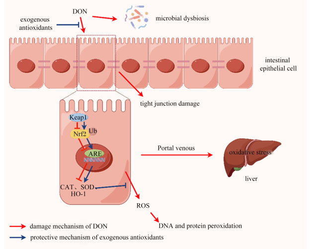
图1 DON对肠道、肝脏的损伤及外源抗氧化剂的保护作用(用Figdraw绘制)

DON: 脱氧雪腐镰刀菌烯醇 deoxynivalenol;exogenous antioxidants:外源抗氧化剂;microbial dysbiosis:菌群失调;intestinal epithelial cell:肠上皮细胞;tight junction damage:紧密连接受损;Keap1:Kelch样ECH相关蛋白1 Kelch-like ECH associated protein;Nrf2:核因子E2相关因子2 nuclear factor-erythroid 2-related factor 2;Ub:泛素 ubiquitin;ARE:抗氧化反应元件 antioxidant response element;CAT:过氧化氢酶 catalase;SOD:超氧化物歧化酶 superoxide dismutase;HO-1:血红素加氧酶-1 heme oxygenase-1;ROS:活性氧 reactive oxygen species;Portal venous:门静脉;DNA and protein peroxidation:DNA和蛋白质过氧化;oxidative stress:氧化应激;liver:肝脏;damage mechanism of DON:DON的损伤机制;protective mechanism of exogenous antioxidants:外源抗氧化剂的保护机制。

Fig.1 Damage of DON to intestine and liver and protective effect of exogenous antioxidants (drew by Figdraw)

2.1 抗氧化酶

当DON诱导机体产生氧化应激时,内源性抗氧化酶的表达受到抑制,外源添加CAT、SOD、GSH-Px、HO-1等抗氧化酶或过表达内源抗氧化酶可有效缓解DON造成的氧化损伤。
外源CAT可增加DON处理的肉鸡空肠CAT、GSH-Px活性和总抗氧化能力(total antioxidant capacity,T-AOC),降低肝脏ROS、8-羟基脱氧鸟苷、MDA含量和半胱氨酸天冬酶-9 mRNA相对表达量,减少肠道中变形菌、肠杆菌等有害菌丰度,增加酸杆菌等有益菌丰度,从而缓解DON诱导的肝脏氧化应激和肠道损伤,其机理可能是基于CAT清除ROS的活性和改善肠道菌群的作用[36]。外源SOD可显著抑制DON诱导的斑马鱼ROS水平上升[37];可显著恢复DON诱导小鼠的抗氧化系统、缓解肝脏氧化应激,具体表现为小鼠体重恢复,血清和肝脏中SOD、CAT、GSH-Px活性以及GSH含量显著增加;还可通过抑制RAB5相互作用因子(RAB5 interacting factor,RAB5IF)、核不均一核糖核蛋白AB(heterogeneous nuclear ribonucleoprotein A/B,hnRNPAB)、线粒体核酸内切酶G(endonuclease G,EndoG)基因的表达,促进干扰素诱导的三十四肽重复蛋白1(interferon-induced protein with tetratricopeptide repeats 1,IFIT1)基因的表达,抑制小鼠肝脏细胞凋亡,从而缓解环磷酰胺和DON共同诱导的肝脏炎症[38]
Ren等[39-40]构建了GPx1敲低和过表达猪脾淋巴细胞,并用DON进行处理,发现敲低GPx1的表达会降低猪脾淋巴细胞的抗氧化能力,且与正常细胞相比,DON对GPx1敲低的猪脾淋巴细胞造成的氧化损伤更严重,而GPx1过表达减少了DON诱导猪脾淋巴细胞的氧化损伤,降低了DNA甲基化的程度,减少了细胞凋亡。Peng等[41-42]则构建了HO-1敲低和过表达小鼠,结果发现,DON可诱导HO-1敲低小鼠肝脏产生严重的病变,显著提高谷丙转氨酶、谷草转氨酶活性,而HO-1过表达可有效保护肝脏正常的结构,通过产生一氧化碳、上调自噬能力来改善DON诱导的肝脏损伤,降低氧化应激和炎症损伤的程度。Meng等[43]也发现,HO-1过表达可维持DON处理下Hepa 1-6细胞的自噬能力,有效激活抗氧化酶/DNA修复系统,缓解肝脏损伤,但HO-1的敲低则会导致抗氧化酶/DNA修复系统的紊乱和肝脏严重损伤。
以上结果说明,抗氧化酶可能是一种缓解DON诱导的氧化应激的作用靶点,但其作用机制仍需要深入研究。

2.2 维生素

维生素是机体最重要的非酶类抗氧化剂。补充适量维生素可显著提高畜禽的抗氧化能力,进而提高其抵抗应激的能力[44]。β-胡萝卜素是维生素A的前体物,可直接有效地清除超氧阴离子,提高GSH-Px的活性,阻止自由基损伤DNA和蛋白质[45]。维生素E是最重要的脂溶性抗氧化剂,具有自由基清除能力,可使细胞膜免受ROS的损伤[46-47]。维生素C可通过维生素E依赖途径清除ROS,防止脂质过氧化和蛋白质烷基化,保护细胞免受氧化损伤[48]
周炳娟等[49]研究发现,用维生素C预处理人外周血单核细胞(HPBMC)可减少DON诱导的细胞凋亡,缓解其对细胞增殖的抑制作用。Shieh等[50]同样发现,用维生素E预处理HBEC-5i细胞可减少DON诱导的细胞凋亡和氧化损伤。维生素A、维生素C和维生素E对YAC-1小鼠淋巴瘤细胞的预处理均可显著减少DON诱导的蛋白质、脂质过氧化,缓解DON对细胞活力和增殖的抑制作用[51]。Van Le Thanh等[52]研究发现,在DON污染饲粮中组合添加维生素A、维生素C和维生素E可显著降低仔猪血浆和肝脏中MDA含量和肝脏SOD活性。叶黄素是一种非维生素A类胡萝卜素,研究发现叶黄素对HT-29细胞的预处理可缓解DON诱导的氧化应激,下调炎症因子核因子-κB(nuclear factor-κB,NF-κB)和环氧合酶-2(cyclooxygenase-2,COX-2)的基因表达水平,阻止NF-κB从细胞质易位到细胞核,并阻止细胞凋亡[53]
适量的维生素可缓解DON诱导的氧化应激和细胞凋亡,但应注意维生素的添加剂量。Sayin等[54]使用剂量为日常建议的5~50倍的维生素E和N-乙酰半胱氨酸(N-acetylcysteine,NAC)饲喂肺癌模型小鼠,结果发现,与对照组相比,维生素E和NAC均可促进肿瘤的发生,且显著提前小鼠因肿瘤死亡的时间。

2.3 微量元素

体内自由基的清除主要是通过抗氧化酶进行,而抗氧化酶发挥抗氧化功能需要微量元素的参与。硒是GSH-Px的重要成分,可提高GSH-Px活性以发挥其抗氧化作用。硒的预处理可通过提高仔猪脾脏淋巴细胞GSH-Px活性,减轻DON对抗氧化酶的损伤程度,缓解DON造成的氧化应激和细胞因子分泌紊乱[55-56]。硒纳米颗粒(SeNP)可通过调节IPEC-J2细胞的内质网应激,减少氧化损伤,以缓解DON诱导的肠上皮屏障功能障碍[57]。硒代蛋氨酸通过提高肝脏抗氧化能力,调节Nrf2/PPARγ-GPX4-铁死亡通路,有效缓解DON诱导的C57BL/6小鼠肝脏氧化损伤、代谢功能障碍[23]。硒代蛋氨酸还可通过调节Keap1/Nrf2信号通路,维持C57BL/6小鼠空肠结构的完整性,缓解DON诱导的氧化损伤[58]。在断奶仔猪的饲粮中添加黄芩苷-铜和黄芩苷-锌可显著降低空肠HO-1的蛋白表达水平,显著增加回肠p-Nrf2、p-NF-κB的蛋白表达水平,提高仔猪的抗氧化能力,从而缓解DON诱导的氧化损伤[59-60]。因此,硒、铜、锌等微量元素具有提高抗氧化酶活性的作用,是体内抗氧化系统中重要酶的活性成分。

2.4 功能性氨基酸

谷氨酰胺(glutamine,Gln)是一种条件性必需氨基酸。Gln作为氮在组织间运输的载体,是核苷酸和其他氨基酸合成的前体[61];同时,Gln也是快速分化细胞和肠黏膜细胞主要的能源物质,参与体内GSH的合成。Gln可降低IPEC-J2细胞的ROS水平,下调促炎症、促凋亡相关基因、蛋白的表达,从而缓解DON诱导的肠道上皮细胞损伤[62]。饲粮中添加精氨酸(arginine,Arg)和Gln可显著降低DON诱导的肝脏重量增加,显著提高血清中SOD活性和降低血清中IL-1β、IL-2、TNF-α含量,有效缓解DON诱导的生长猪氧化应激和免疫损伤[63]
其他氨基酸如蛋氨酸(methionine,Met)、半胱氨酸(cysteine,Cys)也具有抗氧化的作用,Met和Cys是GSH合成的前体,参与体内的氧化还原反应[64-65]。Met可通过改善肠道肝细胞扩张,重新激活Wnt/β-连环蛋白(β-catenin)信号通路来缓解DON诱导的肠道损伤[66]。NAC是Cys的N-乙酰化衍生物,可为细胞内GSH的合成提供底物,并促进其合成[67]。研究发现,NAC可阻止DON诱导的ROS生成增加,降低细胞凋亡和炎症相关基因、蛋白表达水平,从而缓解DON对IPEC-J2细胞造成的氧化损伤[68-69]

2.5 植物提取物

植物提取物中存在多种具有抗氧化功能的生物活性物质,可通过清除体内的自由基在抵抗氧化应激中发挥重要的作用,其中多酚类化合物近年来被广泛应用于抵抗霉菌毒素的毒性[70]
白藜芦醇(resveratrol,RES)是一种二苯乙烯类多酚化合物,在桑葚、蓝莓等食品中多有分布,是研究最多的多酚类物质之一[16,71]。RES能够提高肠道屏障功能,缓解断奶仔猪肠道炎症和氧化应激,减少细胞凋亡,调节肠道菌群,从而有效缓解DON诱导的仔猪肠道损伤[72]。Ling等[73]研究发现,RES可通过缓解DON诱导的细胞跨上皮电阻减低、细胞旁通透性增加,减少细菌易位,增强肠道屏障功能。RES通过Nrf2信号通路缓解DON对IPEC-J2细胞造成的氧化损伤[74],显著增加DON处理组仔猪空肠黏膜SOD活性和T-AOC,显著降低线粒体ROS水平,有效缓解DON诱导的氧化应激[75]。RES还可显著降低DON处理组仔猪回肠中IL-1βIL-6和TNF-α mRNA和蛋白表达水平以及MDA含量,有效减轻肠道炎症和氧化应激,并改善肠道菌群,缓解DON诱导的肠道屏障损伤[76]
除了RES,懈皮素、姜黄素、甘草酸、绿原酸等植物提取物也可防治动物受到DON诱导的毒性损伤[25,77-79]。自然界中存在着大量的植物提取物,只有少部分被研究报道,其中对RES的研究较多,仍有较多植物提取物可深入探索和挖掘[80]

3 小结与展望

DON具有神经毒性、肠道毒性、生殖毒性,会降低畜禽生长性能和繁殖性能,影响免疫功能,造成肠道和肝脏的氧化损伤。DON较重要的致病机制之一是破坏氧化还原平衡,造成氧化应激。通过外源添加抗氧化剂可缓解氧化应激,有效降低DON对畜禽的毒性作用。但是,目前关于外源抗氧化剂缓解DON诱导畜禽氧化应激的研究主要集中在表面,其具体的作用机制包括如何影响机体氧化还原平衡和激活抗氧化系统尚不明确。因此,还需进一步通过动物试验和细胞试验针对外源抗氧化剂的添加在DON诱导的畜禽氧化应激上开展机制研究,为外源抗氧化剂在生产上的精准应用提供理论基础和实践依据。
[1]
张养东, 杨军香, 王宗伟, 等. 饲料中主要霉菌毒素的种类、含量及其对奶牛生理功能的影响[J]. 中国奶牛, 2016(7):1-4.

ZHANG Y D, YANG J X, WANG Z W, et al. Research progress of mycotoxins on physiological function of dairy cows[J]. China Dairy Cattle, 2016(7):1-4. (in Chinese)

[2]
MA R, ZHANG L, LIU M, et al. Individual and combined occurrence of mycotoxins in feed ingredients and complete feeds in China[J]. Toxins, 2018, 10(3):113.

[3]
BENNETT J W, KLICH M. Mycotoxins[J]. Clinical Microbiology Reviews, 2003, 16(3):497-516.

DOI PMID

[4]
LI X Y, ZHAO L H, FAN Y, et al. Occurrence of mycotoxins in feed ingredients and complete feeds obtained from the Beijing region of China[J]. Journal of Animal Science and Biotechnology, 2014, 5(1):37.

[5]
张雷. 呕吐毒素危害断奶仔猪生长性能和肠道健康的机制研究[D].博士学位论文. 武汉: 华中农业大学,2022:26-34.

ZHANG L. Mechanism of DON’s damage to growth performance and intestinal health of weaned piglets[D].Ph.D.Thesis. Wuhan: Huazhong Agricultural University,2022:26-34. (in Chinese)

[6]
嘉吉微营养与健康科技业务. 嘉吉2023年全球霉菌毒素报告[DB/OL].(2024-04-01) [2024-05-01]. https://mp.weixin.qq.com/s/39viZhm2OxIqY8Xuy9SJ_w

Cargill Micronutrition and Health Technologies Business. Cargill global mycotoxin report 2023[DB/OL].(2024-04-01) [2024-05-01]. https://mp.weixin.qq.com/s/39viZhm2OxIqY8Xuy9SJ_win Chinese)

[7]
AL-JAAL B A, JAGANJAC M, BARCARU A, et al. Aflatoxin,fumonisin,ochratoxin,zearalenone and deoxynivalenol biomarkers in human biological fluids:a systematic literature review,2001-2018[J]. Food and Chemical Toxicology, 2019, 129:211-228.

[8]
PARASKEUAS V, GRIELA E, BOUZIOTIS D, et al. Effects of deoxynivalenol and fumonisins on broiler gut cytoprotective capacity[J]. Toxins, 2021, 13(10):729.

[9]
MENG X Y, YU W Y, DUAN N, et al. Protective effects of ferulic acid on deoxynivalenol-induced toxicity in IPEC-J2 cells[J]. Toxins, 2022, 14(4):275.

[10]
史姗姗, 沈飞, 刘晓庚. 中国谷物真菌毒素降解的研究现状与发展趋势[J]. 粮食科技与经济, 2021, 46(2):84-87.

SHI S S, SHEN F, LIU X G. Research status and development trend of mycotoxin degradation of Chinese cereals[J]. Grain Science and Technology and Economy, 2021, 46(2):84-87. (in Chinese)

[11]
邓诣群, 林如琴, 吴思婷, 等. 呕吐毒素的毒理机制及防治策略研究进展[J]. 华南农业大学学报, 2022, 43(6):87-96.

DENG Y Q, LIN R Q, WU S T, et al. Research progess in toxicological mechanism and prevention strategy of deoxynivalenol[J]. Journal of South China Agricultural University, 2022, 43(6):87-96. (in Chinese)

[12]
HOOFT J M, BUREAU D P. Deoxynivalenol:mechanisms of action and its effects on various terrestrial and aquatic species[J]. Food and Chemical Toxicology, 2021, 157:112616.

[13]
PINTON P, OSWALD I P. Effect of deoxynivalenol and other type B trichothecenes on the intestine:a review[J]. Toxins, 2014, 6(5):1615-1643.

[14]
FROBOSE H L, ERCEG J A, FOWLER S Q, et al. The progression of deoxynivalenol-induced growth suppression in nursery pigs and the potential of an algae-modified montmorillonite clay to mitigate these effects[J]. Journal of Animal Science, 2016, 94(9):3746-3759.

DOI PMID

[15]
WU Q H, WANG X, NEPOVIMOVA E, et al. Antioxidant agents against trichothecenes:new hints for oxidative stress treatment[J]. Oncotarget, 2017, 8(66):110708-110726.

[16]
TU Y A, LIU S Q, CAI P R, et al. Global distribution,toxicity to humans and animals,biodegradation,and nutritional mitigation of deoxynivalenol:a review[J]. Comprehensive Reviews in Food Science and Food Safety, 2023, 22(5):3951-3983.

[17]
CHAUDHARI M, JAYARAJ R, SANTHOSH S R, et al. Oxidative damage and gene expression profile of antioxidant enzymes after T-2 toxin exposure in mice[J]. Journal of Biochemical and Molecular Toxicology, 2009, 23(3):212-221.

DOI PMID

[18]
GE M, YAO W F, YUAN D D, et al. Brg1-mediated Nrf2/HO-1 pathway activation alleviates hepatic ischemia-reperfusion injury[J]. Cell Death & Disease, 2017, 8(6):e2841.

[19]
YAMADA M M, IWATA M, WARABI E, et al. p62/SQSTM1 and Nrf2 are essential for exercise-mediated enhancement of antioxidant protein expression in oxidative muscle[J]. The FASEB Journal, 2019, 33(7):8022-8032.

[20]
YU M, CHEN L K, PENG Z, et al. Embryotoxicity caused by DON-induced oxidative stress mediated by Nrf2/HO-1 pathway[J]. Toxins, 2017, 9(6):188.

[21]
YU M, WEI Z Y, XU Z H, et al. Oxidative damage and Nrf2 translocation induced by toxicities of deoxynivalenol on the placental and embryo on gestation day 12.5 d and 18.5 d[J]. Toxins, 2018, 10(9):370.

[22]
渠欢. 孤儿核受体RORγ调控霉菌毒素诱导仔猪肝肠细胞铁死亡的作用机制研究[D].博士学位论文. 扬州: 扬州大学,2023:38-59.

QU H. Mechanism analysis of orphan nuclear receptor RORγ in regulating ferroptosis in porcine hepatic and intestinal cells induced by mycotoxins[D].Ph.D.Thesis. Yangzhou: Yangzhou University,2023:38-59. (in Chinese)

[23]
FAN S J, LIN L X, LI P Y, et al. Selenomethionine protects the liver from dietary deoxynivalenol exposure via Nrf2/PPARγ-GPX4-ferroptosis pathway in mice[J]. Toxicology, 2024, 501:153689.

[24]
LIU M, ZHANG L, MO Y X, et al. Ferroptosis is involved in deoxynivalenol-induced intestinal damage in pigs[J]. Journal of Animal Science and Biotechnology, 2023, 14(1):29.

DOI PMID

[25]
YE Y R, JIANG M Z, HONG X Y, et al. Quercetin alleviates deoxynivalenol-induced intestinal damage by suppressing inflammation and ferroptosis in mice[J]. Journal of Agricultural and Food Chemistry, 2023, 71(28):10761-10772.

DOI PMID

[26]
YU M, PENG Z, LIAO Y X, et al. Deoxynivalenol-induced oxidative stress and Nrf2 translocation in maternal liver on gestation day 12.5 d and 18.5 d[J]. Toxicon, 2019, 161:17-22.

[27]
赵呈祥. 呕吐毒素诱导猪小肠上皮细胞转录组和全基因组DNA甲基化分析[D].硕士学位论文. 扬州: 扬州大学,2019:30-38.

ZHAO C X. Transcriptome and genome-wide DNA methylome analysis of porcine intestinal epithelial cells upon deoxynivalenol exposure[D].Master's Thesis. Yangzhou: Yangzhou University,2019:30-38. (in Chinese)

[28]
XU A A, YAN G R, CHANG J, et al. Astilbin ameliorates deoxynivalenol-induced oxidative stress and apoptosis in intestinal porcine epithelial cells (IPEC-J2)[J]. Journal of Applied Toxicology, 2020, 40(10):1362-1372.

DOI PMID

[29]
HASSAN M, WANG Y N, RAJPUT S A, et al. Ameliorative effects of luteolin and activated charcoal on growth performance,immunity function,and antioxidant capacity in broiler chickens exposed to deoxynivalenol[J]. Toxins, 2023, 15(8):478.

[30]
MATES J M, PEREZ G C, DE CASTRO I N. Antioxidant enzymes and human diseases[J]. Clinical Biochemistry, 1999, 32(8):595-603.

DOI PMID

[31]
ZHANG J S, MA K Y, WANG H L. Cyclophosphamide suppresses thioredoxin reductase in bladder tissue and its adaptive response via inductions of thioredoxin reductase and glutathione peroxidase[J]. Chemico-biological Interactions, 2006, 162(1):24-30.

DOI PMID

[32]
HAQUE R, BIN-HAFEEZ B, PARVEZ S, et al. Aqueous extract of walnut (Juglans regia L.) protects mice against cyclophosphamide-induced biochemical toxicity[J]. Human & Experimental Toxicology, 2003, 22(9):473-480.

[33]
SABI E M, ALAFALEQ N O, MUJAMAMMI A H, et al. Gramine exerts cytoprotective effects and antioxidant properties against H2O2-induced oxidative stress in HEK 293 cells[J]. Applied Biochemistry and Biotechnology, 2024, 196(6):3471-3487.

[34]
REN Z H, HE H Y, ZUO Z C, et al. ROS:trichothecenes’ handy weapon?[J]. Food and Chemical Toxicology, 2020, 142:111438.

[35]
YOU L, ZHAO Y Y, KUCA K, et al. Hypoxia,oxidative stress,and immune evasion:a trinity of the trichothecenes T-2 toxin and deoxynivalenol (DON)[J]. Archives of Toxicology, 2021, 95(6):1899-1915.

[36]
WANG W W, ZHU J Q, CAO Q Y, et al. Dietary catalase supplementation alleviates deoxynivalenol-induced oxidative stress and gut microbiota dysbiosis in broiler chickens[J]. Toxins, 2022, 14(12):830.

[37]
DONG X, WANG W, JIANG T, et al. Construction and potential application of bacterial superoxide dismutase expressed in Bacillus subtilis against mycotoxins[J]. PLoS One, 2021, 16(11):e0260047.

[38]
CHEN L Y, LIU Y, ZHANG Y G, et al. Superoxide dismutase ameliorates oxidative stress and regulates liver transcriptomics to provide therapeutic benefits in hepatic inflammation[J]. PeerJ, 2023, 11:e15829.

[39]
REN Z H, FAN Y, ZHANG Z, et al. Sodium selenite inhibits deoxynivalenol-induced injury in GPX1-knockdown porcine splenic lymphocytes in culture[J]. Scientific Reports, 2018, 8(1):17676.

[40]
REN Z H, CHEN C H, FAN Y, et al. Toxicity of DON on GPx1-overexpressed or knockdown porcine splenic lymphocytes in vitro and protective effects of sodium selenite[J]. Oxidative Medicine and Cellular Longevity, 2019, 2019:5769752.

[41]
PENG Z, LIAO Y X, CHEN L K, et al. Heme oxygenase-1 attenuates low-dose of deoxynivalenol-induced liver inflammation potentially associating with microbiota[J]. Toxicology and Applied Pharmacology, 2019, 374:20-31.

DOI PMID

[42]
PENG Z, LIAO Y X, WANG X Q, et al. Heme oxygenase-1 regulates autophagy through carbon-oxygen to alleviate deoxynivalenol-induced hepatic damage[J]. Archives of Toxicology, 2020, 94(2):573-588.

DOI PMID

[43]
MENG Z T, WANG L L, LIAO Y X, et al. The protective effect of heme oxygenase-1 on liver injury caused by DON-induced oxidative stress and cytotoxicity[J]. Toxins, 2021, 13(10):732.

[44]
CHEN P, MA Q G, JI C, et al. Dietary lipoic acid influences antioxidant capability and oxidative status of broilers[J]. International Journal of Molecular Sciences, 2011, 12(12):8476-8488.

DOI PMID

[45]
SAINI R K, PRASAD P, LOKESH V, et al. Carotenoids:dietary sources,extraction,encapsulation,bioavailability,and health benefits-a review of recent advancements[J]. Antioxidants, 2022, 11(4):795.

[46]
PACKER L, WEBER S U, RIMBACH G. Molecular aspects of alpha-tocotrienol antioxidant action and cell signalling[J]. The Journal of Nutrition, 2001, 131(2):369S-373S.

[47]
NIKI E, TRABER M G. A history of vitamin E[J]. Annals of Nutrition and Metabolism, 2012, 61(3):207-212.

DOI PMID

[48]
TRABER M G, STEVENS J F. Vitamins C and E:beneficial effects from a mechanistic perspective[J]. Free Radical Biology & Medicine, 2011, 51(5):1000-1013.

[49]
周炳娟, 李月红, 张祥宏, 等. 维生素C对脱氧雪腐镰刀菌烯醇诱导的单个核细胞凋亡及增殖抑制作用的影响[J]. 中华预防医学杂志, 2006, 40(5):309-313.

ZHOU B J, LI Y H, ZHANG X H, et al. Effects of vitamin C on apoptosis and proliferation inhibition of human peripheral blood mononuclear cells induced by deoxynivalenol in vitro[J]. Chinese Journal of Preventive Medicine, 2006, 40(5):309-313.

[50]
SHIEH P, HSU S S, LIANG W Z. Mechanisms underlying protective effects of vitamin E against mycotoxin deoxynivalenol-induced oxidative stress and its related cytotoxicity in primary human brain endothelial cells[J]. Environmental Toxicology, 2021, 36(7):1375-1388.

[51]
STRASSER A, CARRA M, GHAREEB K, et al. Protective effects of antioxidants on deoxynivalenol-induced damage in murine lymphoma cells[J]. Mycotoxin Research, 2013, 29(3):203-208.

DOI PMID

[52]
VAN LE THANH B, LEMAY M, BASTIEN A, et al. The potential effects of antioxidant feed additives in mitigating the adverse effects of corn naturally contaminated with fusarium mycotoxins on antioxidant systems in the intestinal mucosa,plasma,and liver in weaned pigs[J]. Mycotoxin Research, 2016, 32(2):99-116.

[53]
KRISHNASWAMY R, DEVARAJ S N, PADMA V V. Lutein protects HT-29 cells against deoxynivalenol-induced oxidative stress and apoptosis:prevention of NF-kappaB nuclear localization and down regulation of NF-kappaB and cyclo-oxygenase-2 expression[J]. Free Radical Biology & Medicine, 2010, 49(1):50-60.

[54]
SAYIN V I, IBRAHIM M X, LARSSON E, et al. Antioxidants accelerate lung cancer progression in mice[J]. Science Translational Medicine, 2014, 6(221):221ra15.

[55]
WANG X M, ZUO Z C, ZHAO C P, et al. Protective role of selenium in the activities of antioxidant enzymes in piglet splenic lymphocytes exposed to deoxynivalenol[J]. Environmental Toxicology and Pharmacology, 2016, 47:53-61.

DOI PMID

[56]
WANG X M, ZUO Z C, DENG J L, et al. Protective role of selenium in immune-relevant cytokine and immunoglobulin production by piglet splenic lymphocytes exposed to deoxynivalenol[J]. Biological Trace Element Research, 2018, 184(1):83-91.

DOI PMID

[57]
SONG X F, QIAO L, DOU X N, et al. Selenium nanoparticles alleviate deoxynivalenol-induced intestinal epithelial barrier dysfunction by regulating endoplasmic reticulum stress in IPEC-J2 cells[J]. Toxicology, 2023, 494:153593.

[58]
ZHU C, LIANG S J, ZAN G X, et al. Selenomethionine alleviates DON-induced oxidative stress via modulating Keap1/Nrf2 signaling in the small intestinal epithelium[J]. Journal of Agricultural and Food Chemistry, 2023, 71(1):895-904.

[59]
ZHA A D, CUI Z J, QI M, et al. Dietary baicalin zinc supplementation alleviates oxidative stress and enhances nutrition absorption in deoxynivalenol challenged pigs[J]. Current Drug Metabolism, 2020, 21(8):614-625.

[60]
ZHA A D, YUAN D X, CUI Z J, et al. The evaluation of the antioxidant and intestinal protective effects of baicalin-copper in deoxynivalenol-challenged piglets[J]. Oxidative Medicine and Cellular Longevity, 2020, 2020:5363546.

[61]
YOO H C, YU Y C, SUNG Y, et al. Glutamine reliance in cell metabolism[J]. Experimental & Molecular Medicine, 2020, 52(9):1496-1516.

[62]
李若楠, 康瑞芬, 沈丹, 等. 谷氨酰胺对呕吐毒素诱导IPEC-J2细胞凋亡和炎症的影响[J]. 南京农业大学学报, 2020, 43(4):740-747.

LI R N, KANG R F, SHEN D, et al. Effects of glutamine on deoxynivalenol induced apoptosis and inflammation of IPEC-J2 cells[J]. Journal of Nanjing Agricultural University, 2020, 43(4):740-747. (in Chinese)

[63]
WU L, WANG W C, YAO K, et al. Effects of dietary arginine and glutamine on alleviating the impairment induced by deoxynivalenol stress and immune relevant cytokines in growing pigs[J]. PLoS One, 2013, 8(7):e69502.

[64]
YIN J, REN W K, YANG G, et al. L-cysteine metabolism and its nutritional implications[J]. Molecular Nutrition & Food Research, 2016, 60(1):134-146.

[65]
MARTÍNEZ Y, LI X, LIU G, et al. The role of methionine on metabolism,oxidative stress,and diseases[J]. Amino Acids, 2017, 49(12):2091-2098.

[66]
ZHOU J Y, WANG Z, ZHANG S W, et al. Methionine and its hydroxyl analogues improve stem cell activity to eliminate deoxynivalenol-induced intestinal injury by reactivating Wnt/β-catenin signaling[J]. Journal of Agricultural and Food Chemistry, 2019, 67(41):11464-11473.

[67]
SAMUNI Y, GOLDSTEIN S, DEAN O M, et al. The chemistry and biological activities of N-acetylcysteine[J]. Biochimica et Biophysica Acta, 2013, 1830(8):4117-4129.

DOI PMID

[68]
KANG R F, LI R N, DAI P Y, et al. Deoxynivalenol induced apoptosis and inflammation of IPEC-J2 cells by promoting ROS production[J]. Environmental Pollution, 2019, 251:689-698.

DOI PMID

[69]
QU J, ZHANG S, HE W, et al. Crucial function of caveolin-1 in deoxynivalenol-induced enterotoxicity by activating ROS-dependent NLRP3 inflammasome-mediated pyroptosis[J]. Journal of Agricultural and Food Chemistry, 2022, 70(40):12968-12981.

[70]
AHMED O S, TARDIF C, ROUGER C, et al. Naturally occurring phenolic compounds as promising antimycotoxin agents:where are we now?[J]. Comprehensive Reviews in Food Science and Food Safety, 2022, 21(2):1161-1197.

[71]
陈小林, 张子龙, 高天慧, 等. 白藜芦醇药理作用及机制研究进展[J]. 中国野生植物资源, 2022, 41(12):67-76.

CHEN X L, ZHANG Z L, GAO T H, et al. Research progress on pharmacological action and mechanism of resveratrol[J]. Chinese Wild Plant Resources, 2022, 41(12):67-76. (in Chinese)

[72]
QIU Y Q, YANG J, WANG L, et al. Dietary resveratrol attenuation of intestinal inflammation and oxidative damage is linked to the alteration of gut microbiota and butyrate in piglets challenged with deoxynivalenol[J]. Journal of Animal Science and Biotechnology, 2021, 12(1):71.

DOI PMID

[73]
LING K H, WAN M L Y, EL-NEZAMI H, et al. Protective capacity of resveratrol,a natural polyphenolic compound,against deoxynivalenol-induced intestinal barrier dysfunction and bacterial translocation[J]. Chemical Research in Toxicology, 2016, 29(5):823-833.

[74]
YANG J, ZHU C, YE J L, et al. Protection of porcine intestinal-epithelial cells from deoxynivalenol-induced damage by resveratrol via the Nrf2 signaling pathway[J]. Journal of Agricultural and Food Chemistry, 2019, 67(6):1726-1735.

DOI PMID

[75]
HONG Q H, LI X, LIN Q, et al. Resveratrol improves intestinal morphology and anti-oxidation ability in deoxynivalenol-challenged piglets[J]. Animals, 2022, 12(3):311.

[76]
QIU Y Q, NIE X Z, YANG J, et al. Effect of resveratrol supplementation on intestinal oxidative stress,immunity and gut microbiota in weaned piglets challenged with deoxynivalenol[J]. Antioxidants, 2022, 11(9):1775.

[77]
XU A A, CHANG J, WANG P, et al. Effect of chlorogenic acid on alleviating inflammation and apoptosis of IPEC-J2 cells induced by deoxyniyalenol[J]. Ecotoxicology and Environmental Safety, 2020, 205:111376.

[78]
XU A A, YAN G R, CHANG J, et al. Comparative transcriptome analysis reveals the protective mechanism of glycyrrhinic acid for deoxynivalenol-induced inflammation and apoptosis in IPEC-J2 cells[J]. Oxidative Medicine and Cellular Longevity, 2020, 2020:5974157.

[79]
CAO Z, GAO J S, HUANG W Y, et al. Curcumin mitigates deoxynivalenol-induced intestinal epithelial barrier disruption by regulating Nrf2/p53 and NF-κB/MLCK signaling in mice[J]. Food and Chemical Toxicology, 2022, 167:113281.

[80]
刘林, 王昕璐, 赵海平, 等. 天然酚类化合物对呕吐毒素诱导毒性损伤保护作用的研究进展[J]. 食品安全质量检测学报, 2023, 14(13):211-220.

LIU L, WANG X L, ZHAO H P, et al. Research progress of protection of natural phenolic compounds against deoxynivalenol-induced toxicity[J]. Journal of Food Safety & Quality, 2023, 14(13):211-220. (in Chinese)

Outlines

/