RESEARCH PAPER

Isolation, Culture and Identification of Endometrial Epithelial Cells of Laying Hens and Establishment of Its Senescence Model

  • QU Yuan , 1, 2 ,
  • SHAO Dan 2 ,
  • YANG Haiming , 1, * ,
  • TONG Haibing , 2, *
Expand
  • 1 College of Animal Science and Technology,Yangzhou University, Yangzhou 225009, China
  • 2 Jiangsu Institute of Poultry Science, Yangzhou 225125, China
*YANG Haiming, professor, E-mail: ;
TONG Haibing, professor, E-mail:

Received date: 2024-05-31

  Online published: 2024-12-12

Abstract

This study aimed to establish a primary culture method for uterine epithelial cells of laying hens and to construct an in vitro senescence model of uterine epithelial cells of laying hens using D-galactose (D-gal). Uterine tissues from egg-laying hens (35 weeks of age) were used as the research object, the uterine epithelial cells of laying hens were isolated and using type Ⅰ collagenase for digestion method, the endometrial epithelial cells from laying hens were isolated and cultured in vitro, the cells were identified by morphological observation and immunofluorescence (IF) staining. The uterine epithelial cells were intervened with different concentrations of D-gal (0, 8, 16, 32, 64 mg/mL), by observed cell morphology and measuring cell viability after 24 hours of intervention for 24 h, and the positive rate of cellular senescence were detected by senescence-associated β-galactosidase (SA-β-gal) staining, to screen the optimal D-gal intervention concentration. The results showed as follows: 1) the cultured uterine epithelial cells of laying hens using scraping combined with enzyme digestion method exhibited a cobblestone-like morphology. 2) Immunofluorescence staining using an anti-cytokeratin 18 (CK18) antibody confirmed the uterine epithelial cells of laying hens, the result was positive, the purity was over 90%. 3) Compared with the control group, the cell viability of 8, 16, 32, 64 mg/mL D-gal groups was significantly decreased (P<0.05), and the cell viability of 32 mg/mL D-gal groups was only 47.87%. 4) Compared with the control group, the positive rates of cellular senescence of different concentrations of D-gal groups were significantly increased (P<0.05), and the positive rates of cellular senescence of 16 mg/mL D-gal group was 41.08%. In conclusion, the method used in this study successfully isolated purify and identify the primary chicken uterine epithelial cells, treatment with 16 mg/mL D-gal for 24 h can successfully construct a senescence model of uterine epithelial cells of laying hens.

Cite this article

QU Yuan , SHAO Dan , YANG Haiming , TONG Haibing . Isolation, Culture and Identification of Endometrial Epithelial Cells of Laying Hens and Establishment of Its Senescence Model[J]. Chinese Journal of Animal Nutrition, 2024 , 36(12) : 8041 -8048 . DOI: 10.12418/CJAN2024.686

产蛋后期鸡蛋蛋壳质量下降,使得鸡蛋在运输和储存过程中极易破损,不仅给蛋鸡生产带来巨大的经济损失,也带来了食品安全的风险[1]。据报道,在产蛋后期,破损蛋占鸡蛋总产量的12%~20%[2]。已有研究表明,衰老是导致蛋壳破裂的主要因素之一,在衰老状态下,子宫容易引发慢性炎症等生理变化,造成子宫组织功能受损,增加了蛋壳破裂的风险[3-4]。目前有关家禽生殖系统的衰老研究集中于卵巢,因为卵巢和卵泡的老化被认为是影响哺乳动物以及蛋鸡生育能力下降的主导因素,而输卵管子宫部的衰老常常被忽视[5-6]。输卵管子宫部在蛋壳形成过程中发挥着重要作用,整个产蛋过程需要20~22 h,经过漏斗部、膨大部、峡部、子宫及阴道才能形成高度组织化、钙化的蛋壳,其中15~17 h在子宫中完成蛋壳的形成和矿化,是整个产蛋过程中最关键且时间最长的阶段[7]
蛋鸡子宫内膜上皮细胞是研究子宫调节蛋壳矿化机制的重要材料,但目前国内外在蛋鸡子宫内膜上皮细胞上的研究还鲜见报道,有待进一步研究[8]。衰老模型的建立是研究蛋鸡输卵管子宫部衰老与蛋壳质量相关性的有利手段。现阶段,大多数衰老研究主要集中在啮齿动物上,D-半乳糖(D-galactose,D-gal)会升高半乳糖含量,致使自由基的进一步积累和细胞损伤,加速细胞衰老过程,这种衰老与自然衰老相似,因此D-gal被认为是构建衰老机制的适当模型[9]。本研究旨在通过机械刮取结合酶消化法分离培养蛋鸡子宫内膜上皮细胞,使用稀释后的细胞外基质对细胞培养板进行包被,克服现有方法培养的蛋鸡子宫内膜上皮细胞贴壁效果不理想、细胞生长缓慢的问题,并利用D-gal建立子宫内膜上皮细胞衰老模型,以期为蛋壳形成相关机制的研究提供试验材料和细胞模型。

1 材料与方法

1.1 试验材料

1.1.1 子宫组织

本试验选取产蛋高峰期(35周龄)的海兰褐商品代蛋鸡子宫组织,由镇江丹徒区上党凌塘蛋鸡家禽养殖场提供。

1.1.2 主要试剂

DMEM低糖培养基、胎牛血清(FBS)购自美国Gibco公司,0.2% Ⅰ型胶原酶、磷酸盐缓冲液(PBS)、青-链霉素、牛血清白蛋白(BSA)和抗荧光衰减封片剂购自北京索莱宝科技有限公司,细胞外基质(GFR)购自美国Corning公司,兔抗细胞角蛋白18(cytokeratin 18,CK18)、异硫氰酸荧光素(FITC)标记兔抗免疫球蛋白G(IgG)购自北京博奥森生物技术有限公司,CCK-8试剂购自武汉亚科因生物技术有限公司,4%多聚甲醛、曲拉通X-100(Triton X-100)购自北京兰杰柯科技有限公司,4',6-二脒基-2-苯基吲哚(DAPI)染色液、β-半乳糖苷酶染色试剂盒购自上海碧云天生物技术有限公司,D-gal购自上海阿拉丁生化科技股份有限公司。

1.1.3 主要仪器

主要仪器包括:超净工作台(北京斯尔福仪器设备有限公司)、倒置荧光显微镜(德国Sigma公司)、高速离心机(德国Eppendorf公司)、微量移液枪(德国Eppendorf公司)、LDZX-50KBS型立式压力蒸汽灭菌锅(上海申安医疗器械厂)、电子天平(江苏海门其林贝尔公司)、恒温水浴锅(北京科学仪器厂)、二氧化碳(CO2)培养箱(美国Thermo公司)、多功能酶标仪(美国SpectraMax公司)。

1.1.4 主要试剂配制

完全培养基:每50 mL完全培养基含10 mL FBS,500 μL青-链霉素,其余为DMEM培养基,现用现配。
0.2% Ⅰ型胶原酶:50 mL DMEM低糖培养基充分溶解100 mg Ⅰ型胶原酶。
细胞外基质:将细胞外基质与低糖DMEM培养基按1∶24(V/V)的比例进行稀释,加入细胞培养板中于室温下薄层包被1~2 h,使用低糖DMEM培养基润洗后,即用。

1.2 蛋鸡子宫内膜上皮细胞的体外分离、培养与鉴定

选用产蛋高峰期(35周龄)的海兰褐商品代蛋鸡,颈部放血处死后,在无菌条件下,取出子宫,放置于装有预冷的(4 ℃)PBS的烧杯中,用锡箔纸封好,转移至细胞培养超净工作台内。

1.2.1 子宫内膜上皮细胞的分离、培养

将上述置于预冷PBS烧杯中的子宫组织转移至装有PBS的培养皿中,用眼科剪剔除子宫组织上的脂肪组织和结缔组织筋膜,反复漂洗直至缓冲液澄清,无血水。放置于装有少量DMEM低糖培养基的培养皿中,纵向剖开子宫,展开后使用手术刀刮取子宫内膜,在保证子宫部不破裂的情况下,尽量多刮取。60×g条件下离心1 min,弃去上清后加入0.2% Ⅰ型胶原酶消化液,轻轻吹打混匀,于37 ℃水浴锅中消化40 min,不定时振荡混匀。待大部分组织被消化,加入同等体积完全培养基终止消化,经200目细胞筛过滤后,将过滤液置于15 mL离心管中,160×g离心5 min,弃上清,加入完全培养基再次清洗,获得细胞沉淀。完全培养基重悬细胞,反复吹打混匀后,使用血细胞计数板计数,分别接种于包被后的6孔板、96孔板细胞培养板中,于37 ℃、5% CO2的培养箱中培养24 h。

1.2.2 子宫内膜上皮细胞的鉴定

将上述步骤提取的蛋鸡子宫内膜上皮细胞原代,接种于包被后的6孔板细胞爬片上生长,待细胞贴壁密度达80%~90%后,采用细胞免疫荧光检测子宫内膜上皮细胞的纯度。步骤如下:1)将密度为1×10⁶个/mL的原代子宫内膜上皮细胞接种于包被过的6孔板细胞爬片上,于37 ℃、5% CO2常规培养,待细胞密度达80%左右;2)使用PBS清洗细胞1次,5 min/次;3)4%多聚甲醛固定细胞15 min;4)PBS清洗细胞3次,5 min/次;5)去PBS,加入含0.1% Triton X-100的3%牛血清白蛋白溶液(0.1% Triton X-100的3% BSA溶液),室温孵育30 min;6)去上清,加入兔抗细胞CK18,用PBS-0.1% Triton X-100-3% BSA(稀释1∶250),室温孵育2 h,4 ℃过夜;7)PBS-0.1% TritonX-100-3% BSA清洗2次,5 min/次;8)去上清,加入FITC标记兔抗IgG,用PBS-0.1% Triton X-100-3%BSA(稀释1∶500),室温避光孵育30 min;9)PBS-0.1% Triton X-100清洗4次,5 min/次;10)加入DAPI,用PBS-0.1% Triton X-100-3% BSA 1∶500稀释,室温避光细胞核复染10 min;11)去上清,PBS-0.1% Triton X-100清洗1次,5 min/次;12)用注射器针头取出爬片,使用抗荧光淬灭封片剂封固载玻片,自然干燥后,荧光显微镜下避光,统一激光设置、亮度、对比度收集图像。

1.3 细胞衰老模型的建立

1.3.1 D-gal对诱导衰老细胞存活率的影响

基于Ye等[10]和Remigante等[11]D-gal体外应用模拟细胞早期衰老的研究,本研究选择24 h作为D-gal诱导子宫内膜上皮衰老细胞的作用时间。将子宫内膜上皮细胞悬液以8×10⁴ 个/mL接种于包被后的96孔板中,分为对照组(不做任何处理)、模型组和空白对照组(完全培养基)。模型组首先在培养箱中常规预培养24 h,待其贴壁后,更换为含有不同浓度梯度(8、16、32和64 mg/mL)D-gal培养基诱导24 h后加入20 μL CCK8溶液,孵育1~2 h。酶标仪测定450 nm处的吸光度(OD)值,并计算子宫上皮细胞存活率:
细胞存活率(%)=[(试验组-空白组)/(对照组-空白组)]×100。

1.3.2 基于衰老相关β-半乳糖苷酶(senescence-associated β-galactosidase,SA-β-gal)衰老染色技术检测子宫内膜上皮细胞衰老情况

通过对SA-β-gal活性染色来进行子宫内膜上皮细胞的衰老测定,SA-β-gal染色溶液的pH为6。常规培养24 h至细胞贴壁后分为对照组和模型组,对照组和模型组分别为不同浓度梯度(0、8、16、32和64 mg/mL)D-gal培养基培养24 h。根据衰老β-半乳糖苷酶试剂盒方案,除去细胞培养基并用PBS洗涤1次,将培养的子宫内膜上皮细胞在室温下固定15 min,固定完成后,移除固定液,PBS洗涤3次,5 min/次。每孔加入1 mL染色工作液,为避免染色工作液蒸发,使用保鲜膜封边后,放置于37 ℃,无CO2培养箱中孵育过夜,次日在倒置显微镜下观察并拍摄染色图像。

1.4 统计学分析

通过GraphPad Prism 8.0.2统计分析、作图,进行单因素方差分析(one-way ANOVA)和Tukey氏法多重比较分析,结果以平均值±标准误的形式表现。P<0.05为差异显著。

2 结果与分析

2.1 子宫内膜上皮细胞的分离、培养
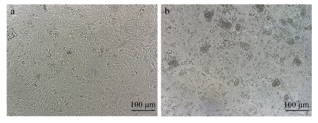
分离培养的原代子宫内膜上皮细胞在常规培养24 h后,约有80%的细胞贴壁,细胞边界清晰,以单层和铺路石的形状生长(图1-a);继续培养至48 h,少部分区域可见重叠现象(图1-b)。
图1 蛋鸡子宫内膜上皮细胞

a:培养24 h后的蛋鸡子宫内膜上皮细胞;b:培养48 h后的蛋鸡子宫内膜上皮细胞。

Fig.1 Morphology of endometrial epithelial cells in laying hens

a: endometrial epithelial cells of laying hens 24 h after inoculation; b: endometrial epithelial cells of laying hens 48 h after inoculation.

2.2 子宫内膜上皮细胞鉴定

利用细胞特异性细胞骨架蛋白细胞CK18的表达来证明上皮细胞的纯度。如图2所示,CK18可以在子宫内膜上皮细胞中高效表达,且杂质较少,纯度达93.99%。另外,仅用二抗对细胞进行染色,结果完全呈阴性。证实了通过机械刮取结合酶消化法分离的原代细胞为子宫内膜上皮细胞。
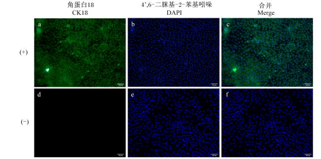
图2 蛋鸡子宫内膜上皮细胞免疫荧光染色鉴定

(+):一抗为CK18;(-):阴性对照,一抗为3%BSA。

Fig.2 Identification of endometrial epithelial cells in laying hens by immunofluorescence staining

(+): primary antibody is CK18; (-): negative control, primary antibody is 3% BSA.

2.3 蛋鸡子宫内膜上皮细胞衰老模型

2.3.1 不同浓度D-gal对细胞存活率的影响

通过CCK-8检测法评估不同浓度D-gal干预24 h的子宫内膜上皮细胞存活率,由图3所示,随着D-gal浓度的增加,子宫内膜上皮细胞的存活率呈现剂量依赖性降低。与对照组相比,8、16、32、64 mg/mL D-gal组的细胞存活率均显著降低(P<0.05);8 mg/mL D-gal组的细胞存活率为73.86%;16 mg/mL D-gal组的细胞存活率为60.88%;32 mg/mL D-gal组视野中可见大量细胞出现脱壁现象,过半数细胞死亡,存活率仅剩47.87%。综合细胞存活率结果,后续研究建议D-gal浓度为16 mg/mL。
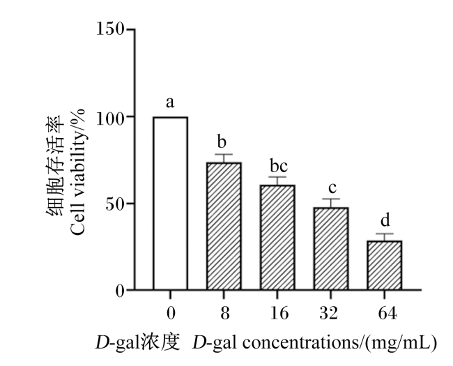
图3 不同浓度D-gal对子宫内膜上皮细胞存活率的影响

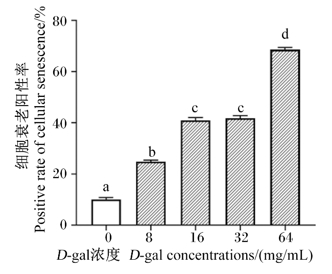
数据柱标相同小写字母表示差异不显著(P>0.05),不同小写字母表示差异显著(P<0.05)。图5同。

Fig.3 Effects of different concentrations of D-gal on viability of endometrial epithelial cells

Value columns with the same small letter mean no significant difference (P>0.05), while with different small letters mean significant difference (P<0.05). The same as Fig.5.

2.3.2 SA-β-gal活性

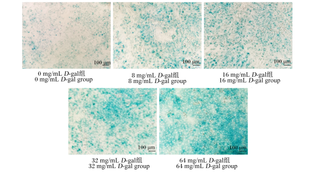
为进一步证实子宫内膜上皮细胞中的衰老是由D-gal诱导的,观察并统计对照组与不同浓度D-gal组中的蓝染细胞率。如图4所示,与对照组相比,不同浓度D-gal组SA-β-gal阳性细胞数量增加,提示D-gal干预24 h可诱导子宫内膜上皮细胞过早衰老。如图5所示,与对照组相比,不同浓度D-gal组细胞衰老阳性率显著增加(P<0.05);8 mg/mL D-gal组的细胞衰老阳性率为24.93%,16 mg/mL D-gal组的细胞衰老阳性率为41.08%,因此建议采用16 mg/mL D-gal建立细胞衰老模型。
图4 β-半乳糖苷酶染色结果

Fig.4 β-galactosidase staining results

图5 细胞衰老β-半乳糖苷酶染色阳性率

Fig.5 Positive rate of β-galactosidase staining for cellular senescence

3 讨论

3.1 子宫内膜上皮细胞分离培养与鉴定

蛋壳在子宫内形成,子宫的结构和功能变化对蛋壳质量有着至关重要的影响。创建一个体外的动态模型系统来复制其生长的过程为研究子宫内蛋壳的形成机制和改善蛋壳质量提供了有力的研究基础。在先前的研究中,已经发表了多种用于上皮细胞分离和培养的方案,包括组织块法和酶消化法[12-13]。本研究参考李兰兰[14]的方法,采用机械刮取结合酶消化法获取体外子宫内膜上皮细胞。然而,细胞的贴壁效果并不理想,细胞生长缓慢,需要培养72 h,才能够达到70%~80%的细胞密度。此外,原代上皮细胞是一种完全分化的细胞,寿命短,子宫内膜上皮原代细胞存活时间大约为7 d,细胞在培养72 h时可能已经开始出现自然衰老的现象[8]。因此,考虑后续需要构建细胞衰老模型,本研究在胶原酶浓度和消化时长上进行了调整,并使用细胞外基质对细胞培养板进行包被,以改善子宫内膜上皮细胞附着能力。在常规培养24 h时内,细胞密度即可达到80%左右,且细胞边界清晰,与相邻的细胞形成紧密的连接,与Sun等[15]利用Ⅰ型胶原酶消化60 min提取的蛋鸡子宫内膜上皮细胞的形态相一致。常见的CK18是一种细胞特异性蛋白标记物,主要在上皮细胞中表达,并与多种细胞内信号通路偶联[16]。通过对CK18表达的免疫荧光测定来验证培养的原代子宫内膜上皮细胞纯度,本试验结果显示,从蛋鸡子宫内膜分离的细胞几乎全部呈现绿色荧光,表明从子宫内膜分离的细胞为上皮细胞,且纯度较高(>90%),符合试验要求。

3.2 子宫内膜上皮细胞衰老模型的建立

衰老是所有细胞、组织和器官都会发生的自然现象,D-gal被广泛应用于体内和体外衰老模型的探索。多项研究表明,在暴露于高水平D-gal的动物模型中观察到与氧化应激介导的损伤,如衰老、认知障碍等的关联[16-17]。长期注射D-gal会导致半乳糖氧化酶形成大量的活性氧(ROS),氧化应激促进促炎因子的过度表达,反之,炎症也加速了ROS的积累,导致恶性循环和氧化应激-炎症轴的形成,加速衰老进程[18-19]。现阶段缺少诱导蛋鸡子宫内膜上皮细胞建立衰老模型的相关研究,本研究选用D-gal诱导蛋鸡子宫内膜上皮细胞,构建体外衰老模型,与自然衰老相似,具有现实意义。当D-gal干预剂量不足时,尽管细胞活率些许降低,但不足以诱导子宫内膜上皮细胞发生明显的实质性衰老,D-gal浓度较高时,对细胞的损伤较大,导致细胞大量脱壁死亡,无法满足后续试验需求。因此,本研究重点关注D-gal处理后子宫内膜上皮细胞的存活率并结合SA-β-gal染色阳性率,确定了D-gal作用适宜的干预浓度。

3.2.1 不同浓度D-gal对子宫内膜上皮细胞存活率的影响

现阶段研究建立体外衰老或氧化应激等模型时,通常选用处于60%~70%的细胞存活率水平,相较于过低的存活率水平,此范围内的细胞损伤较小,同时又能够保持足够的细胞生理活性,反之,存活率过高则可能掩盖衰老表型,不利于观察和分析衰老相关机制[20-21]。Xu等[22]研究发现,随着D-gal浓度由10 mg/mL升至60 mg/mL,大鼠小肠隐窝上皮细胞(IEC-6)存活率显著降至45%左右,推荐40 mg/mL(存活率为70%左右)为适宜浓度。本研究中的D-gal组以浓度依赖性的方式降低细胞存活率,尤其是64 mg/mL的D-gal组,细胞大量死亡,存活率仅剩28.77%,8 mg/mL组的细胞存活率为73.86%,当D-gal浓度增加至16 mg/mL时,细胞存活率降至60.88%,32 mg/mL D-gal诱导细胞衰老,视野内可见半数细胞出现死亡现象,存活率为47.87%。因此,本研究初步判断以16 mg/mL D-gal为蛋鸡子宫内膜上皮细胞衰老模型的最佳造模浓度。

3.2.2 不同浓度D-gal诱导子宫内膜上皮细胞衰老的程度

当细胞因各种因素而衰老时,细胞中会积累溶酶体,并开始丧失功能[23]。SA-β-gal是一种溶酶体水解酶,能够水解末端β-D-半乳糖残基,用于检测不同条件或化合物是否能诱导或抑制衰老细胞的出现[24]。在最适pH(pH=6.0)下,使用X-gal作为显色底物,通过检测衰老细胞中总溶酶体蛋白的活性,可将衰老细胞与正常细胞区分开,二聚形成蓝色沉淀[25]。Wang等[26]研究以20 mg/mL D-gal为适宜浓度,诱导成肌细胞(C2C12)衰老,观察到衰老模型组中的SA-β-gal细胞数量显著增加。尽管特定的细胞类型对D-gal的反应敏感程度不一致,但多数研究均证实D-gal增加了衰老标志物的表达,加速氧化应激,诱导细胞衰老。本研究采用SA-β-gal对D-gal诱导的子宫内膜上皮细胞衰老进行验证,在D-gal组衰老染色结果中可观察到,细胞胞浆内出现细小均匀的蓝色颗粒,为SA-β-gal阳性信号。与对照组相比,D-gal组子宫内膜上皮细胞SA-β-gal染色阳性细胞数显著提高,其中16 mg/mL D-gal处理24 h的SA-β-gal阳性率为41.08%,较对照组显著增加,证实了D-gal可诱导子宫内膜上皮细胞发生衰老,模型建立成功。

4 结论

本研究以产蛋高峰期的蛋鸡子宫为材料,采用机械刮取结合酶消化酶法成功分离培养蛋鸡子宫内膜上皮细胞,通过形态学观察与细胞免疫荧光鉴定表明细胞为子宫内膜上皮细胞。利用D-gal诱导成功建立蛋鸡子宫内膜上皮细胞衰老模型,适宜作用浓度为16 mg/mL,作用时间为24 h。
[1]
ZHANG Y N, DENG Y Z, JIN Y Y, et al. Age-related changes in eggshell physical properties,ultrastructure,calcium metabolism-related serum indices,and gene expression in eggshell gland during eggshell formation in commercial laying ducks[J]. Poultry Science, 2022, 101(2):101573.

[2]
FENG J, LU M, MA L, et al. Uterine inflammation status modulates eggshell mineralization via calcium transport and matrix protein synthesis in laying hens[J]. Animal Nutrition, 2023, 13:411-425.

DOI PMID

[3]
FU Y, ZHOU J M, SCHROYEN M, et al. Decreased eggshell strength caused by impairment of uterine calcium transport coincide with higher bone minerals and quality in aged laying hens[J]. Journal of Animal Science and Biotechnology, 2024, 15(1):37.

DOI PMID

[4]
YANG L B, LI S M, MO C H, et al. Transcriptome analysis and identification of age-associated fertility decreased genes in hen uterovaginal junction[J]. Poultry Science, 2021, 100(3):100892.

[5]
SALEMI S, ZAMANIAN M Y, GIMÉNEZ-LLORT L, et al. Distinct signatures on d-galactose-induced aging and preventive/protective potency of two low-dose vitamin D supplementation regimens on working memory,muscular damage,cardiac and cerebral oxidative stress,and SIRT1 and calstabin2 downregulation[J]. Food Science&Nutrition, 2023, 11(9):5050-5062.

[6]
SONG L N, WENG K Q, BAO Q, et al. TMT-based quantitative proteomic analysis unveils uterine fluid difference in hens producing normal and pimpled eggs[J]. Poultry Science, 2023, 102(11):103081.

[7]
ZHANG J C, WANG Y N, ZHANG C, et al. The differences of gonadal hormones and uterine transcriptome during shell calcification of hens laying hard or weak-shelled eggs[J]. BMC Genomics, 2019, 20(1):707.

DOI PMID

[8]
LI L L, ZHOU W T, MIAO S S, et al. Effects of zinc on cell proliferation,zinc transport,and calcium deposition in primary endometrial epithelial cells of laying hens in vitro[J]. Biological Trace Element Research, 2021, 199(11):4251-4259.

[9]
AZMAN K F, ZAKARIA R. D-galactose-induced accelerated aging model:an overview[J]. Biogerontology, 2019, 20(6):763-782.

[10]
YE P T, FENG L, ZHANG D, et al. Metformin ameliorates D-galactose-induced senescent human bone marrow-derived mesenchymal stem cells by enhancing autophagy[J]. Stem Cells International, 2023, 2023:1429642.

[11]
REMIGANTE A, SPINELLI S, TRICHILO V, et al. D-galactose induced early aging in human erythrocytes: role of band 3 protein[J]. Journal of Cellular Physiology, 2022, 237(2):1586-1596.

[12]
凌芳. 永生化绵羊子宫内膜上皮细胞系的建立与鉴定[D].硕士学位论文. 石河子: 石河子大学, 2022.

LING F. Establishment and identification of immortem sheep endometlial epithelial cell lines[D].Master's Thesis. Shihezi: Shihezi University, 2022. (in Chinese)

[13]
黄选洋, 王建萍, 丁雪梅, 等. 钒诱导蛋鸡输卵管膨大部上皮细胞氧化应激模型的建立[J]. 畜牧兽医学报, 2017, 48(2):340-350.

HUANG X Y, WANG J P, DING X M, et al. Establishment of an oxidative stress model induced by vanadium in oviduct magnum epithelial cell of laying hens[J]. Acta Veterinaria et Zootechnica Sinica, 2017, 48(2):340-350. (in Chinese)

DOI

[14]
李兰兰. 蛋氨酸锌调控蛋鸡蛋壳品质的机理研究[D].博士学位论文. 杭州: 浙江大学, 2020.

LI L L. A study on the mechanism of zinc-methionine to regulate eggshell quality in laying hens[D].Ph.D.Thesis. Hangzhou: Zhejiang University, 2020. (in Chinese)

[15]
SUN J, YE Y, LIAO H, et al. Effects of steroid hormones on epithelial and stromal cells cultureand proliferation of laying hens,in vitro[J]. Kafkas Universitesi Veteriner Fakultesi Dergisi, 2018, 24(3):401-407.

[16]
SHEPHERD M C, RADNAA E, TANTENGCO O A, et al. Extracellular vesicles from maternal uterine cells exposed to risk factors cause fetal inflammatory response[J]. Cell Communication and Signaling, 2021, 19(1):100.

DOI PMID

[17]
RAHMAN M A, SHUVO A A, BEPARI A K, et al. Curcumin improves D-galactose and normal-aging associated memory impairment in mice: in vivo and in silico-based studies[J]. PLoS One, 2022, 17(6):e0270123.

[18]
UMBAYEV B, ASKAROVA S, ALMABAYEVA A, et al. Galactose-induced skin aging:the role of oxidative stress[J]. Oxidative Medicine and Cellular Longevity, 2020, 2020:7145656.

[19]
DE MERA-RODRÍGUEZ J A, ÁLVAREZ-HERNÁN G, GAÑÁN Y, et al. Is senescence-associated β-galactosidase a reliable in vivo marker of cellular senescence during embryonic development?[J]. Frontiers in cell and developmental biology, 2021, 9:623175.

[20]
YIN L L, XU L H, CHEN B, et al. SRT1720 plays a role in oxidative stress and the senescence of human trophoblast HTR8/svneo cells induced by D-galactose through the SIRT1/FOXO3a/ROS signalling pathway[J]. Reproductive Toxicology, 2022, 111:1-10.

[21]
CHEN H, DONG L, CHEN X, et al. Anti-aging effect of phlorizin on D-galactose-induced aging in mice through antioxidant and anti-inflammatory activity,prevention of apoptosis,and regulation of the gut microbiota[J]. Experimental Gerontology, 2022, 163:111769.

[22]
XU W, XIANG X, LIN L, et al. Theanine delays d-galactose-induced senescence by regulating the cell cycle and inhibiting apoptosis in rat intestinal cells[J]. Journal of the Science of Food and Agriculture, 2024, 104(4):2073-2084.

[23]
LEE J, KIM Y S, KIM E, et al. Curcumin and hesperetin attenuate D-galactose-induced brain senescence in vitro and in vivo[J]. Nutrition Research and Practice, 2020, 14(5):438-452.

[24]
HEIDARI S, MEHRI S, SHARIATY V, et al. Preventive effects of crocin on neuronal damages induced by D-galactose through AGEs and oxidative stress in human neuroblastoma cells (SH-SY5Y)[J]. J Pharmacopuncture, 2018, 21(1):18-25.

[25]
VALIEVA Y, IVANOVA E, FAYZULLIN A, et al. Senescence-associated β-galactosidase detection in pathology[J]. Diagnostics, 2022, 12(10):2309.

[26]
WANG H H, SUN Y N, QU T Q, et al. Nobiletin prevents D-galactose-induced C2C12 cell aging by improving mitochondrial function[J]. International Journal of Molecular Sciences, 2022, 23(19):11963.

Outlines

/