RESEARCH PAPER

Study on Sensitization Mechanism of Soybean Antigen Proteins on Intestinal Cells of Litopenaeus vannamei

  • TIAN Renhong , 1, 2 ,
  • QIU Jianqiang 2, 3, * ,
  • PENG Kai , 2, ** ,
  • SUN Wenhao 1, 2 ,
  • DONG Ruiqi 2, 4 ,
  • LU Huijie 2 ,
  • ZHAO Hongxia 2 ,
  • CHEN Bing 2 ,
  • CHEN Wenchun 1, 2 ,
  • GUO Hui 1 ,
  • ZHU Xifeng 1, 5 ,
  • LI Guoli 5 ,
  • HUANG Wen , 2, **
Expand
  • 1 College of Fisheries, Guangdong Ocean University, Zhanjiang 524088, China
  • 2 Key Laboratory of Animal Nutrition and Feed Science in South China, Ministry of Agriculture in Rural Affairs, Guangdong Key Laboratory of Animal Breeding and Nutrition, Collaborative Innovation Center of Aquatic Sciences, Institute of Animal Science, Guangdong Academy of Agricultural Sciences, Guangzhou 510640, China
  • 3 College of Fisheries, Huazhong Agricultural University, Wuhan 430070, China
  • 4 College of Fisheries and Life, Shanghai Ocean University, Shanghai 201306, China
  • 5 Guangzhou Fishtech Biotechnology Co., Ltd., Guangzhou 510640, China
** PENG Kai, professor, E-mail: ;
HUANG Wen, professor, E-mail:

* Contributed equally

Received date: 2024-06-06

  Online published: 2025-01-10

Abstract

This experiment was conducted to explore the sensitization mechanism of soybean antigen proteins 7S and 11S on intestinal cells of Litopenaeus vannamei. The intestinal cells of Litopenaeus vannamei were divided into control group (NC group), 7S group and 11S group with three replicates in each group. After 48 h of cell culture, the cell activity, cell membrane integrity-related indexes, cell antioxidant indexes, cell inflammatory factor contents, apoptosis rate, and apoptosis and tight junction protein-related gene expression were determined in each group. The results showed that compared with NC group, the alkaline phosphatase(AKP), lactate dehydrogenase (LDH) activities and reactive oxygen species (ROS), malondialdehyde (MDA), tumor necrosis factor-α (TNF-α),interferon-γ (IFN-γ) contents of 7S group and 11S group were significantly increased (P<0.05), the total antioxidant capacity (T-AOC), superoxide dismutase (SOD), catalase (CAT) activities and interleukin-10 content were significantly decreased (P<0.05), the apoptosis rate was significantly increased (P<0.05), the mRNA relative expression levels of cysteine aspartate protease-3 (Caspase-3), B-lymphoblastoma/leukaemia-2 related X protein (Bax), c-Jun amino-terminal kinase (JNK) and p38 mitogen-activated protein kinase (p38 MAPK) were significantly up-regulated (P<0.05), and the mRNA relative expression levels of tight junction protein-3 (Claudin-3), Occludin and zonula occludens-1 (ZO-1) was significantly down-regulated (P<0.05). The soybean antigen protein 7S induced ROS accumulation, exacerbated the inflammatory response, promoted apoptosis and disrupted the cellular barrier more strongly than soybean antigen protein 11S. In summary, soybean antigenic proteins 7S and 11S inhibit cellular activity and lead to cellular anaphylaxis by disrupting cell membrane integrity, releasing AKP and LDH, inducing the accumulation of ROS, promoting cellular pro-inflammatory factor secretion in cells, and up-regulating the expression levels of genes associated with apoptotic factors and tight junction proteins.

Cite this article

TIAN Renhong , QIU Jianqiang , PENG Kai , SUN Wenhao , DONG Ruiqi , LU Huijie , ZHAO Hongxia , CHEN Bing , CHEN Wenchun , GUO Hui , ZHU Xifeng , LI Guoli , HUANG Wen . Study on Sensitization Mechanism of Soybean Antigen Proteins on Intestinal Cells of Litopenaeus vannamei[J]. Chinese Journal of Animal Nutrition, 2025 , 37(1) : 536 -549 . DOI: 10.12418/CJAN2025.046

豆粕是大豆经浸提和脱油后得到的副产品,其氨基酸组成良好、蛋白质含量高,是水产饲料重要的植物蛋白质资源[1-2]。然而,饲料中添加高水平的豆粕会引起水产动物肠道损伤[3],诱发肠炎[4-5],导致水产动物生长性能下降[6]。豆粕含有的抗营养因子是限制其在水产饲料中广泛应用的主要瓶颈[7]。大豆抗原蛋白是豆粕主要的抗营养因子,具有较高的热稳定性和较强的免疫原性,用常规方法难以失活[7-8]。按超速离心沉降系数分类,大豆抗原蛋白可分为2S、7S、11S和15S[9],其中7S和11S约占大豆总蛋白的70%[10],是引起过敏反应的主要致敏原[7]。大豆抗原蛋白引起过敏反应造成的免疫损伤主要在肠道[11]。大豆抗原蛋白进入动物机体后,大部分大豆抗原蛋白可被降解,以肽和氨基酸形式存在,少部分未被降解的大豆抗原蛋白则通过肠上皮细胞及其间隙穿过肠道,进入血液和淋巴,刺激动物机体产生免疫应答,导致动物机体发生过敏反应[11],从而造成动物机体肠黏膜上皮细胞通透性增加、肠黏膜水肿、肠黏膜形态结构与完整性受损、肠绒毛萎缩缩短[12-13]
凡纳滨对虾(Litopeaneus vannamei)具有生长快、抗应激能力强、肉质鲜美等特点,是对虾集约化养殖的优良品种。据《2024中国渔业统计年鉴》数据显示,2023年,我国凡纳滨对虾的养殖产量近224万t,约占对虾总产量的87%,并呈逐年增长趋势[14]。肠道作为水产动物重要的消化和免疫器官,是营养物质消化吸收的场所,也是防止细菌等有害物质入侵的屏障。目前,大豆抗原蛋白7S和11S在水产动物上的研究主要聚焦于动物的生长性能及肠道健康[15-20],而关于大豆抗原蛋白7S和11S对水产动物肠道细胞致敏机制的研究尚未见报道。因此,本试验通过体外提取和培养凡纳滨对虾肠道细胞,评价大豆抗原蛋白7S和11S对凡纳滨对虾肠道细胞活性、细胞膜完整性、抗氧化能力、炎症因子含量、细胞凋亡和紧密连接蛋白相关基因表达的影响,旨在探究其对凡纳滨对虾肠道细胞的致敏机制,为大豆副产物在凡纳滨对虾饲料中的合理应用及对虾健康养殖提供理论依据。

1 材料与方法

1.1 肠道细胞的分离与培养

本试验方案及动物试验伦理审查由广东省农业科学院动物福利伦理委员会批准,批准号:GDAAS2022037。
试验所用的凡纳滨对虾来源于广州市某虾苗孵化场,平均初始体重为(15.2±0.5) g。对虾肠道细胞的提取和培养方法参考本实验室发明专利(CN115261300A),具体操作如下:对虾在正式试验前禁食24 h,禁食结束后随机取20尾虾,立即在75%乙醇中浸泡5 min(用于对虾表面消毒)。将虾转移至无菌操作台中,快速解剖并取出肠道,采用8 mL四抗液(5×,青霉素:链霉素:庆大霉素:支原体清除剂=1:1:1:1)的Dhanks溶液(含氯化钠、氯化钾、磷酸氢二钾、碳酸氢钠)冲洗4次(用于抑制肠道内容物和肠道细菌的感染)。
收集肠道样品,置于细胞筛中,加入消化液(胶原蛋白酶Ⅳ:胶原蛋白酶Ⅰ=1:1)进行研磨(用于减少细胞碎片和提高虾肠道组织的消化效率),吸取滤过细胞筛中的消化液于EP管中,放入200 r/min的摇床(28 ℃)消化4 min,将消化处理得到的消化液加入含胎牛血清(FBS)的M199完全培养基(M199+15% FBS+5 g/L氯化钠+1 g/L葡萄糖+1%四抗液)终止消化。转移消化液,先32×g离心5 min,然后50×g离心8 min,用含FBS的M199完全培养基进行细胞重悬。重悬后的细胞铺于提前用鼠尾胶原Ⅰ型蛋白(北京索莱宝科技有限公司,货号:C8062)包被好的96孔培养板中,置于含6% CO2培养箱(27 ℃)中培养,48 h后进行换液处理,得到对虾肠道细胞。

1.2 试验设计

试验分为对照组(NC组)、7S组和11S组,每组3个重复,每个重复2个孔。将对虾肠道细胞接种于96孔板中培养(1×10⁴个/mL),将不同浓度(1、5、10 mg/mL)的大豆抗原蛋白7S和11S(由中国农业大学食品学院提供,其分离纯化步骤参考罗明昌等[21]的方法)添加至细胞培养基中,置于6%二氧化碳(CO2)的培养箱(27 ℃)中培养48 h,随后终止培养。所得细胞一部分用于细胞活性、细胞膜完整性相关指标的分析,一部分用于细胞抗氧化指标、炎症因子含量、凋亡(凋亡率、凋亡相关基因表达)和细胞紧密连接蛋白相关基因表达的分析。

1.3 指标分析

采用CCK-8细胞增殖毒性检测试剂盒(上海东仁化学科技有限公司,货号:CK04)并依照说明书步骤测定细胞存活率,以细胞存活率表示细胞活性,筛选大豆抗原蛋白7S和11S的适宜作用浓度。细胞存活率计算公式如下:
细胞存活率(%)=(试验孔450 nm吸光度值/对照孔450 nm吸光度值)×100。
采用商业试剂盒(南京建成生物工程研究所)并依照说明书步骤测定细胞膜完整性相关指标,如碱性磷酸酶(AKP,货号:A059-2)和乳酸脱氢酶(LDH,货号:A020-2)。
采用商业试剂盒(北京索莱宝科技有限公司,货号:CA1410)并依照说明书步骤检测活性氧(ROS)含量(荧光探针法)。采用商业试剂盒(南京建成生物工程研究所)并依照说明书步骤测定细胞抗氧化指标,如总抗氧化能力(T-AOC,货号:A015-2-1)、超氧化物歧化酶(SOD,货号:A001-1)、过氧化氢酶(CAT,货号:A007-1-1)、丙二醛(MDA,货号:A003-1)。
采用酶联免疫吸附测定(ELISA)试剂盒(上海优选生物科技有限公司)并依照说明书步骤测定细胞炎症因子含量,如干扰素-γ(IFN-γ,货号:YX-090617S)、白细胞介素-10(IL-10,货号:YX-091210S)、肿瘤坏死因子-α(TNF-α,货号:YX-201407S)。采用商业试剂盒(南京建成生物工程研究所)并依照说明书步骤测定一氧化氮(NO,货号:A013-2-1)含量。
采用膜联蛋白V-荧光素异硫氰酸酯(Annexin V-FITC)/碘化丙啶(PI)细胞凋亡检测试剂盒(美国BD公司,货号:556547)并依照说明书步骤测定细胞凋亡率。
采用TRNzol试剂盒(广州兴誉生物科技有限公司,货号:XY-001)提取细胞总RNA,然后用cDNA反转录试剂盒[翌圣生物科技(上海)股份有限公司,货号:11141]将RNA逆转录为cDNA。以β-肌动蛋白(β-actin)作为内参基因,采用实时荧光定量PCR方法测定半胱氨酸天冬氨酸蛋白酶-3(Caspase-3)、B淋巴细胞瘤/白血病-2相关X蛋白(Bax)、c-Jun氨基末端激酶(JNK)、p38丝裂原活化蛋白激酶(p38 MAPK)、紧密连接蛋白-3(Claudin-3)、闭合蛋白(Occludin)、闭锁小带蛋白-1(ZO-1)等基因表达,并按照2-△△Ct法计算目的基因mRNA相对表达量。PCR循环条件为:95 ℃预变性5 min,1个循环;95 ℃变性10 s,60 ℃复性34 s,72 ℃延伸30 s,40个循环。引物序列见表1
表1 引物序列

Table 1 Primer sequences

目的基因
Target genes
引物序列
Primer sequences(5'—3')
β-肌动蛋白
β-actin
F:GAGCAACACGGAGTTCGTTGT
R:CATCACCAACTGGGACGACATGGA
半胱氨酸天冬氨酸蛋白酶-3
Caspase-3
F:GACGCCCTTGCCGTAGTG
R:CTCAGCCGTGAAGTTTATCCA
B淋巴细胞瘤/白血病-2相关X蛋白
Bax
F:AAGAACCAGATGCAGCGACT
R:GAGAGAAGCAGGCAAACACC
c-Jun氨基末端激酶
JNK
F:AGGTTCCCTGTTGTGACTGG
R:TGCATGCATTCTCACACTCA
p38丝裂原活化蛋白激酶
p38 MAPK
F:AATGCTGACCTTGGATCCTG
R:TGCATAGCCTGTTCTGTTGC
紧密连接蛋白-3
Claudin-3
F:ATGCTTCACCGACTCTGCTT
R:ACGCACAGATCGGTTCTTCT
闭合蛋白
Occludin
F:TACCATTACTGCGTGGTGGA
R:TCACTCTGCGCCATAAGATG
闭锁小带蛋白-1
ZO-1
F:ATTTGTAGCTTCCCCTCGGC
R:GCGACGCATTCGTCAAAAGT

1.4 数据分析

采用SPSS 20.0统计分析软件进行单因素方差分析(one-way ANOVA)和独立t检验,采用GraphPad prism 8.0.2软件作图,试验结果用平均值±标准误(mean±SEM)表示,P<0.05表示差异显著。

2 结果与分析

2.1 大豆抗原蛋白7S和11S对凡纳滨对虾肠道细胞活性的影响

图1可知,细胞活性随着大豆抗原蛋白7S和11S浓度的升高而降低。与0 mg/mL的大豆抗原蛋白7S和11S相比,添加1、5和10 mg/mL的大豆抗原蛋白7S和11S显著降低了细胞活性(P<0.05);与1 mg/mL的大豆抗原蛋白7S和11S相比,添加5和10 mg/mL的大豆抗原蛋白7S和11S显著降低了细胞活性(P<0.05);添加5和10 mg/mL的大豆抗原蛋白7S和11S之间的细胞活性差异不显著(P>0.05)。因此,筛选出大豆抗原蛋白7S和11S的适宜作用浓度均为5 mg/mL。
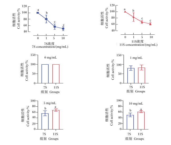
图1 大豆抗原蛋白7S和11S对凡纳滨对虾肠道细胞活性的影响

数据柱标相同小写字母表示差异不显著(P>0.05),不同小写字母表示差异显著(P<0.05)。下图同。

Fig.1 Effects of soybean antigen proteins 7S and 11S on intestinal cell viability of Litopenaeus vannamei

Value columns with the same small letter mean no significant difference (P>0.05), while with different small letters mean significant difference (P<0.05). The same as below.

在0和1 mg/mL浓度下,7S组和11S组的细胞活性均差异不显著(P>0.05)。在5和10 mg/mL浓度下,11S组的细胞活性显著高于7S组(P<0.05)。

2.2 大豆抗原蛋白7S和11S对凡纳滨对虾肠道细胞膜完整性的影响

图2可知,与NC组相比,7S和11S组细胞AKP和LDH活性均显著升高(P<0.05)。与7S组相比,11S组细胞AKP和LDH活性均差异不显著(P>0.05)。
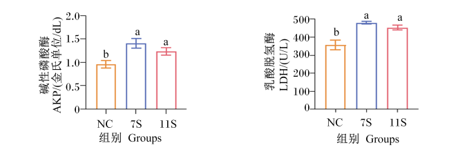
图2 大豆抗原蛋白7S和11S对凡纳滨对虾肠道细胞膜完整性的影响

Fig.2 Effects of soybean antigen proteins 7S and 11S on integrity of intestinal cell membranes of Litopenaeus vannamei

2.3 大豆抗原蛋白7S和11S对凡纳滨对虾肠道细胞抗氧化指标的影响

图3可知,与NC组相比,7S和11S组细胞T-AOC、SOD和CAT活性均显著降低(P<0.05),细胞ROS和MDA含量均显著升高(P<0.05)。与7S组相比,11S组细胞T-AOC和SOD活性均显著升高(P<0.05),细胞ROS含量显著降低(P<0.05),细胞CAT活性及MDA含量均差异不显著(P>0.05)。
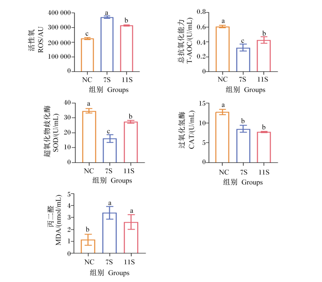
图3 大豆抗原蛋白7S和11S对凡纳滨对虾肠道细胞抗氧化指标的影响

Fig.3 Effects of soybean antigen proteins 7S and 11S on intestinal cells antioxidant indexes of Litopenaeus vannamei

2.4 大豆抗原蛋白7S和11S对凡纳滨对虾肠道细胞炎症因子含量的影响

图4可知,与NC组相比,7S和11S组细胞TNF-α和IFN-γ含量均显著升高(P<0.05),细胞IL-10含量显著降低(P<0.05);7S组细胞NO含量显著升高(P<0.05),11S组细胞NO含量差异不显著(P>0.05)。与7S组相比,11S组细胞IL-10含量显著升高(P<0.05),细胞TNF-α含量显著降低(P<0.05),细胞IFN-γ和NO含量均差异不显著(P>0.05)。
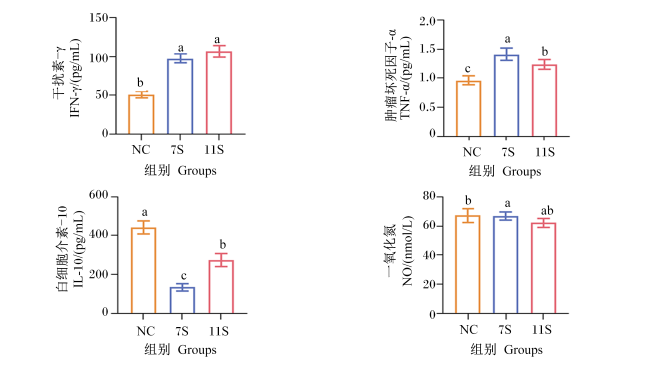
图4 大豆抗原蛋白7S和11S对凡纳滨对虾肠道细胞炎症因子含量的影响

Fig.4 Effects of soybean antigen proteins 7S and 11S on contents of inflammatory factors in intestinal cells of Litopenaeus vannamei

2.5 大豆抗原蛋白7S和11S对凡纳滨对虾肠道细胞凋亡率的影响

图5可知,与NC组相比,7S和11S组凋亡细胞数量迅速增长,细胞呈现明显损伤,细胞凋亡率显著升高(P<0.05)。与7S组相比,11S组细胞凋亡率显著降低(P<0.05)。
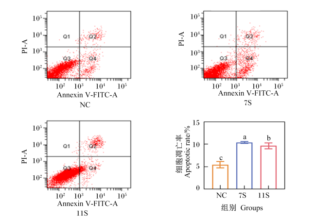
图5 大豆抗原蛋白7S和11S对凡纳滨对虾肠道细胞凋亡率的影响

PI-A:碘化丙啶区 propidium iodide area;Annexin V-FITC:膜联蛋白V-荧光素异硫氰酸酯区 Annexin V-fluorescein isothiocyanate area;NC:对照组 control group;7S:7S组 7S group;11S:11S组 11S group。

Fig.5 Effects of soybean antigen proteins 7S and 11S on intestinal cells apoptosis rate of Litopenaeus vannamei

2.6 大豆抗原蛋白7S和11S对凡纳滨对虾肠道细胞凋亡相关基因表达的影响

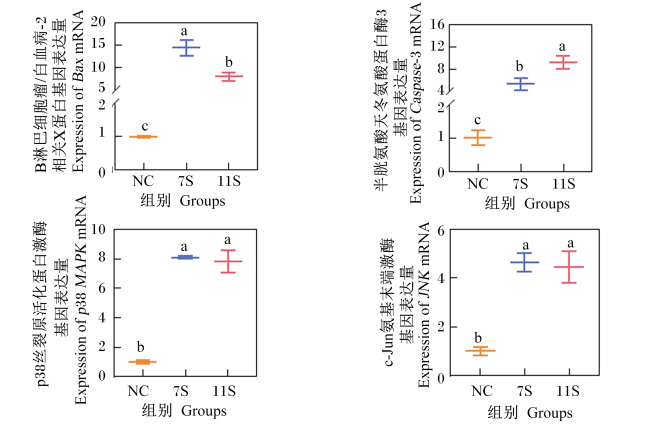
图6可知,与NC组相比,7S和11S组细胞Caspase-3、BaxJNKp38 MAPK mRNA相对表达量均显著上调(P<0.05)。与7S组相比,11S组细胞Caspase-3 mRNA相对表达量显著上调(P<0.05),细胞Bax mRNA相对表达量显著下调(P<0.05),细胞JNKp38 MAPK mRNA相对表达量差异不显著(P>0.05)。
图6 大豆抗原蛋白7S和11S对凡纳滨对虾肠道细胞凋亡相关基因表达的影响

Fig.6 Effects of soybean antigen proteins 7S and 11S on expression of apoptosis-related genes in intestinal cells of Litopenaeus vannamei

2.7 大豆抗原蛋白7S和11S对凡纳滨对虾肠道细胞紧密连接蛋白相关基因表达的影响

图7可知,与NC组相比,7S和11S组细胞Claudin-3、OccludinZO-1 mRNA相对表达量均显著下调(P<0.05)。与7S组相比,11S组细胞Claudin-3和ZO-1 mRNA相对表达量均显著上调(P<0.05),细胞Occludin mRNA相对表达量显著下调(P<0.05)。
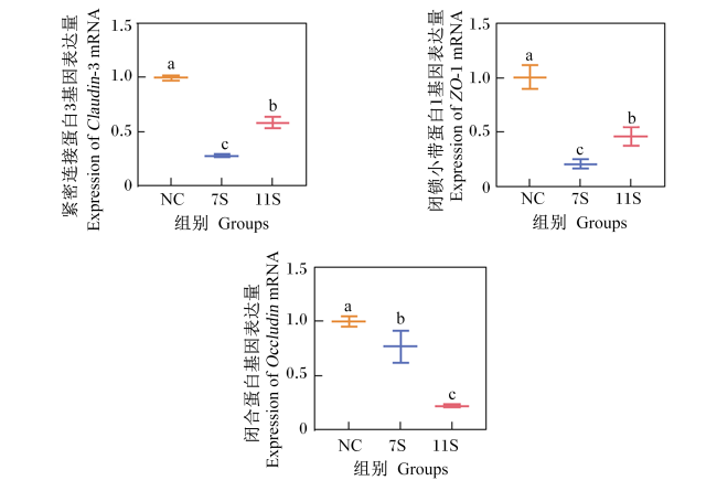
图7 大豆抗原蛋白7S和11S对凡纳滨对虾肠道细胞紧密连接蛋白相关基因表达的影响

Fig.7 Effects of soybean antigen proteins 7S and 11S on expression of tight junction protein-related genes in intestinal cells of Litopenaeus vannamei

3 讨论

3.1 大豆抗原蛋白7S和11S对凡纳滨对虾肠道细胞活性及细胞膜完整性的影响

细胞活性反映了细胞健康的总体情况[22]。Jia等[23]通过原代培养仔猪肠上皮细胞研究发现,浓度为1.2和2.4 mg/mL的大豆抗原蛋白7S均可显著降低仔猪肠上皮细胞活性。彭成璐[13]报道,浓度为1、5和10 mg/mL的大豆抗原蛋白7S和11S处理猪小肠上皮细胞(IPEC-J2细胞)均会显著降低细胞活性。本试验结果表明,浓度为1、5和10 mg/mL的大豆抗原蛋白7S和11S均能显著降低凡纳滨对虾肠道细胞活性,这与Jia等[23]和彭成璐[13]的研究结果一致。AKP是一种同源二聚体去磷酸化酶,主要位于细胞膜和细胞质[24-25]。LDH是一类相对稳定的四聚体氧化还原酶,广泛存在于细胞质[26]。当细胞受到损伤、细胞膜完整性被破坏时,AKP和LDH会被大量释放至细胞外,使细胞外的AKP和LDH活性急剧上升。因此,AKP和LDH活性可作为评价细胞膜完整性和损伤程度的重要指标[27]。本研究中,大豆抗原蛋白7S和11S显著提高了凡纳滨对虾肠道细胞AKP和LDH活性,这与在IPEC-J2细胞[13]和小鼠肠上皮细胞[28]中的研究结果一致,说明大豆抗原蛋白7S和11S会破坏细胞膜完整性,导致凡纳滨对虾肠道细胞受损。

3.2 大豆抗原蛋白7S和11S对凡纳滨对虾肠道细胞抗氧化指标的影响

机体在氧化应激情况下会产生大量的ROS,过量的ROS会攻击抗氧化防御系统,造成机体氧化损伤,其中包括DNA氧化损伤、蛋白质降解以及脂质过氧化[29-31]。T-AOC、SOD和CAT是机体抗氧化系统的重要组成部分,T-AOC反映机体总体的抗氧化能力,SOD可清除机体自由基,CAT能够清除机体产生的过氧化氢[32-34]。MDA是脂质过氧化的终产物,可反映细胞氧化受损程度[35-36]。本研究中,大豆抗原蛋白7S和11S显著降低了凡纳滨对虾肠道细胞T-AOC、SOD和CAT活性,显著提高了ROS、MDA含量,这与在中华绒螯蟹[19]、草鱼[37-38]、建鲤[39]和黄金鲫[40]中的报道相似,这可能是由于大豆抗原蛋白通过激活核因子E2相关因子2(Nrf2)信号通路来抑制肠道细胞ROS清除能力[20,37],诱导ROS累积,造成细胞抗氧化防御系统失衡,从而引起对虾肠道细胞氧化损伤。此外,本研究结果表明,与7S组相比,11S组细胞T-AOC和SOD活性显著提高,细胞ROS含量显著降低,表明大豆抗原蛋白7S对凡纳滨对虾肠道细胞的氧化损伤效果要高于大豆抗原蛋白11S,这与李宝等[41]和Miller等[42]的研究结果一致。

3.3 大豆抗原蛋白7S和11S对凡纳滨对虾肠道细胞炎症因子含量的影响

辅助性T细胞1(Th1细胞)和辅助性T细胞2(Th2细胞)能够调节炎症反应、免疫反应和能量代谢[43-45]。Th1细胞主要分泌TNF-α和IFN-γ等促炎因子,介导与细胞免疫有关的免疫应答,参与迟发型超敏反应[46-47]。Th2细胞主要分泌IL-10和白细胞介素-4(IL-4)等抗炎因子,参与体液免疫,与过敏性炎症有关[48-49]。本研究中,大豆抗原蛋白7S和11S显著提高了凡纳滨对虾肠道细胞TNF-α和IFN-γ含量,显著降低了肠道细胞IL-10含量,这与Yang等[18]在石斑鱼、Zhang等[50]在草鱼幼鱼、徐君[28]在小鼠以及张瑜[51]和孙智峰等[52]在断奶仔猪中的研究结果相似,说明大豆抗原蛋白7S和11S可通过促进促炎因子的分泌和抑制抗炎因子的分泌,加剧对虾肠道细胞炎症反应。本研究中,与7S组相比,11S组凡纳滨对虾肠道细胞TNF-α含量显著降低,肠道细胞IL-10含量显著提高,说明大豆抗原蛋白7S对凡纳滨对虾肠道细胞的免疫原性强于大豆抗原蛋白11S。

3.4 大豆抗原蛋白7S和11S对凡纳滨对虾肠道细胞凋亡的影响

细胞凋亡是为了维持细胞或机体内环境稳定而发生的一种程序性死亡[53]。Caspase-3处于细胞凋亡级联反应的核心位置,是细胞凋亡的执行者[54]。Bax作为B淋巴细胞瘤/白血病-2(Bcl-2)家族重要的促凋亡蛋白,是细胞凋亡过程的关键调节因子[55]。Bax是线粒体膜上离子通道的组成部分,可以使细胞色素C穿过线粒体膜,激活Caspase-3,促进细胞凋亡[56-57]。本研究结果表明,大豆抗原蛋白7S和11S显著上调了线粒体凋亡通路相关的促凋亡分子BaxCaspase-3 mRNA相对表达量,表明大豆抗原蛋白7S和11S可能是通过激活Caspase-3/线粒体调节的凋亡途径,引起凡纳滨对虾肠道细胞凋亡,这与Peng等[58]在IPEC-J2细胞、Duan等[59]在草鱼中肠细胞中的研究结果相似。细胞凋亡率可以反映氧化应激的程度[60]。已有研究发现,高浓度ROS能够改变线粒体通透性,从而激活Caspase-3,诱导线粒体凋亡途径激活[61]。本研究中,大豆抗原蛋白7S和11S显著提高了凡纳滨对虾肠道细胞ROS含量和细胞凋亡率,显著上调了Caspase-3 mRNA相对表达量,说明大豆抗原蛋白7S和11S可能是通过提高ROS含量,诱导线粒体凋亡途径启动,从而促进凡纳滨对虾肠道细胞凋亡,但该推测需要进一步研究。p38 MAPK和JNK 2个亚家族蛋白属于丝裂原活化蛋白激酶(MAPKs)家族蛋白,可调控细胞凋亡[62-63]。氧化应激与细胞因子能选择性地激活p38 MAPK和JNK蛋白[63-64]。在胡子鲶头肾巨噬细胞中研究发现,ROS能够通过激活 JNK/p38 MAPK信号通路,导致细胞凋亡[65]。本研究中,大豆抗原蛋白7S和11S显著提高了凡纳滨对虾肠道细胞ROS和TNF-α含量,显著上调了JNKp38 MAPK mRNA相对表达量。这表明大豆抗原蛋白7S和11S可能是通过提高ROS和TNF-α含量来激活JNK/p38 MAPK信号通路,从而促进凡纳滨对虾肠道细胞凋亡,类似于草鱼后肠细胞的研究结果[59]。此外,本研究结果表明,与7S组相比,11S组凡纳滨对虾肠道细胞凋亡率显著降低,细胞Caspase-3 mRNA相对表达量显著上调,细胞Bax mRNA相对表达量显著下调,表明大豆抗原蛋白7S诱导凡纳滨对虾肠道细胞凋亡的效果强于大豆抗原蛋白11S。

3.5 大豆抗原蛋白7S和11S对凡纳滨对虾肠道细胞紧密连接蛋白相关基因表达的影响

肠道紧密连接能够封闭肠道上皮细胞之间的缝隙,是维持肠道物理屏障完整性的关键。紧密连接受损可增强肠道上皮细胞通透性及病原体敏感性[66]。跨膜蛋白紧密连接蛋白(Claudin)、Occludin和胞质蛋白ZO-1是肠紧密连接的重要组成部分[67]。Claudin-3调控肠道上皮细胞的屏障功能,Occludin调控细胞膜的通透性,ZO-1直接与F-肌动蛋白结合,参与细胞骨架的形成[66,68]。本研究结果表明,大豆抗原蛋白7S和11S显著下调了凡纳滨对虾肠道细胞Claudin-3、OccludinZO-1 mRNA相对表达量,表明大豆抗原蛋白7S和11S可通过破坏紧密连接结构成分,增加细胞膜的通透性,进而破坏对虾肠道细胞的屏障功能。这与在石斑鱼[18]、建鲤[39]、杂交黄颡鱼幼鱼[16]和草鱼幼鱼[20]中的研究结果相似。本研究中,与7S组相比,11S组凡纳滨对虾肠道细胞Claudin-3和ZO-1 mRNA相对表达量显著上调,细胞Occludin mRNA相对表达量显著下调,表明大豆抗原蛋白7S对凡纳滨对虾肠道细胞屏障功能的破坏效果强于大豆抗原蛋白11S。

4 结论

本试验条件下,大豆抗原蛋白7S和11S通过破坏细胞膜完整性,释放AKP和LDH,诱导ROS累积,促进细胞促炎因子分泌,上调细胞凋亡因子和紧密连接蛋白基因表达水平,抑制细胞活性并导致细胞过敏反应。
[1]
杨小佳, 王金水, 管军军, 等. 豆粕的营养价值及影响因素[J]. 粮食与饲料工业, 2013(3):44-46.

YANG X J, WANG J S, GUAN J J, et al. Nutritional value and influencing factors of soybean meal[J]. Cereal & Feed Industry, 2013(3):44-46. (in Chinese)

[2]
范泽, 李晨辉, 吴迪, 等. 两种新型植物蛋白替代豆粕在大规格鲤饲料中的应用效果研究[J]. 水产学杂志, 2023, 36(4):20-28,37.

FAN Z, LI C H, WU D, et al. Application effect of two new plant protein substitutes for soybean meal in large-size common carp feed[J]. Chinese Journal of Fisheries, 2023, 36(4):20-28,37. (in Chinese)

[3]
吴莉芳, 瞿子惠, 周锴, 等. 豆粕替代鱼粉对黄金鲈生长及肠道组织的影响[J]. 西北农林科技大学学报(自然科学版), 2017, 45(6):1-8.

WU L F, QU Z H, ZHOU K, et al. Effects of replacing fish meal with soybean meal on growth and intestinal tissue of Perca flavescens[J]. Journal of Northwest A&F University (Natural Science Edition), 2017, 45(6):1-8. (in Chinese)

[4]
王晓艳, 李宝山, 李璐, 等. 患豆粕型肠炎对许氏平鲉生长性能及肠道健康的影响[J]. 海洋渔业, 2023, 45(6):719-728.

WANG X Y, LI B S, LI L, et al. Effects of soybean meal-induced enteritis on growth performance and intestinal health of Sebastes schlegelii[J]. Marine Fisheries, 2023, 45(6):719-728. (in Chinese)

[5]
何远法. 谷氨酰胺对β-伴大豆球蛋白和大豆球蛋白诱导珍珠龙胆石斑鱼肠炎的保护机制研究[D]. 博士学位论文. 湛江: 广东海洋大学, 2021.

HE Y F. Study on the protective mechanism of glutamine against β-conglycinin and glycinin induced enteritis in pearl gentian grouper (Epinephelus fuscogutta♀×Epinephelus lanceolatus♂)[D].Ph.D.Thesis. Zhenjiang: Guangdong Ocean University, 2021. (in Chinese)

[6]
刘洋, 孟连仲, 张建雄. 豆粕替代鱼粉对褐点石斑鱼生长、营养组成及血液指标影响[J]. 中国饲料, 2018(4):60-64.

LIU Y, MENG L Z, ZHANG J X. Effect of partial fish meal replacement by soybean meal on the growth performance,nutritional composition and blood biochemical indices of juvenile Epinephelus fuscoguttatus[J]. China Feed, 2018(4):60-64. (in Chinese)

[7]
游金明, 李德发. 大豆抗营养因子研究进展[J]. 饲料与畜牧, 2006(9):40-43.

YOU J M, LI D F. Research progress of anti-nutrient factors in soybean[J]. Animal Agriculture, 2006(9):40-43. (in Chinese)

[8]
李德发. 大豆抗营养因子[M]. 北京: 中国科学技术出版社, 2003.

LI D F. Soy anti-nutritional Factor[M].Science and Technology of China Press, 2003 (in Chinese)

[9]
李堂昊, 布冠好, 陈复生. 大豆主要过敏原β-伴大豆球蛋白及其抗原表位的研究进展[J]. 大豆科学, 2019, 38(5):806-812.

LI T H, BU G H, CHEN F S. Development in major allergen β-conglycinin and its antigen epitopes of soybean[J]. Soybean Science, 2019, 38(5):806-812. (in Chinese)

[10]
THANH V H, SHIBASAKI K. Major proteins of soybean seeds.Subunit structure of beta-conglycinin[J]. Journal of Agricultural and Food Chemistry, 2002, 26(3):692-695.

[11]
常美楠. 果寡糖缓解大豆抗原诱导仔猪过敏反应的研究[D]. 硕士学位论文. 长春: 吉林农业大学, 2019.

CHANG M N. The effects of fructo-oligosaccharide alleviates soybean-induced anaphylaxis in piglets[D]. Master’s Thesis. Changchun: Jilin Agricultural University, 2019. (in Chinese)

[12]
PENG C L, CAO C M, HE M C, et al. Soybean glycinin- and β-conglycinin-induced intestinal damage in piglets via the p38/JNK/NF-κB signaling pathway[J]. Journal of Agricultural and Food Chemistry, 2018, 66(36):9534-9541.

DOI PMID

[13]
彭成璐. β-伴大豆球蛋白和大豆球蛋白诱导IPEC-J2细胞损伤的机制研究[D]. 硕士学位论文. 合肥: 安徽农业大学, 2021.

PENG C L. The mechanism of β-conglycinin and glycinin induced IPEC-J2 cell damage[D]. Master’s Thesis. Hefei: Anhui Agricultural University, 2021. (in Chinese)

[14]
农业农村部渔业渔政管理局, 全国水产技术推广总站,中国水产学会. 2024中国渔业统计年鉴[M]. 北京: 中国农业出版社, 2024.

Bureau of Fisheries and Fishery Administration of the Ministry of Agriculture and Rural Affairs,National Fisheries Technology Extension Station, Chinese Fisheries Society. 2024 China fisheries statistical yearbook[M]. Beijing: China Agriculture Press,2024. (in Chinese)

[15]
HE Y F, LIANG J F, DONG X H, et al. Soybean β-conglycinin and glycinin reduced growth performance and the intestinal immune defense and altered microbiome in juvenile pearl gentian groupers Epinephelus fuscoguttatus♀×Epinephelus lanceolatus♂[J]. Animal Nutrition, 2022,9:193-203.

[16]
YI L Y, LIU J W, YANG H J, et al. Effects of dietary glycinin on oxidative damage,apoptosis and tight junction in the intestine of juvenile hybrid yellow catfish,Pelteobagrus fulvidraco♀×Pelteobaggrus vachelli ♂[J]. International Journal of Molecular Sciences, 2022, 23(19):11198.

[17]
YIN B, LIU H Y, TAN B P, et al. MHC II-PI3K/Akt/mTOR signaling pathway regulates intestinal immune response induced by soy glycinin in hybrid grouper:protective effects of sodium butyrate[J]. Frontiers in Immunology, 2020,11:615980.

[18]
YANG L L, ZHAO X Q, YIN Y X, et al. Dietary high β-conglycinin reduces the growth through enhancing hepatic lipid peroxidation and impairing intestinal barrier function of orange-spotted grouper (Epinephelus coioides)[J]. Frontiers in Marine Science, 2023,10:1237387.

[19]
HAN F L, WANG X D, GUO J L, et al. Effects of glycinin and β-conglycinin on growth performance and intestinal health in juvenile Chinese mitten crabs (Eriocheir sinensis)[J]. Fish & Shellfish Immunology, 2019,84:269-279.

[20]
张亚林. 大豆球蛋白对幼草鱼生长和肠道结构完整性的影响及机制[D]. 博士学位论文. 雅安: 四川农业大学, 2021.

ZHANG Y L. Effect of glycinin on the growth and intestinal structural integrity of juvenile grass carp (Ctenopharyngodon idella) and involved mechanism[D]. Ph.D.Thesis. Ya’an: Sichuan Agricultural University, 2021. (in Chinese)

[21]
罗明昌, 张昱格, 朱宝燕, 等. 基于动力学分析β-伴大豆球蛋白和大豆球蛋白抑制淀粉酶活性机制[J]. 食品安全质量检测学报, 2022, 13(2):536-543.

LUO M C, ZHANG Y G, ZHU B Y, et al. Mechanism of inhibitory on porcine pancreatic α-amylase activity by β-conglycinin and glycinin based on kinetic analysis[J]. Journal of Food Safety & Quality, 2022, 13(2):536-543. (in Chinese)

[22]
王雪纯. 牙髓卟啉单胞菌对根尖牙乳头干细胞细胞活性影响的体外研究[D]. 硕士学位论文. 沈阳: 中国医科大学, 2023.

WANG X C. The effect of Porphyromonas endodontalis on the cell viability of stem cells from apical papilla:an in vitro study[D]. Master’s Thesis. Shenyang: China Medical University, 2023. (in Chinese)

[23]
JIA G, JIANG R C, YAN J Y, et al. Cytoprotection and cytothetic effects of GLP-2 on enterocytes from a weaned piglet injured by β-conglycinin in vitro[J]. Chinese Journal of Animal and Veterinary Sciences, 2010, 41(S1):51-57.

[24]
MA C Y, HE N, ZHAO Y Y, et al. Antimicrobial mechanism of hydroquinone[J]. Applied Biochemistry and Biotechnology, 2019, 189(4):1291-1303.

DOI PMID

[25]
WU C, WANG C L, ZHENG Y Y, et al. Triple enzyme-regulated molecular hydrogels for carrier-free delivery of lonidamine[J]. Advanced Functional Materials, 2021, 31(42):2104418.

[26]
赵盈雪, 蒋晓宏. LDH、CK-MB对新生儿呼吸窘迫综合征严重程度的预测价值[J]. 临床医学研究与实践, 2023, 8(7):105-108.

ZHAO Y X, JIANG X H. Predictive value of LDH and CK-MB for the severity of neonatal respiratory distress syndrome[J]. Clinical Research and Practice, 2023, 8(7):105-108. (in Chinese)

[27]
HAWKINS D H, ABRAHAMSE H. The role of laser fluence in cell viability,proliferation,and membrane integrity of wounded human skin fibroblasts following helium-neon laser irradiation[J]. Lasers in Surgery and Medicine, 2006, 38(1):74-83.

[28]
徐君. 大豆球蛋白和β-伴球蛋白对小鼠肠上皮细胞营养生理功能的影响与分子机理研究[D]. 博士学位论文. 雅安: 四川农业大学, 2009.

XU J. The Influence of the extracted soybean glycinin and β-conglycinin on the function and molecular mechanism of mice Intestinal-epithelial cells in primary cultivation[D]. Ph.D.Thesis. Ya’an: Sichuan Agricultural University, 2009. (in Chinese)

[29]
NOVAIS A K, DESCHÊNE K, MARTEL-KENNES Y, et al. Weaning differentially affects mitochondrial function,oxidative stress,inflammation and apoptosis in normal and low birth weight piglets[J]. PloS One, 2021, 16(2):e0247188.

[30]
SOŁTAN M, BARTUSIK-AEBISHER D, AEBISHER D. The potential of oxygen and nitrogen species-regulating drug delivery systems in medicine[J]. Frontiers in Bioengineering and Biotechnology, 2022,10:973080.

[31]
ZHAO H Q, ZHANG R F, YAN X Y, et al. Superoxide dismutase nanozymes:an emerging star for anti-oxidation[J]. Journal of Materials Chemistry B, 2021, 9(35):6939-6957.

[32]
薛萌晓, 王传龙, 张卫云, 等. 饲粮添加不同形态锌对肉仔鸡血浆生化指标、抗氧化能力及免疫功能的影响[J]. 动物营养学报, 2024, 36(5):2982-2991.

DOI

XUE M X, WANG C L, ZHANG W Y, et al. Effects of dietary different forms of zinc on plasma biochemical indices,antioxidant capacity and immune function of broilers[J]. Chinese Journal of Animal Nutrition, 2024, 36(5):2982-2991. (in Chinese)

DOI

[33]
蔡思琳, 汪彬. 白藜芦醇对糖尿病心脏微血管损伤的作用及机制研究[J]. 汕头大学医学院学报, 2024, 37(1):7-11.

CAI S L, WANG B. The effect and mechanism of resveratrol on diabetic cardiac microvascular injury[J]. Journal of Shantou University Medical College, 2024, 37(1):7-11. (in Chinese)

[34]
徐自强, 刘金松, 刘玉兰, 等. 月桂酸替代金霉素对肉鸡腿肌肉品质和抗氧化功能的影响[J/OL]. 中国粮油学报,1-15[2024-04-11].https://doi.org/10.20048/j.cnki.issn.1003-0174.000762.

XU Z Q, LIU J S, LIU Y L, et al. Effect of lauric acid substitution of chlortetracycline on muscle quality and antioxidant function in broilers[J/OL]. Journal of the Chinese Cereals and Oils Association,1-15[2024-04-11].https://doi.org/10.20048/j.cnki.issn.1003-0174.000762. (in Chinese)

[35]
龙锐, 马小雪, 聂存喜, 等. 围产期饲粮中添加异位酸对奶牛生产性能、营养物质表观消化率、瘤胃发酵参数和奶牛、新生犊牛血清指标的影响[J]. 动物营养学报, 2023, 35(10):6423-6437.

DOI

LONG R, MA X X, NIE C X, et al. Effects of adding isoacids in perinatal diets on performance,apparent digestibility of nutrients,rumen fermentation parameters of dairy cows,and serum indicators of dairy cows and newborn calves[J]. Chinese Journal of Animal Nutrition, 2023, 35(10):6423-6437. (in Chinese)

DOI

[36]
SUN Y, TSAO R, CHEN F, et al. The phenolic profiles of Radix tetrastigma after solid phase extraction (SPE) and their antitumor effects and antioxidant activities in H22 tumor-bearing mice[J]. Food & Function, 2017, 8(11):4014-4027.

[37]
DUAN X D, JIANG W D, WU P, et al. Soybean β-conglycinin caused intestinal inflammation and oxidative damage in association with NF-κB,TOR and Nrf2 in juvenile grass carp (Ctenopharyngodon idella):varying among different intestinal segments[J]. Fish & Shellfish Immunology, 2019,95:105-116.

[38]
ZHANG Y L, JIANG W D, DUAN X D, et al. Soybean glycinin caused NADPH-oxidase-regulated ROS overproduction and decreased ROS elimination capacity in the mid and distal intestine of juvenile grass carp (Ctenopharyngodon idella)[J]. Aquaculture, 2020,516:734651.

[39]
JIANG W D, HU K, ZHANG J X, et al. Soyabean glycinin depresses intestinal growth and function in juvenile Jian carp (Cyprinus carpio var Jian):protective effects of glutamine[J]. British Journal of Nutrition, 2015, 114(10):1569-1583.

[40]
LI M, LI L, KONG Y D, et al. Effects of glycinin on growth performance,immunity and antioxidant capacity in juvenile golden crucian carp,Cyprinus carpio×Carassius auratus[J]. Aquaculture Research, 2020, 51(2):465-479.

[41]
李宝, 李玉, 马良友, 等. 大豆抗原蛋白对断奶仔猪细胞因子及肠上皮紧密连接蛋白Claudin-1 mRNA表达的影响[J]. 中国兽医学报, 2015, 35(9):1511-1517.

LI B, LI Y, MA L Y, et al. Effects of soybean antigen protein on cytokines and the mRNA expression of tight junction protein Claudin-1 in intestinal epithelium of weaning piglets[J]. Chinese Journal of Veterinary Science, 2015, 35(9):1511-1517. (in Chinese)

[42]
MILLER B G, NEWBY T J, STOKES C R, et al. Influence of diet on postweaning malabsorption and diarrhoea in the pig[J]. Research in Veterinary Science, 1984, 36(2):187-193.

PMID

[43]
LIANG P, PENG S, ZHANG M, et al. Huai Qi huang corrects the balance of Th1/Th2 and Treg/Th17 in an ovalbumin-induced asthma mouse model[J]. Bioscience Reports, 2017, 37(6):BSR20171071.

[44]
CHANG H, ZHANG Q Y, LIN Y, et al. Correlation of TLR2 and TLR4 expressions in peripheral blood mononuclear cells to Th1- and Th2-type immune responses in children with henoch-schonlein purpura[J]. International Journal of Clinical and Experimental Medicine, 2015, 8(8):13532-13539.

[45]
LI R J, KOU X J, TIAN J J, et al. Effect of Sulfur dioxide on inflammatory and immune regulation in asthmatic rats[J]. Chemosphere, 2014,112:296-304.

[46]
郎佳佳, 王克华. 基于T淋巴细胞探究补肾法治疗卵巢功能下降的研究进展[J]. 湖南中医杂志, 2023, 39(7):206-212.

LANG J J, WANG K H. T-lymphocyte-based investigation of the progress of kidney tonic method for treating decreased ovarian function[J]. Hunan Journal of Traditional Chinese Medicine, 2023, 39(7):206-212. (in Chinese)

[47]
MIAO J F, ZHANG Y S, HUANG G Q, et al. Polysaccharide nucleic acid of bacillus calmette guerin modulates Th1/Th2 cytokine gene expression in lipopolysaccharide-induced mastitis in rats[J]. Agricultural Sciences in China, 2009, 8(8):1010-1018.

[48]
刘明美, 齐斌, 占今舜, 等. 大豆低丰度蛋白提取物对小鼠免疫功能及抗氧化能力的影响[J]. 中国农业大学学报, 2018, 23(2):57-63.

LIU M M, QI B, ZHAN J S, et al. Effects of low-abundant soybean protein extracts on the immune function and antioxidant capacity of mice[J]. Journal of China Agricultural University, 2018, 23(2):57-63. (in Chinese)

[49]
彭勤, 曹伟军, 杨帆, 等. STING agonist 22免疫增强剂配伍ASFV P30蛋白对猪免疫效果的评价[J/OL]. 中国兽医科学,1-13[2024-06-05].https://doi.org/10.16656/j.issn.1673-4696.2024.0147.

PENG Q, CAO W J, YANG F, et al. Evaluation of the immune efficacy of STING agonist 22 immune enhancer combined with ASFV P30 protein in pigs[J/OL]. Chinese Veterinary Science, 1-13[2024-06-05].https://doi.org/10.16656/j.issn.1673-4696.2024.0147. (in Chinese)

[50]
ZHANG Y L, DUAN X D, FENG L, et al. Soybean glycinin impaired immune function and caused inflammation associated with PKC-ζ/NF-κb and mTORC1 signaling in the intestine of juvenile grass carp (Ctenopharyngodon idella)[J]. Fish & Shellfish Immunology, 2020,106:393-403.

[51]
张瑜. 大豆原蛋白引起猪小肠上皮细胞损伤的机制研究[D]. 硕士学位论文. 合肥: 安徽农业大学, 2018.

ZHANG Y. The mechanism of porcine small intestinal epithelial cells damage induced by soybean antigen protein[D]. Master’s Thesis. Hefei: Anhui Agricultural University, 2018. (in Chinese)

[52]
孙智峰, 王蕾, 刘羽佳, 等. β-伴大豆球蛋白对IPEC-J2细胞损伤的作用机制研究[J]. 西北农林科技大学学报(自然科学版), 2022, 50(1):27-35.

SUN Z F, WANG L, LIU Y J, et al. Mechanism of β-conglycinin on IPEC-J2 cell injury[J]. Journal of Northwest A&F University (Natural Science Edition), 2022, 50(1):27-35. (in Chinese)

[53]
欧小燕, 柳刚, 杨丽芳, 等. 基于Mas/PKA/CREB通路探讨颈椎横突尖针刺法治疗后循环缺血性眩晕模型大鼠的作用机制[J]. 北京中医药大学学报, 2024, 47(2):288-296.

OU X Y, LIU G, YANG L F, et al. Exploration of the mechanism of action of cervical transverse process tip acupuncture for the treatment of rats with posterior circulation ischemic vertigo based on the Mas/PKA/CREB pathway[J]. Journal of Beijing University of Traditional Chinese Medicine, 2024, 47(2):288-296. (in Chinese)

[54]
田颐, 董肖, 刘晓庆, 等. 海藻玉壶汤加减海藻甘草反药组合对甲状腺肿大大鼠p53/p21/Caspase-3通路表达的影响[J]. 中国实验方剂学杂志, 2024, 30(16):68-75.

TIAN Y, DONG X, LIU X Q, et al. Effect of seaweed Yuhu decoction combined with reduced seaweed licorice on expression of p53/p21/Caspase-3 pathway in goiter rats[J]. Chinese Journal of Experimental Traditional Medical Formulae, 2018, 30(16):68-75. (in Chinese)

[55]
CAMPBELL K J, TAIT S W G. Targeting BCL-2 regulated apoptosis in cancer[J]. Open Biology, 2018, 8(5):180002.

[56]
王晓燕, 孟建辉. miR-29a调控细胞凋亡因子影响糖尿病心肌病大鼠心功能的机制[J]. 河北医药, 2023, 45(20):3061-3065,3070.

WANG X Y, MENG J H. MiR-29a affects the cardiac function of rats with diabetic cardiomyopathy by regulating cardiomyocyte apoptosis factors[J]. Hebei Medical Journal, 2023, 45(20):3061-3065,3070. (in Chinese)

[57]
魏科东, 吴婉婉, 丁芮, 等. 基于网络药理学与实验验证探讨肉桂酸治疗慢性心力衰竭的作用机制[J]. 安徽中医药大学学报, 2024, 43(3):65-73.

WEI K D, WU W W, DING R, et al. Exploring the mechanism of action of cinnamic acid in the treatment of chronic heart failure based on network pharmacology and experimental validation[J]. Journal of Anhui University of Chinese Medicine, 2024, 43(3):65-73. (in Chinese)

[58]
PENG C L, SUN Z F, WANG L, et al. Soybean antigen protein induces caspase-3/mitochondrion-regulated apoptosis in IPEC-J2 cells[J]. Food and Agricultural Immunology, 2020, 31(1):100-119.

[59]
DUAN X D, FENG L, JIANG W D, et al. Dietary soybean β-conglycinin suppresses growth performance and inconsistently triggers apoptosis in the intestine of juvenile grass carp (Ctenopharyngodon idella) in association with ROS-mediated MAPK signalling[J]. Aquaculture Nutrition, 2019,25:770-782.

[60]
NAMAZI SARVESTANI N, SABERI FIROUZI S, FALAK R, et al. Phosphodiesterase 4 and 7 inhibitors produce protective effects against high glucose-induced neurotoxicity in PC12 cells via modulation of the oxidative stress,apoptosis and inflammation pathways[J]. Metabolic Brain Disease, 2018, 33(4):1293-1306.

[61]
HSU W H, HSIEH Y S, KUO H C, et al. Berberine induces apoptosis in SW620 human colonic carcinoma cells through generation of reactive oxygen species and activation of JNK/p38 MAPK and FasL[J]. Archives of Toxicology, 2007, 81(10):719-728.

[62]
OKADA T, OTANI H, WU Y, et al. Role of F-actin organization in p38 MAP kinase-mediated apoptosis and necrosis in neonatal rat cardiomyocytes subjected to simulated ischemia and reoxygenation[J]. American Journal of Physiology:Heart and Circulatory Physiology, 2005, 289(6):H2310-H2318.

[63]
PERFETTINI J L, CASTEDO M, NARDACCI R, et al. Essential role of p53 phosphorylation by p38 MAPK in apoptosis induction by the HIV-1 envelope[J]. The Journal of Experimental Medicine, 2005, 201(2):279-289.

[64]
SHARAR G M, HEIF H M A, ASHRAM Y A. Phospho-p38 mitogen activated protein kinase (phospho-p38 MAPK) and oxidative stress in cerebral ischemia/reperfusion in rats and the neuroprotective potential of omega-3 fatty acids[J]. Journal of American Science, 2012, 8(3):723-733.

[65]
DATTA S, MAZUMDER S, GHOSH D, et al. Low concentration of arsenic could induce caspase-3 mediated head kidney macrophage apoptosis with JNK-p38 activation in Clarias batrachus[J]. Toxicology and Applied Pharmacology, 2009, 241(3):329-338.

[66]
ULLUWISHEWA D, ANDERSON R C, MCNABB W C, et al. Regulation of tight junction permeability by intestinal bacteria and dietary components[J]. The Journal of Nutrition, 2011, 141(5):769-776.

[67]
FANNING A S, JAMESON B J, JESAITIS L A, et al. The tight junction protein ZO-1 establishes a link between the transmembrane protein Occludin and the actin cytoskeleton[J]. Journal of Biological Chemistry, 1998, 273(45):29745-29753.

DOI PMID

[68]
FURUSE M, HATA M, FURUSE K, et al. Claudin-based tight junctions are crucial for the mammalian epidermal barrier:a lesson from claudin-1-deficient mice[J]. The Journal of Cell Biology, 2002, 156(6):1099-1111.

Outlines

/