RESEARCH PAPER

Analysis of Alleviating Effect of Quercetin Against High Copper Induced Ovarian Oxidative Damage in Mice

  • XING Wenwen , 1, 2 ,
  • QI Nannan 1, 2, * ,
  • GE Fangcai 1, 2 ,
  • WANG Binbin 1, 2 ,
  • LI Mengxuan 1, 2 ,
  • LIU Jiying , 1, 2, **
Expand
  • 1 Jiangsu Key Laboratory of Sericultural and Animal Biotechnology, School of Biotechnology, Jiangsu University of Science and Technology, Zhenjiang 212000, China
  • 2 Key Laboratory of Silkworm and Mulberry Genetic Improvement, Ministry of Agriculture and Rural Affairs, Sericultural Scientific Research Center, Chinese Academy of Agricultural Sciences, Zhenjiang 212000, China
**associate professor, E-mail:

*Contributed equally

Received date: 2024-07-30

  Online published: 2025-02-16

Abstract

This experiment aimed to investigate the alleviating effect of quercetin on high copper-induced oxidative damage in mouse ovaries. Female ICR mice were used and randomly divided into six groups, and each group had seven mice. Mice in the control group were gavaged with 0.5% carboxymethylcellulose sodium (CMC-Na)+orally administered purified water every day, mice in the high copper group were orally administered 200 mg/kg copper sulfate pentahydrate (CuSO4·5H2O) every day, mice in the quercetin group were gavaged with 100 mg/kg quercetin+orally administered purified water every day, mice in the low concentration of quercetin protection group were gavaged with 25 mg/kg quercetin+orally administered 200 mg/kg CuSO4·5H2O every day, mice in the medium concentration of quercetin protection group were gavaged with 50 mg/kg quercetin+orally administered 200 mg/kg CuSO4·5H2O every day, and mice in the high concentration of quercetin protection group were gavaged with 100 mg/kg quercetin+orally administered 200 mg/kg CuSO4·5H2O every day. During the experiment period, all mice were fed a basal diet for 7 days in the preliminary experiment and 28 days in the formal experiment. After the experiment, the body weight of the mice was recorded, the ovaries were weighed to calculate the ovarian coefficient, blood samples were collected to measure serum antioxidant stress indicators, the expression of oxidative stress-related genes in ovarian tissue was detected by real-time fluorescence quantitative PCR, the histological changes of the ovaries were observed by hematoxylin and eosin (HE) staining, and the ovarian tissue were observed for apoptosis by Tunel staining. The results showed as follows: 1) compared with the control group, the ovarian coefficient of the high copper group decreased significantly (P<0.05); compared with the high copper group, the ovarian coefficient was significantly increased after supplementation with 50 and 100 mg/kg of quercetin (P<0.05). 2) Histopathological changes in ovarian tissues showed that follicular atresia and apoptotic cell number were obviously increased, and the connection between granulosa cells and oocytes was looser in the high copper group, and that after quercetin supplementation, the number of apoptotic cells and atretic follicles were decreased, especially mice supplemented with 50 mg/kg quercetin, the connection between granulosa cells and oocytes became tighter, and apoptosis cell number was obviously reduced. 3) Compared with the control group, the serum malondialdehyde (MDA) content of mice in the high copper group was extremely significantly increased (P<0.01), while the serum total superoxide dismutase (T-SOD) and catalase (CAT) activities were significantly or extremely significantly decreased (P<0.05 or P<0.01); the supplementation of 50 mg/kg quercetin extremely significantly reduced the serum MDA content which was increased by high copper (P<0.01), and significantly or extremely significantly increased the serum CAT and T-SOD activities (P<0.05 or P<0.01). 4) Compared with the control group, the mRNA relative expression levels of CAT (P>0.05), superoxide dismutase 1 (SOD1) (P>0.05) and superoxide dismutase 2 (SOD2) (P<0.05) in ovarian tissues of the high copper group were decreased, whereas the mRNA relative expression level of heme oxygenase-1 (HO-1) was increased (P>0.05); the mRNA relative expression levels of CAT, SOD1, SOD2 and HO-1 did not change significantly (P>0.05), but returned to normal level after supplementation with 50 or 100 mg/kg quercetin. In conclusion, the supplementation of 50 or 100 mg/kg quercetin can alleviate the oxidative damage induced by high copper in the ovaries of mice.

Cite this article

XING Wenwen , QI Nannan , GE Fangcai , WANG Binbin , LI Mengxuan , LIU Jiying . Analysis of Alleviating Effect of Quercetin Against High Copper Induced Ovarian Oxidative Damage in Mice[J]. Chinese Journal of Animal Nutrition, 2025 , 37(2) : 1286 -1297 . DOI: 10.12418/CJAN2025.111

铜是机体必需的微量元素之一,其作为多种酶的重要组成部分,参与多种生化功能如线粒体呼吸、抗氧化防御和铁代谢等[1],在提高机体免疫力、生长发育、机体造血以及调节新陈代谢等方面均发挥重要的作用[2-3]。硫酸铜(CuSO4)因有良好的促生长、杀菌、驱虫等作用被作为动物饲料添加剂广泛使用。已有研究证实仔猪断奶过程中添加适宜高剂量的铜能够增加仔猪采食量、提高仔猪生长性能与抗氧化能力等[4-5]。然而,在实际生产中由于养殖管理不当导致铜作为饲料添加剂被滥用[6],引起畜禽摄入铜的途径与含量增多,超过机体本身对铜需要的阈值,进而导致铜中毒,对机体的生长性能产生抑制作用[7]。摄入机体的铜通过血液循环到达全身各个脏器,因此长期铜暴露会对肝脏[8]、肾脏[3]、脑[9]、卵巢[10]等器官组织造成不可逆的毒性作用。铜的摄入过量与不足均会引起组织器官的异常发育,而胚胎发育异常会对繁殖性能产生直接或间接影响。Gou等[11]研究发现,饲粮中铜不足(3.5 mg/kg)和过量(83.5 mg/kg)铜均可诱导母鸡氧化应激,引起母鸡后代的孵化率、生长性能及蛋品质降低。Babaei等[10]研究发现,低剂量短期(100 mg/kg,14 d)给予硫酸铜会对小鼠卵巢细胞的胞内细胞器产生有害影响;给予高剂量(200 mg/kg)硫酸铜的小鼠卵巢中所有类别卵泡的数量均显著减少。高铜还会扰乱生殖激素的分泌,诱导细胞的氧化应激与凋亡,进而对动物的繁殖性能产生副作用。Zhang等[12]研究发现,纳米铜可显著损伤线粒体膜电位(MMP),破坏性激素平衡,诱导小鼠卵巢和胎盘生理和功能障碍。Roychoudhury等[13]研究发现,铜通过激素和细胞内肽周期蛋白B1干扰猪卵巢颗粒细胞的增殖。Wang等[14]研究发现,长期暴露于高铜水平的膳食会影响肠道微生物群的组成,引起炎症和氧化应激,进而干扰激素信号,最终影响卵巢卵泡发育。铜中毒的机制相关研究显示,高铜主要通过引起机体氧化应激诱导细胞凋亡发挥作用,暴露于高铜环境下,机体内的铜稳态无法维持,氧化还原的动态平衡被破坏,导致活性氧(ROS)的积累,从而引起氧化损伤,进而对机体各器官造成损害。因此,筛选可以缓解高铜诱导的氧化应激的天然抗氧化物对缓解高铜诱发的生殖毒性非常重要。
槲皮素是一种具有抗氧化、抗凋亡、抗炎等作用的天然黄酮类化合物[15-17],已逐渐被作为替代抗生素的绿色饲料添加剂应用于畜牧生产中[18]。已有研究显示,饲粮中添加槲皮素能够改善仔猪应激综合征,降低断奶仔猪腹泻率,提高其生长性能[19];饲粮中添加1 600 mg/kg槲皮素能显著提高母猪常乳的抗氧化能力[18];在高精饲粮中添加槲皮素可显著提高山羊血清中总抗氧化能力(T-AOC)与谷胱甘肽(GSH)含量,增强山羊的抗氧化能力[20];槲皮素可通过升高核因子E2相关因子2(Nrf2)和血红素氧合酶-1(HO-1)的mRNA表达扭转硫酸铜诱导的大鼠肾脏毒性[3]。此外,还有一些研究报道了槲皮素对不同动物模型中卵巢功能的作用。例如,在饲粮中添加槲皮素能够改善热应激对兔卵泡发育的影响,减少颗粒细胞凋亡[21];槲皮素能够提高小鼠卵巢重量,促进卵泡的生成[22],增加健康卵泡的数量;槲皮素通过其抗氧化和抗凋亡作用缓解氯化镉(CdCl2)诱导的大鼠子宫和卵巢毒性。一些体外研究也证实了槲皮素对卵巢细胞的良好保护作用。槲皮素能够减轻老年小鼠卵母细胞的氧化应激[23],改善卵母细胞的质量[17],提高猪[24]和大鼠[25]卵巢颗粒细胞的抗氧化能力。本实验室前期研究发现,槲皮素可以通过清除ROS和改善线粒体功能来减轻高铜诱导的猪卵巢颗粒细胞毒性[26]。而槲皮素能否缓解高铜诱导的小鼠卵巢氧化损伤,需要进一步探讨。
因此,本文通过构建小鼠高铜体内损伤模型,探讨槲皮素对高铜诱导的小鼠卵巢氧化损伤的缓解作用,为减少氧化应激诱导的母畜卵泡闭锁,进而提高雌性动物的繁殖能力提供理论基础。

1 材料与方法

1.1 动物试验伦理声明

该研究得到了江苏科技大学伦理委员会的批准(B2024SJ26)。

1.2 试验材料

试验动物为4周龄16~18 g的雌性ICR小鼠,由江苏大学提供;五水硫酸铜(CuSO4·5H2O)购自南京都莱生物技术有限公司;槲皮素购自上海源叶生物科技有限公司;Trizol、PrimeScriptTM RT reagent Kit with gDNA Eraser试剂盒购自TaKaRa公司;ChamQ SYBR qPCR Master Mix购自南京诺威赞生物科技股份有限公司;丙二醛(MDA)检测试剂盒、总超氧化物歧化酶(T-SOD)检测试剂盒和过氧化氢酶(CAT)检测试剂盒购自南京建成生物工程研究所。本试验所用小鼠基础饲粮为市售产品,其营养指标为生产企业测定值,具体含量如下:水分10.0%、粗蛋白质20.0%、粗脂肪4.0%、粗纤维5.0%、粗灰分8.0%、钙1.5%、总磷1.0%、赖氨酸1.32%、蛋氨酸+胱氨酸0.78%。

1.3 试验设计

所有小鼠在试验前适应期为1周。在试验过程中,所有小鼠给予足够的食物和水,并饲养在明暗节律12 h/12 h、相对湿度(50±10)%、温度(22±2) ℃的条件下。试验开始前对小鼠进行称重,并将小鼠分为6组,每组7只,分别为对照组、槲皮素组、高铜组、槲皮素低浓度保护组、槲皮素中浓度保护组、槲皮素高浓度保护组。6组分别进行如下处理:对照组,每天灌胃0.5%羧甲基纤维素钠(CMC-Na)+口服纯水;高铜组,每天口服200 mg/kg CuSO4·5H2O;槲皮素组,每天灌胃100 mg/kg槲皮素+口服纯水;槲皮素低浓度保护组,每天灌胃25 mg/kg槲皮素+口服200 mg/kg CuSO4·5H2O;槲皮素中浓度保护组,每天灌胃50 mg/kg槲皮素+口服200 mg/kg CuSO4·5H2O;槲皮素高浓度保护组,每天灌胃100 mg/kg槲皮素+口服200 mg/kg CuSO4·5H2O。在试验期间小鼠均饲喂基础饲粮,将CuSO4·5H2O溶解于纯水中,槲皮素溶解在0.5% CMC-Na中,每日按照相应剂量灌胃,预试验7 d,正式试验持续28 d。

1.4 样品采集

在试验结束后,小鼠禁食12 h,称量体重并记录。摘取眼球采集血液并置于1.5 mL离心管中,室内静置2 h,956×g离心10 min后收集上清,保存于-80 ℃冰箱,用于氧化应激指标的测定;采血后的小鼠快速解剖取出卵巢,用生理盐水清洗后称重并记录;将左侧卵巢放置到1.5 mL离心管中,放入液氮中速冻30 s,随后-80 ℃保存,用于氧化应激相关基因表达的检测;将右侧卵巢放入4%多聚甲醛中,4 ℃保存,用于切片制作。

1.5 卵巢组织苏木精-伊红(HE)染色切片观察

卵巢组织用生理盐水清洗后,用4%多聚甲醛固定24 h以上,随后取出依次进行脱水浸蜡-石蜡包埋与切片-烤片-脱蜡-脱水与清洗-苏木精染色-伊红染色-脱水-透明-封片。将制作好的切片在倒置显微镜(IX73,Olympus)下观察,采集图像并分析。

1.6 卵巢组织Tunel染色切片观察

将制备的石蜡包埋卵巢组织切片脱蜡并脱水,自来水下冲洗2 min;将蛋白酶K修复工作液(按照原液∶磷酸盐缓冲液=1∶9配制)覆盖组织,37 ℃恒温箱孵育20 min,孵育完成后用磷酸盐缓冲液清洗5 min,重复清洗3次;用破膜液覆盖组织,室温孵育20 min,清洗操作同上;将Buffer覆盖组织,室温平衡孵育10 min;按照末端脱氧核苷酸转移酶(TdT)∶脱氧尿嘧啶三磷酸(dUTP)∶Buffer=1∶5∶50配制Tunel反应混合液,37 ℃恒温箱孵育2 h,清洗操作同上;将DAPI染液覆盖组织,室温避光孵育10 min,清洗操作同上;用含DAPI抗荧光淬灭封片剂封片。将制作好的切片在倒置显微镜下(IX73,Olympus)观察,采集图像并分析。

1.7 血清中氧化应激指标测定

将血清解冻后置于冰上,检测氧化应激指标,包括MDA含量与CAT、T-SOD活性,具体操作见相应试剂盒说明书。

1.8 实时荧光定量PCR(RT-qPCR)检测

采用TRIzol法提取小鼠卵巢的总RNA,参照PrimeScriptTM RT reagent Kit with gDNA Eraser试剂盒说明书,加入逆转录试剂将总RNA合成cDNA。采用RT-qPCR技术检测CAT、超氧化物歧化酶1(SOD1)、超氧化物歧化酶2(SOD2)和HO-1的mRNA相对表达量,RT-qPCR引物序列见表1。PCR的反应体系为10 μL:模板cDNA 1 μL(200 ng/μL),上、下游引物各0.3 μL(10 μmol/L),ChamQ SYBR qPCR Master Mix 5 μL,DEPC处理水3.4 μL。反应条件:95 ℃ 30 s;95 ℃ 10 s,60 ℃ 30 s,共40个循环。以β-肌动蛋白(β-actin)为内参基因,采用2-△△Ct法计算目的基因mRNA相对表达量。
表1 RT-qPCR引物序列

Table 1 Primer sequences used to RT-qPCR

基因
Genes
引物序列
Primer sequences (5'—3')
产物大小
Product size/bp
β-肌动蛋白β-actin F:CCTAGGCACCAGGGTGTGAT
R:GCACAGGGTGCTCCTCAG
199
过氧化氢酶CAT F:ATGGTCACCGGCACATGAAT
R:GCCCTGGTCGGTCTTGTAAT
104
超氧化物歧化酶1 SOD1 F:CGGTGAACCAGTTGTGTTGTC
R:GGTCTCCAACATGCCTCTCTT
176
超氧化物歧化酶2 SOD2 F:AGGAGAGTTGCTGGAGGCTA
R:AGCGGAATAAGGCCTGTTGTT
121
血红素氧合酶-1 HO-1 F:GCTAGCCTGGTGCAAGATACT
R:AAGCTGAGAGTGAGGACCCA
110

1.9 数据统计与分析

采用SPSS 26.0统计学软件对试验数据进行独立样本t检验,所有数值均以平均值±标准误表示。运用GraphPad Prism 9.0对数据进行可视化。P<0.05表示差异显著,P<0.01表示差异极显著。

2 结果与分析

2.1 槲皮素对高铜处理小鼠卵巢系数的影响

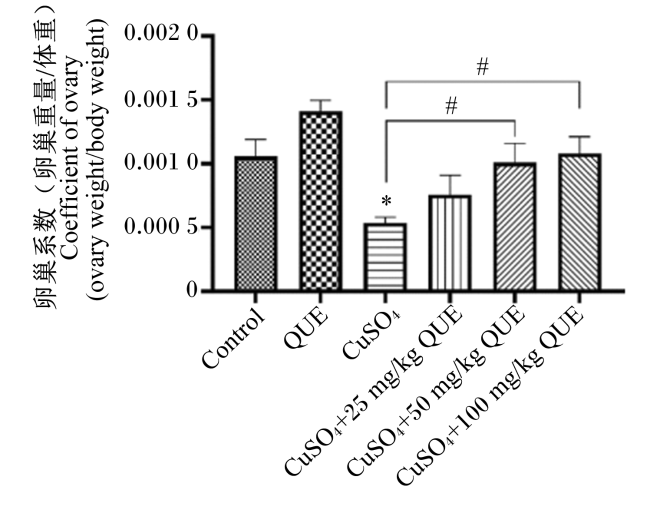
卵巢系数是卵巢重量与小鼠体重的比值,能够直观地反映出卵巢的相对重量变化。如图1所示,与对照组相比,高铜组的卵巢系数显著下降(P<0.05),表明饲喂高铜不利于小鼠卵巢的生长发育。然而,在补充槲皮素后,卵巢系数明显上升,其中补充50和100 mg/kg槲皮素的效果显著(P<0.05)。
图1 小鼠卵巢系数

Control:对照组;QUE:槲皮素组;CuSO4:高铜组;CuSO4+25 mg/kg QUE:槲皮素低浓度保护组;CuSO4+50 mg/kg QUE:槲皮素中浓度保护组;CuSO4+100 mg/kg QUE:槲皮素高浓度保护组。高铜组和槲皮素组数据柱标注“*”表示与对照组相比差异显著(P<0.05),标注“**”表示与对照组相比差异极显著(P<0.01);槲皮素保护组数据柱标注“#”表示与高铜组相比差异显著(P<0.05),标注“##”表示与高铜组相比差异极显著(P<0.01)。图4图5同。

Fig.1 Ovarian coefficient of mice

Control: control group; QUE: quercetin group; CuSO4: high copper group; CuSO4+25 mg/kg QUE: low concentration of quercetin protection group; CuSO4+50 mg/kg QUE: medium concentration of quercetin protection group; CuSO4+100 mg/kg QUE: high concentration of quercetin protection group. Compared with the control group, data columns of high copper group and quercetin group with “*” indicates a significant difference (P<0.05), and with “**” indicates an extremely significant difference (P<0.01); compared with the high copper group, data columns of quercetin protection groups with “#” indicates a significant difference (P<0.05), and with “##” indicates an extremely significant difference (P<0.01). The same as Fig.4 and Fig.5.

2.2 槲皮素对高铜处理小鼠卵巢组织的影响

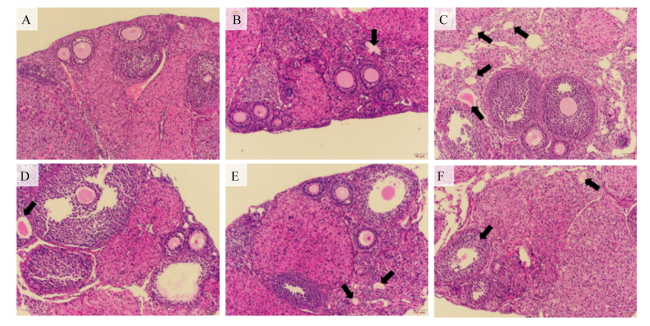
卵巢组织病理学变化如图2所示,对照组小鼠(图2-A)与单独补充槲皮素组小鼠(图2-B)的卵巢颗粒细胞发育状况良好,紧密排列在卵泡内膜周围,与卵母细胞之间的连接紧密,表明在正常情况下或100 mg/kg槲皮素的作用下,卵巢颗粒细胞的发育正常、排列有序。然而,在高铜组小鼠卵巢中可观察到卵泡闭锁的数量明显增多(图2-C),表明卵泡发育受到高铜的影响;此外,高铜组小鼠卵巢颗粒细胞松散并且在卵泡中有明显的扩散现象(图2-C)。在3个槲皮素保护组中,小鼠卵巢中闭锁卵泡数量均比高铜组减少,其中补充50 mg/kg槲皮素的槲皮素中浓度保护组的小鼠卵巢颗粒细胞与卵母细胞的连接较另外2组更加紧密(图2-D~F)。
图2 小鼠卵巢的代表性组织学切片

A:对照组;B:槲皮素组;C:高铜组;D:槲皮素低浓度保护组;E:槲皮素中浓度保护组;F:槲皮素高浓度保护组。黑色箭头指示的是闭锁卵泡。比例尺为50 μm。

Fig.2 Representative histological sections of mouse ovaries

A: control group; B: quercetin group; C: high copper group; D: low concentration of quercetin protection group; E: medium concentration of quercetin protection group; F: high concentration of quercetin protection group. The black arrow points to an atretic follicle. The scale bar is 50 μm.

2.3 槲皮素对高铜处理小鼠卵巢颗粒细胞凋亡的影响

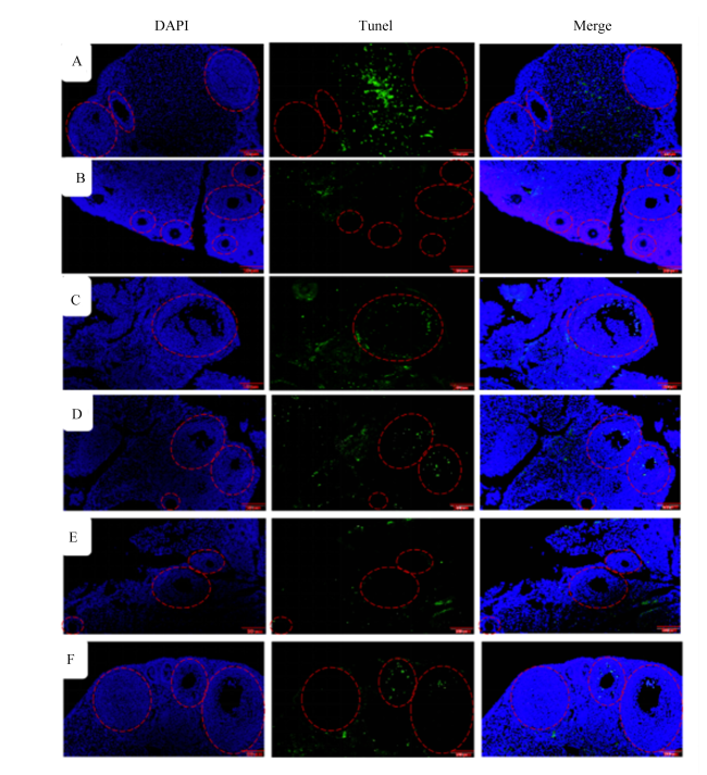
颗粒细胞凋亡是引起卵泡闭锁的主要原因,因此本试验采用Tunel染色法分析小鼠卵巢颗粒细胞凋亡情况。如图3所示,与对照组相比,高铜组小鼠卵泡颗粒细胞Tunel阳性信号增多,代表高铜引起凋亡细胞增多;与高铜组相比,给予25、50或100 mg/kg槲皮素后,小鼠卵泡颗粒细胞Tunel阳性信号减少,表明槲皮素降低了高铜引起的小鼠卵泡颗粒细凋亡,尤其以补充50 mg/kg槲皮素缓解高铜引起卵泡颗粒细胞凋亡效果最为显著。综上所述,槲皮素能够缓解高铜引起的小鼠卵泡颗粒细胞凋亡。
图3 小鼠卵巢Tunel染色图

A:对照组;B:槲皮素组;C:高铜组;D:槲皮素低浓度保护组;E:槲皮素中浓度保护组;F:槲皮素高浓度保护组。红色虚线指示不同卵泡;绿色代表凋亡细胞,蓝色代表细胞核。比例尺为50 μm。

Fig.3 Tunel staining images of mouse ovaries

A: control group; B: quercetin group; C: high copper group; D: low concentration of quercetin protection group; E: medium concentration of quercetin protection group; F: high concentration of quercetin protection group. Red dashed line indicates different follicles; green represents apoptotic cells, blue represents nucleus. The scale bar is 50 μm.

2.4 槲皮素对高铜处理小鼠血清氧化应激指标的影响

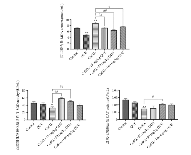
为了深入探究小鼠体内的氧化还原平衡状态,对小鼠血清中的氧化应激相关指标进行检测,结果如图4所示。与对照组相比,槲皮素组小鼠血清中MDA含量极显著降低(P<0.01),高铜组小鼠血清中MDA含量极显著升高(P<0.01)。各槲皮素保护组血清中MDA含量与高铜组相比均显著或极显著下降(P<0.05或P<0.01),表明槲皮素能够降低小鼠体内的脂质过氧化水平。与对照组相比,高铜组小鼠血清中T-SOD与CAT活性显著或极显著下降(P<0.05或P<0.01);与高铜组相比,各槲皮素保护组血清中CAT、T-SOD活性均上升,其中,补充50 mg/kg槲皮素的小鼠血清中CAT活性显著上升(P<0.05),补充25和50 mg/kg槲皮素的小鼠血清中T-SOD活性极显著上升(P<0.01)。以上结果表明,槲皮素能够恢复高铜诱导的血清CAT、T-SOD活性的降低,降低小鼠体内的脂质过氧化水平。
图4 槲皮素对高铜处理小鼠血清氧化应激指标的影响

Fig.4 Effects of quercetin on oxidative stress indicators in serum of mice treated with high copper

2.5 槲皮素对高铜处理小鼠卵巢组织中氧化应激相关基因表达的影响

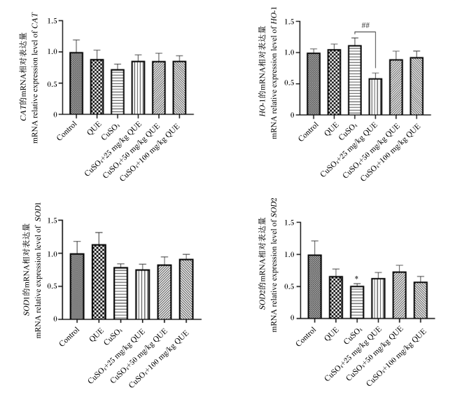
为了进一步分析小鼠体内的氧化还原状态,利用RT-qPCR检测小鼠卵巢组织中氧化应激相关基因的表达情况,结果如图5所示。与对照组相比,高铜组小鼠卵巢组织中氧化应激相关基因CATSOD1、SOD2的mRNA相对表达量均下降,其中SOD2的mRNA相对表达量显著下降(P<0.05);与对照组相比,高铜组小鼠卵巢组织中HO-1的mRNA相对表达量有所上升,但差异未达显著水平(P>0.05)。与高铜组相比,补充50或100 mg/kg槲皮素后小鼠卵巢组织中CATSOD1、SOD2、HO-1的mRNA相对表达量虽未产生显著变化(P>0.05),但均向着正常水平恢复。以上结果表明,槲皮素能够缓解高铜诱导的小鼠卵巢中氧化应激相关基因表达的异常变化。
图5 槲皮素对高铜处理小鼠卵巢组织中氧化应激相关基因表达的影响

Fig.5 Effects of quercetin on expression of oxidative stress-related genes in ovarian tissue of mice treated with high copper

3 讨论

本试验利用灌服槲皮素的方式研究其对高铜处理小鼠卵巢损伤的保护作用。尽管没有直接的证据表明槲皮素在肠道中以哪种形式直接作用于卵巢,但槲皮素能够通过多种途径对肠道黏膜屏障功能产生积极影响,可能间接影响卵巢健康。例如,通过改善肠道屏障功能,槲皮素可能有助于减少炎症和氧化应激[27-28],进而影响卵巢功能。此外,槲皮素在动物模型中显示出能够通过降低晚期糖基化终末产物(AGEs)/晚期糖基化终末产物受体(RAGE)通路活性,促进多囊卵巢综合征大鼠卵巢颗粒细胞的增殖,并抑制细胞凋亡,从而改善多囊卵巢综合征症状[25,29]。因此,槲皮素对肠道屏障功能的保护作用和对卵巢健康的潜在益处表明槲皮素可能通过多种机制在全身范围内发挥作用。然而,槲皮素在肠道中的具体作用形式尚不完全清楚,其在体内的具体作用途径还需要更多的研究来明确。
铜是动物机体内一种必需微量元素,在机体内发挥重要作用。然而,过量的铜造成机体多种器官损伤。已有研究证明,纳米铜能够在大鼠卵巢中聚集,诱导细胞凋亡,并调节细胞内氧化应激水平介导卵巢功能障碍,导致性激素水平降低、卵巢系数降低[30]。本研究结果显示,高铜处理后小鼠的卵巢系数显著下降,而槲皮素单独处理能够升高卵巢系数,并且能够缓解铜造成的卵巢系数下降。此外,Elkady等[31]的试验结果显示,槲皮素能够增加小鼠原始卵泡数量并减少卵泡闭锁。Bolouki等[32]的试验结果显示,槲皮素能够增加卵巢和生长卵泡的体积,增加生长卵泡和黄体的数量,显著减少闭锁卵泡的数量。本研究中的组织切片结果显示高铜导致小鼠卵巢颗粒细胞松散,并诱导卵泡闭锁,这可能是由于高铜处理对卵巢组织产生了负面影响,导致卵泡颗粒细胞的正常结构和功能受到了破坏。卵泡发育的任何阶段都可能发生闭锁,尽管各阶段闭锁卵泡的形态结构各异,但最终都将遭到中性粒细胞和巨噬细胞的清除。早期的卵泡闭锁后,逐渐消失,不留任何痕迹。当闭锁发生于次级卵泡或成熟卵泡时,其内部的初级卵母细胞将逐渐退化并最终消失。与此同时,透明带会先皱缩成一个不规则的嗜酸性环状物,随后也会退化消失。卵泡壁层的颗粒细胞会变得松散并脱落至卵泡腔,这些细胞最终会经历凋亡过程[33]。卵泡膜细胞逐渐增大,形态类似于膜黄体细胞,它们被结缔组织和血管分隔成独立的细胞团索。当单独补充槲皮素时,小鼠卵巢中的颗粒细胞的连接比对照组更加紧密,卵泡质量有所提升;当小鼠摄入过量的铜时,补充槲皮素能够部分缓解高铜诱导的卵巢损伤,并减少高铜诱导的卵泡闭锁。研究发现,颗粒细胞凋亡是引起卵泡闭锁的主要原因之一[34]。本研究的Tunel染色结果显示高铜引起小鼠卵巢颗粒细胞凋亡增多,闭锁卵泡增多,当补充槲皮素时,能够缓解高铜诱导的小鼠卵巢颗粒细胞凋亡。以上结果表明,槲皮素具有缓解卵巢颗粒细胞凋亡,减少卵泡闭锁,提高母畜繁殖能力的功能。
外源性铜离子可使机体产生过量的ROS,从而导致脂质过氧化、DNA断裂和蛋白质氧化,打破氧化-抗氧化系统的平衡,诱发氧化应激。研究发现,暴露于CuSO4或氯化铜(CuCl2)可诱导HEK293细胞、小鼠肝细胞或神经元细胞产生过量的ROS,进而诱发细胞氧化应激[35-37]。同样,过量铜离子会引起大鼠脑和肝脏组织中的MDA含量升高、超氧化物歧化酶(SOD)活性降低,诱发大鼠氧化应激[38]。研究表明,卵巢氧化应激损伤通过下调抗氧化基因的表达来诱导脂质过氧化和DNA氧化损伤,进而导致卵巢功能减退和卵母细胞质量下降[39]。相反,补充天然抗氧化剂可以减少颗粒细胞氧化应激的发生,可以减轻氧化应激对颗粒细胞的损伤,从而减少卵泡闭锁[40]。槲皮素作为自然界中广泛存在的具有生物活性的黄酮类化合物,现已证明是一种有效的抗凋亡和抗氧化剂[16]。有研究发现,槲皮素能够发挥其抗氧化和抗凋亡的特性抵抗铜诱导的神经毒性[41]。Jia等[42]研究表明,槲皮素能够激活细胞的抗氧化保护颗粒细胞免受氧化应激的损伤。Wang等[25]研究发现,低剂量的槲皮素能够升高大鼠卵巢中SOD1、CAT和谷胱甘肽过氧化物酶(GSH-Px)的mRNA相对表达量。本研究发现,与对照组相比,高铜处理后小鼠血清中脂质过氧化标志物MDA的含量显著升高,抗氧化酶CAT、T-SOD的活性显著降低;小鼠卵巢中抗氧化相关基因SOD1、SOD2、CAT的mRNA相对表达量下降,HO-1的mRNA相对表达量上升;此外,补充槲皮素能够使血清中的氧化应激指标以及卵巢组织中的氧化应激相关基因的mRNA相对表达量向着正常水平恢复。机体脂质过氧化的程度可以利用MDA含量反映,作为氧化应激的终产物,其可以导致核酸和蛋白质等大分子的交联和聚合,进而诱发细胞组织细胞毒性,因此MDA含量是常用的反映机体氧化损伤的指标。SOD作为一种抗氧化酶,其主要功能在于催化超氧阴离子自由基转化为过氧化氢和氧气,从而在生物体内发挥其抗氧化作用。与此同时,CAT则进一步分解过氧化氢为水和氧气,共同维持细胞环境的氧化还原平衡。HO-1催化分解血红素,当细胞受到氧化应激和损伤时,其mRNA和蛋白表达水平会上调。抗氧化酶通过互相协作将ROS转化为对细胞组分没有损伤作用的物质,保护细胞免受氧化损伤。这些结果表明,高铜可导致小鼠体内的氧化还原平衡失调,进而引发卵巢组织的氧化应激反应,造成氧化损伤;而槲皮素能够发挥其优越的抗氧化作用保护小鼠卵巢免于氧化损伤。综上所述,槲皮素对因高铜摄入造成的小鼠卵巢毒性具有保护作用。该研究结果可为减少雌性动物的卵泡闭锁、促进卵巢发育以及提高雌性动物的繁殖能力提供新的思路和理论依据。

4 结论

补充50或100 mg/kg槲皮素可以减少卵泡闭锁与卵巢颗粒细胞凋亡,促进卵泡发育,升高高铜降低的小鼠体内的抗氧化酶活性,降低高铜诱导的脂质过氧化水平,进而缓解高铜诱导的小鼠卵巢氧化损伤。
[1]
宋明明, 黄凯. 铜的生物学功能及其应用[J]. 饲料博览, 2014(10):13-17.

SONG M M, HUANG K. Biological function and application of copper[J]. Feed Review, 2014(10):13-17. (in Chinese)

[2]
TSANG T, DAVIS C I, BRADY D C. Copper biology[J]. Current Biology, 2021, 31(9):R421-R427.

[3]
PENG X Y, DAI C S, ZHANG M, et al. Molecular mechanisms underlying protective role of quercetin on copper sulfate-induced nephrotoxicity in mice[J]. Frontiers in Veterinary Science, 2020, 7:586033.

[4]
丰艳平, 刘小飞. 高铜日粮对断奶仔猪生长性能和腹泻率的影响[J]. 湖南畜牧兽医, 2020(5):17-18.

FENG Y P, LIU X F. High copper diet on weaning piglet growth performance and diarrhea rate impact[J]. Hunan Journal of Animal Science & Veterinary Medicine, 2020(5):17-18. (in Chinese)

[5]
王希春, 吴金节, 李义刚, 等. 高铜日粮对早期断奶应激仔猪生长性能和抗氧化酶活性的影响[J]. 安徽农业大学学报, 2004, 31(4):412-416.

WANG X C, WU J J, LI Y G, et al. Effect of high copper diets on growth performance and serum antioxidative enzyme activities of early weanling stress piglets[J]. Journal of Anhui Agricultural University, 2004, 31(4):412-416. (in Chinese)

[6]
ALKHANJAF A A M, SHARMA S, SHARMA M, et al. Microbial strategies for copper pollution remediation:mechanistic insights and recent advances[J]. Environmental Pollution, 2024, 346:123588.

[7]
GAO Y, YANG W Y, CHE D S, et al. Advances in the mechanism of high copper diets in restraining pigs growth[J]. Journal of Animal Physiology and Animal Nutrition, 2020, 104(2):667-678.

DOI PMID

[8]
ZHONG G L, LI Y X, MA F Y, et al. copper exposure induced chicken hepatotoxicity:involvement of ferroptosis mediated by lipid peroxidation,ferritinophagy,and inhibition of FSP1-CoQ10 and Nrf2/SLC7A11/GPX4 axis[J]. Biological Trace Element Research, 2024, 202(4):1711-1721.

[9]
ADELEKE P A, AJAYI A M, BEN-AZU B, et al. Involvement of oxidative stress and pro-inflammatory cytokines in copper sulfate-induced depression-like disorders and abnormal neuronal morphology in mice[J]. Naunyn-Schmiedeberg’s Archives of Pharmacology, 2023, 396(11):3123-3133.

[10]
BABAEI H, ROSHANGAR L, SAKHAEE E, et al. Ultrastructural and morphometrical changes of mice ovaries following experimentally induced copper poisoning[J]. Iranian Red Crescent Medical Journal, 2012, 14(9):558-568.

PMID

[11]
GOU Z Y, FAN Q L, LI L, et al. High dietary copper induces oxidative stress and leads to decreased egg quality and reproductive performance of Chinese yellow broiler breeder hens[J]. Poultry Science, 2021, 100(3):100779.

[12]
ZHANG C H, WANG Y, SUN Q Q, et al. copper nanoparticles show obvious in vitro and in vivo reproductive toxicity via ERK mediated signaling pathway in female mice[J]. International Journal of Biological Sciences, 2018, 14(13):1834-1844.

[13]
ROYCHOUDHURY S, BULLA J, SIROTKIN A V, et al. in vitro changes in porcine ovarian granulosa cells induced by copper[J]. Journal of Environmental Science and Health:Part A, Toxic/Hazardous Substances & Environmental Engineering, 2014, 49(6):625-633.

[14]
WANG Q, SUN Y, ZHAO A L, et al. High dietary copper intake induces perturbations in the gut microbiota and affects host ovarian follicle development[J]. Ecotoxicology and Environmental Safety, 2023, 255:114810.

[15]
BAYAZID A B, LIM B O. Quercetin is an active agent in berries against neurodegenerative diseases progression through modulation of Nrf2/HO1[J]. Nutrients, 2022, 14(23):5132.

[16]
LI F, LIU J A, TANG S F, et al. Quercetin regulates inflammation,oxidative stress,apoptosis,and mitochondrial structure and function in H9C2 cells by promoting PVT1 expression[J]. Acta Histochemica, 2021, 123(8):151819.

[17]
SUL O J, RA S W. Quercetin prevents LPS-Induced oxidative stress and inflammation by modulating NOX2/ROS/NF-κB in lung epithelial cells[J]. Molecules, 2021, 26(22):6949.

[18]
唐军, 王旭东, 方素庭, 等. 妊娠后期日粮中添加槲皮素对母猪繁殖性能、血清生化及抗氧化功能的影响[J]. 中国畜牧杂志, 2024, 60(6):222-226.

TANG J, WANG X D, FANG S T, et al. Add quercetin in late pregnancy diet on reproductive performance,the influence of serum biochemical and antioxidant function[J]. Chinese Journal of Animal Science, 2024, 60(6):222-226. (in Chinese)

[19]
张卫辉, 朱秋凤, 张永静, 等. 槲皮素对仔猪生长性能、肠道抗氧化及免疫相关基因表达的影响[J]. 河南农业大学学报, 2023, 57(1):136-142.

ZHANG W H, ZHU Q F, ZHANG Y J, et al. Effects of quercetin on growth performance and intestinal antioxidant capacity and immune related gene expression in piglets[J]. Journal of Henan Agricultural University, 2023, 57(1):136-142. (in Chinese)

[20]
郭长征, 冯泮飞, 薛春旭, 等. 高精料饲粮添加槲皮素对山羊瘤胃发酵、瘤胃菌群数量及血清指标的影响[J]. 动物营养学报, 2016, 28(9):2839-2846.

DOI

GUO C Z, FENG P F, XUE C X, et al. Effects of quercetin supplemented in high-concentrate diet on rumen fermentation,rumen bacteria counts and serum indices of goats[J]. Chinese Journal of Animal Nutrition, 2016, 28(9):2839-2846. (in Chinese)

[21]
NASEER Z, AHMAD E, EPIKMEN E T, et al. Quercetin-supplemented diet improves follicular development,oocyte quality,and reduces ovarian apoptosis in rabbits during summer heat stress[J]. Theriogenology, 2017, 96:136-141.

[22]
BEAZLEY K E, NURMINSKAYA M. Effects of dietary quercetin on female fertility in mice:implication of transglutaminase 2[J]. Reproduction,Fertility,and Development, 2016, 28(7):974-981.

[23]
NNA V U, USMAN U Z, OFUTET E O, et al. Quercetin exerts preventive,ameliorative and prophylactic effects on cadmium chloride-induced oxidative stress in the uterus and ovaries of female Wistar rats[J]. Food and Chemical Toxicology, 2017, 102:143-155.

[24]
CAPCAROVA M, PETRUSKA P, ZBYNOVSKA K, et al. Changes in antioxidant status of porcine ovarian granulosa cells after quercetin and T-2 toxin treatment[J]. Journal of Environmental Science and Health:Part B, Pesticides,Food Contaminants,and Agricultural Wastes, 2015, 50(3):201-206.

[25]
WANG J, QIAN X, GAO Q, et al. Quercetin increases the antioxidant capacity of the ovary in menopausal rats and in ovarian granulosa cell culture in vitro[J]. Journal of Ovarian Research, 2018, 11(1):51.

[26]
QI N N, XING W W, LI M X, et al. Quercetin alleviates toxicity induced by high levels of copper in porcine follicular granulosa cells by scavenging reactive oxygen species and improving mitochondrial function[J]. Animals, 2023, 13(17):2745.

[27]
梅华迪, 李袁飞, 马现永, 等. 槲皮素对动物肠道黏膜屏障功能的影响及其调控机制[J]. 动物营养学报, 2022, 34(9):5475-5488.

DOI

MEI H D, LI Y F, MA X Y, et al. Effects and regulatory mechanisms of quercetin on animal intestinal mucosal barrier function[J]. Chinese Journal of Animal Nutrition, 2022, 34(9):5475-5488. (in Chinese)

DOI

[28]
GAO F, ZHU F, SHUAI B, et al. Quercetin ameliorates ulcerative colitis by restoring the balance of M2/M1 and repairing the intestinal barrier via downregulating cGAS-Sting pathway[J]. Frontiers in Pharmacology, 2024, 15:1351538.

[29]
江雪娟, 陈晓菲, 俞佳, 等. 槲皮素对多囊卵巢综合征大鼠卵巢颗粒细胞增殖与凋亡的影响研究[J]. 浙江中医药大学学报, 2022, 46(10):1055-1062,1069.

JIANG X J, CHEN X F, YU J, et al. Effect of quercetin on proliferation and apoptosis of ovarian granulosa cells in rats with polycystic ovarian syndrome[J]. Journal of Zhejiang Chinese Medical University, 2022, 46(10):1055-1062,1069. (in Chinese)

[30]
YANG J, HU S F, RAO M, et al. copper nanoparticle-induced ovarian injury,follicular atresia,apoptosis,and gene expression alterations in female rats[J]. International Journal of Nanomedicine, 2017, 12:5959-5971.

[31]
ELKADY M A, SHALABY S, FATHI F, et al. Effects of quercetin and rosuvastatin each alone or in combination on cyclophosphamide-induced premature ovarian failure in female albino mice[J]. Human & Experimental Toxicology, 2019, 38(11):1283-1295.

[32]
BOLOUKI A, ZAL F, BORDBAR H. Ameliorative effects of quercetin on folliculogenesis in diabetic mice:a stereological study[J]. Gynecological Endocrinology, 2020, 36(10):864-868.

[33]
贺名扬. 褪黑素对绵羊卵巢颗粒细胞增殖及类固醇激素分泌的影响[D]. 硕士学位论文. 保定: 河北农业大学, 2023.

HE M Y. Effects of melatonin on granulosa cell proliferation and steroid hormone secretion in sheep ovary[D]. Master’s Thesis. Baoding: Hebei Agricultural University, 2023. (in Chinese)

[34]
ZHOU J W, PENG X W, MEI S Q. Autophagy in ovarian follicular development and atresia[J]. International Journal of Biological Sciences, 2019, 15(4):726-737.

DOI PMID

[35]
DAI C S, LIU Q Q, LI D W, et al. Molecular insights of copper sulfate exposure-induced nephrotoxicity:involvement of oxidative and endoplasmic reticulum stress pathways[J]. Biomolecules, 2020, 10(7):1010.

[36]
PARK K A, YUN N R, OH Y J. Anamorsin attenuates cupric chloride-induced dopaminergic neuronal cell death[J]. Biochemical and Biophysical Research Communications, 2019, 520(1):99-106.

[37]
LIU H, GUO H R, JIAN Z J, et al. copper induces oxidative stress and apoptosis in the mouse liver[J]. Oxidative Medicine and Cellular Longevity, 2020, 2020:1359164.

[38]
OZCELIK D, UZUN H. copper intoxication; antioxidant defenses and oxidative damage in rat brain[J]. Biological Trace Element Research, 2009, 127(1):45-52.

[39]
LIM J, LUDERER U. Oxidative damage increases and antioxidant gene expression decreases with aging in the mouse ovary[J]. Biology of Reproduction, 2011, 84(4):775-782.

DOI PMID

[40]
王赫川, 李天峰, 张涵, 等. 天然抗氧化剂对动物颗粒细胞保护作用的研究进展[J]. 畜牧与兽医, 2023, 55(1):145-150.

WANG H C, LI T F, ZHANG H, et al. Progress in research on the protective effect of natural antioxidants on granule cells in animals[J]. Animal Husbandry & Veterinary Medicine, 2023, 55(1):145-150. (in Chinese)

[41]
CHAKRABORTY J, PAKRASHI S, SARBAJNA A, et al. Quercetin attenuates copper-induced apoptotic cell death and endoplasmic reticulum stress in SH-SY5Y cells by autophagic modulation[J]. Biological Trace Element Research, 2022, 200(12):5022-5041.

DOI PMID

[42]
JIA Y D, LIN J X, MI Y L, et al. Quercetin attenuates cadmium-induced oxidative damage and apoptosis in granulosa cells from chicken ovarian follicles[J]. Reproductive Toxicology, 2011, 31(4):477-485.

DOI PMID

Outlines

/