REVIEW

Progress on Structure-Activity Relationship, Biosynthesis and Application of Microcins

  • YANG Li , 1, 2 ,
  • SU Guoqi 1, 2 ,
  • HUANG Jinxiu 1, 2 ,
  • YANG Feiyun , 1, 2, *
Expand
  • 1 Chongqing Academy of Animal Sciences, Chongqing 402460, China
  • 2 National Center of Technology Innovation for Pigs (Chongqing), Chongqing 402460, China
*professor, E-mail:

Received date: 2024-05-30

  Online published: 2025-02-16

Abstract

The extensive use of antibiotics in livestock farming poses serious threats to the health of both livestock and humans, creating an urgent need for novel antibiotic alternatives. Microcins are a class of small peptides produced by Gram-negative bacteria that mediate interspecies and intraspecies competition among Enterobacteriaceae in the inflamed gut. They can act as narrow-spectrum therapeutics to inhibit gut pathogens and reduce the risk of resistance, highlighting their unique potential in the field of antibiotic alternatives. This article aimed to review the structure-activity relationship and biosynthesis mechanisms of microcins, and to explore their applications in livestock farming, providing a reference for the development and utilization of microcins and their analogs.[Chinese Journal of Animal Nutrition, 2025, 37(2):812-825]

Cite this article

YANG Li , SU Guoqi , HUANG Jinxiu , YANG Feiyun . Progress on Structure-Activity Relationship, Biosynthesis and Application of Microcins[J]. Chinese Journal of Animal Nutrition, 2025 , 37(2) : 812 -825 . DOI: 10.12418/CJAN2025.071

长期以来,抗生素被广泛应用于畜牧业生产中,在治疗疾病、动物保健和促进生长方面发挥着重要作用[1]。但长期或过度不规范使用抗生素会导致病原菌耐药性的产生,并在畜产品中残留,对畜禽生产和人类健康造成严重威胁[2-3]。随着新型抗生素开发数量的减少,寻找抗生素替代品迫在眉睫[4-5]。微菌素(microcins)是一类由革兰氏阴性菌所产生的小分子多肽,能够介导发炎肠道中肠杆菌的种间和种内竞争,可以作为窄谱疗剂来抑制肠道病原体,减少耐药细菌的传播[6-7]。微菌素通过与特定受体结合和干扰细胞代谢途径发挥抗菌作用,其活性和稳定性受氨基酸序列和化学修饰的影响[8]。微菌素对肠杆菌的生长及其在肠道中的定植、侵袭和易位等过程都有重要影响,表现出抗菌和免疫调节的双重功能[9-10]。这些研究表明,微菌素及其类似物具有替代抗生素的功能及其在畜牧业中的应用潜力。本文将从微菌素的构效关系、生物合成机制及其在畜牧业中的应用等方面进行综述,旨在为微菌素及其类似物的合理开发与科学应用提供参考。

1 微菌素的分类及其抗菌谱

Baquero等[11]将微菌素分为2类:第Ⅰ类微菌素是分子质量小于5 ku的由3个质粒编码的肽,普遍具有翻译后修饰骨架,并针对特定的细胞内靶标发挥其抗菌功能,包括微菌素B17[12]、微菌素C(也称为微菌素C7或C51)[13-14]、微菌素J25[15]、微菌素D93[16]和微菌素Y[17]。第Ⅱ类微菌素分子质量为5~10 ku,又细分为Ⅱa类和Ⅱb类,其中Ⅱa类是指由3个质粒编码的微菌素,包括微菌素L[18]、微菌素V[19]、微菌素N(也称为微菌素24)[20]、微菌素S[21]和微菌素PDI(也称为微菌素25)[22],它们没有翻译后的修饰过程;Ⅱb类是由携带C末端铁载体的染色体编码的线性微菌素,包括微菌素E492、微菌素G492、微菌素M、微菌素H47和微菌素I47[23-25]。研究发现,利用信息学的方法在公开的数据库中筛选细菌基因组发现了Ⅱb类中3个新的微菌素分支(微菌素W、微菌素X和微菌素Z),分别来自于Gibbsiella quercinecansBrenneria goodwinii以及解鸟氨酸拉乌尔菌(Raoultella ornithinolytica)菌种,但其生物学特性还未经表征确认[26]
微菌素对某些非近缘物种或特定耐药菌株具有活性[27]。例如,由大肠杆菌产生的微菌素L对铜绿假单胞菌也表现出活性[28]。最近发现的微菌素Y可抑制沙门氏菌、志贺氏菌、假单胞菌、枯草芽孢杆菌和金黄色葡萄球菌等病原菌[17]。此外,微菌素PDI和微菌素24对人类病原菌有抑制作用[22,29];微菌素H47还能抑制多重耐药性的菌株,例如能抑制产生碳青霉烯酶和超广谱β-内酰胺酶的多种耐药菌株[30];微菌素J25对食源性沙门氏菌和大肠杆菌也有抗菌活性[31]。成功纯化的微菌素抑菌活性通常在nmol/L到μmol/L。例如,微菌素N、微菌素L、微菌素J25和微菌素E492对其主要靶标菌株的最小抑菌浓度(minimal inhibit concentration,MIC)在4~150 nmol/L[20,28,32-33],而微菌素H47的MIC在1~12 μmol/L[30]。Naimi等[34]发现,微菌素J25对新港沙门氏菌(Salmonella Newport)的MIC为30 nmol/L,显著优于革兰氏阳性细菌素罗伊氏菌素(reuterin)和小分子抗生素利福平。这些结果表明,微菌素有望成为抗生素的替代品。微菌素的分类及其特性见表1[12,14-21,23-25,35-36]
表1 微菌素的分类及其特性

Table 1 Classification and characteristics of microcins

微菌素
Microcins
菌种来源及质粒
Strain source and
plasmid
微菌素前
体长度
Pre-
microcin
length/
个氨基酸
成熟微菌
素长度
Mature
microcin
length/
个氨基酸
确认成熟
微菌素
分子质量
Confirmed
mature microcin
molecular
mass/ku
甘氨酸残基
Glycine
residues/
%
疏水性残基
Hydrophobic
residues/
%
参考文献
References
Ⅰ类Class Ⅰ
J25 E. coli AY25 pTUC100 58 21 2.107 28.6 71.4 Salomón等[15]
Y S. enterica pAUSMDU
00010532_01
53 21 2.224 23.8 57.1 Li等[17]
B17 E. coli pMccB17 69 43 3.074 60.5 67.4 Asensio等[12]
C7/C51 E. coli pMccC7 7 7 1.177 14.3 42.9 García-
Bustos等[14]
D93 E. coli LP93 pMccD93 <1 Martínez等[16]
Ⅱa类Class Ⅱa
V E. coli pColV-K30 103 88 8.734 17 61.4 Gilson等[19]
L E. coli LR05 plasmid 105 90 8.884 15.6 62.2 Gaillard-
Gendron等[18]
N/24 E. coli 2424 p24-2 89 74 7.224 18.9 71.6 Kaur等[20]
PDI/25 E. coli 25 plasmid 120 103 9.960 22.6 51.2 Vassiliadis等[23]
S E. coli G3/10 pSYM1 120 84 7.957 26.2 54.8 Zschüttig等[21]
Ⅱb类Class Ⅱb
H47 E. coli H47 75 60 5.696 26.7 76.7 Laviña等[25]
I47 E. coli H47 77 62 6.275 12.9 50 Poey等[35]
M E. coli Nissle 1917 92 77 8.115 19.5 55.8 Patzer等[24]
G492 K. pneumoniae RYC492 89 74 7.342 24.3 70.3 Vassiliadis等[23]
E492 K. pneumoniae RYC492 99 84 8.717 22.6 64.3 Destoumieux-
Garzón等[36]

E. coli:大肠杆菌 Escherichia coli;S. enterica:肠道沙门氏菌 Salmonella enterica;K. pneumoniae:肺炎克雷伯菌 Klebsiella pneumoniae;Mcc:微菌素 microcin;plasmid:质粒。

2 微菌素的构效关系

2.1 Ⅰ类微菌素

Ⅰ类微菌素是结构复杂、经过翻译后修饰的抗菌肽,如微菌素C、微菌素J25、微菌素B17及新发现的微菌素Y,具有独特的结构特征和作用机理。
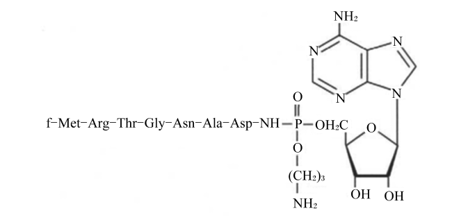
微菌素C由7个氨基酸组成,其结构特征包括1个甲酰化的N端甲硫氨酸,1个C端天冬氨酸,1个连接C端天冬氨酸α羧基的N-酰基氨基磷酸酯键,以及丙胺基修饰的磷酸基团[37]。微菌素C的N端N-甲酰甲硫氨酸(N-formylmethionine,f-Met)先被细菌细胞外膜上的外膜蛋白F(outer membrane protein F,OmpF)识别,然后被细胞内膜上的ABC转运体YejABEF识别进入细胞[38]。N端甲酰基团在细胞内被脱甲酰酶去除之后被3种氨肽酶(aminopeptidase,Pep)(PepA、PepB和PepN)中的1种或多种消化,当第6个与第7个肽残基之间的肽键水解时,就会释放一种不可水解的天冬酰基腺苷酸酯类似物,通过阻止天冬氨酰-tRNA合成酶(aspartyl-tRNA synthetase,Asp-RS)合成氨酰-tRNA,进而有效地阻断细菌的蛋白质合成[39-40]。Kazakov等[41]首次对微菌素C的前体mccA基因进行了定点诱变,替换了mccA基因编码的第2~7个密码子,共产生了114个突变;其中28个变异没有明显改变微菌素C的生物活性,4个取代(R2Y、R2M、A6M和A6F)导致抗菌活性丧失;这些发现表明,Arg2是靶细胞吸收的识别位点,而Ala6负责把微菌素C修饰成有活性的肽。野生型微菌素C多肽序列为MRTGNAN,容易在Arg2位置被胰蛋白酶裂解,因此Yang等[42]对微菌素C的Arg2进行精确的点突变,以抵抗胰蛋白酶的降解,增强其稳定性和抗菌活性。此外,延长微菌素C的肽链并不会提升其抗菌效果,主要是因为延长前体肽的长度会降低迭代合成效率,且N端甲酰化的微菌素C前体微菌素A作为微菌素B酶的底物,完全能满足生物合成的需要[43]。微菌素C的化学结构如图1所示[44]
图1 微菌素C的化学结构

Fig.1 Chemical structure of microcin C[44]

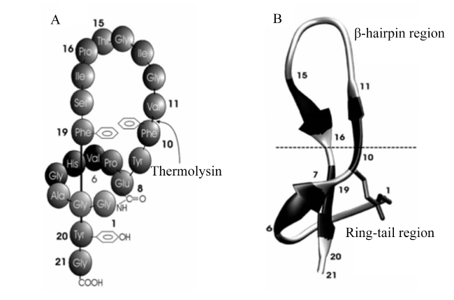
微菌素J25是由21个氨基酸组成的具有独特套索结构的多肽,该肽链包含1个由8个氨基酸构成的未封闭环状结构和1个由13个氨基酸组成的尾部结构,其特点在于Gly1的氨基和Glu8的羧基之间形成了1个骨架——侧链内酰胺键,能够固定环状结构,让肽链在环状结构中紧密穿行,形成大量的链内接触,稳定肽链的结构,防止新形成的肽链段从环中解离;而C端的甘氨酸残基则通过与His5侧链的电荷相互作用进一步增强了结构的稳定性[45]。这种结构特性使得微菌素J25即使在120 ℃高温或肽键断裂的情况下,其N端环状结构和C端尾部仍能维持相互连接,因此微菌素J25具有较高的热稳定性和有机溶剂稳定性,成为抗菌药物设计的重要支架[46-47]。微菌素J25的套索结构可以被细菌外膜的FhuA受体识别并结合,这主要依赖其His5残基的侧链能够插入FhuA腔内,同时His5的咪唑环和主链羰基与FhuA的Phe115主链羰基和Tyr116侧链形成氢键,与其环状结构和C端的下尾部共同结合在FhuA受体上,占据一个与天然配体非常相似的位置[48-49]。微菌素J25通过TonB系统和SbmA蛋白进入细胞质,抑制RNA聚合酶从而获得抗菌活性[50-51]。此外,微菌素J25的抗菌活性与其形成内酰胺连接的关键残基(Gly1和Glu8)、相邻的1个残基(Gly2)以及C端的芳香族残基(Tyr20)密切相关,这些残基形成了一个表面表位,能够被McjB和McjC蛋白识别并修饰,转化为具有活性的微菌素J25[52]。微菌素J25的化学结构及其二级结构如图2所示[53]
图2 微菌素J25的化学结构(A)及其二级结构(B)

Thermolysin:嗜热菌蛋白酶;β-hairpin region:β-发夹区;Ring-tail region:卷尾区。

Fig.2 Chemical structure (A) and secondary structure (B) of microcin J25[53]

微菌素B17是发现首个含有噻唑/恶唑修饰的线性微菌素,成熟序列的长度为43个氨基酸残基[54]。微菌素B17从N端到C端依次包含8个杂环芳香环,分别是:OTZ39(G39S40C41形成)、TAZ47(G47C48形成)、TAZ50(G50C51形成)、TOZ54(G54C55S56形成)、OAZ61(G61S62形成)和OAZ64(G64S65形成)[55]。微菌素B17的抗菌活性源自其恶唑和噻唑环,这些杂环通过捕获DNA旋转酶活动的中间状态,稳定DNA与旋转酶之间的共价断裂复合物,阻止DNA链的重新连接,从而导致DNA损伤累积,引发细菌细胞死亡[56-57]。此外,微菌素B17还能与DNA旋转酶的GyrB亚基C末端结构域相互作用,干扰DNA旋转酶必需ATP的结合和水解,影响细菌的生长和复制[58]。对微菌素B17构效关系的研究发现,OTZ39中恶唑环转变成噻唑环(TTZ39)不影响其抗菌活性,2个杂环位置互换(TOZ39)导致其抗菌活性较野生型微菌素B17下降30%,而去除OTZ39的恶唑环则使其完全丧失抗菌活性;对于TOZ54,其噻唑-恶唑转为噻唑-噻唑(TTZ54)或恶唑-恶唑(OOZ54)后,抗菌活性下降了70%,证实了其活性中心基团是恶唑和噻唑环[59]。微菌素B17的化学结构如图3所示[44]
图3 微菌素B17的化学结构

Fig.3 Chemical structure of microcin B17[44]

2.2 Ⅱ类微菌素

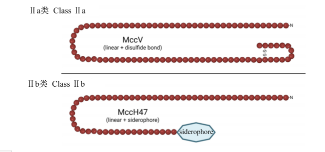
Ⅱ类微菌素通常含有N末端“双甘氨酸”信号序列(15~18个氨基酸),该序列以甘氨酸-甘氨酸(GG)或甘氨酸-丙氨酸(GA)残基对终止,且在信号序列的特定位置含有疏水残基,让微菌素有效分泌[60]。此外,Ⅱ类微菌素的序列富含甘氨酸,占总氨基酸的12%~26%(表1)。目前,已鉴定出10种不同的Ⅱ类微菌素,均是长度为60~100个氨基酸的阳离子肽,这些微菌素包含2个结构区域:高度保守的N端区域和保守程度较低的C端区域[Ⅱb类微菌素在C末端具有独特的类似于嗜铁体(siderophore)的铁载体修饰],其结构示例如图4[61]。Ⅱ类微菌素在水溶液中似乎以非结构化形式存在,但在细胞膜的模拟环境中形成明确的构象,其N端部分形成3股反平行β-片状结构,而C端部分形成1个或2个两亲性螺旋,这说明微菌素与磷脂直接相互作用可能会引起自身结构变化[62]
图4 Ⅱ类微菌素的结构示例

linear:线性结构;disulfide bond:二硫键;siderophore:嗜铁体。

Fig.4 Examples of class Ⅱ microcin structures[61]

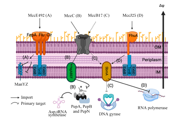
同时,Ⅱa类微菌素在目标细菌的膜中具有特异性受体,可在膜中形成亲水孔导致细胞质成分和离子外排发挥细菌杀伤效应,其跨膜转运机制由EtpM介导,且依赖YidC转运系统[63];而Ⅱb类微菌素的转运依赖儿茶酚盐铁载体受体FepA/Fiu/Cir识别其C末端铁载体基团[36]。其中,微菌素H47进入细胞后通过抑制ATP合成酶的内膜组分F0质子通道,阻碍细菌ATP的合成,达到抑菌作用[64]。微菌素E492通过抑制糖运输蛋白组件ManYZ发挥抑菌作用,阻断细菌对甘露糖和相关戊糖的摄取[65]。此外,结构或序列改造的研究进一步阐明了Ⅱ类微菌素的作用机制及其与靶细胞间复杂的相互作用。例如,通过改变微菌素的C端区域,进行特定的氨基酸替换或插入可以改变微菌素对靶细胞的特异性,调整其对细胞膜的穿透能力,从而影响诱导细胞渗透和死亡的能力[66]。部分微菌素的作用机制如图5所示[8]
图5 部分微菌素的作用机制

Mcc:微菌素 microcin;Δψ:电位差 potential difference;OM:外膜 outer membrane;IM:内膜 inner membrane;Periplasm:胞间质;Asp-tRNA synthetase:天冬氨酰-tRNA合成酶 aspartyl-tRNA synthetase;Pep:氨肽酶 aminopeptidase;DNA gyrase:DNA旋转酶;RNA polymerase:RNA聚合酶;Import:物质进入;Primary target:主要靶向。

Fig.5 Mechanism of action of some microcins[8]

3 微菌素的生物合成机制

3.1 Ⅰ类微菌素

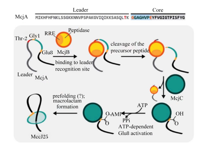
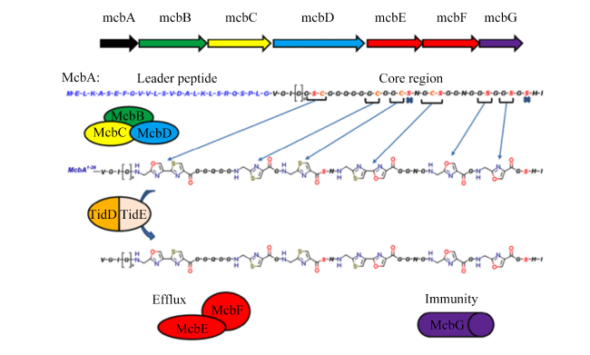
Ⅰ类微菌素主要由质粒编码,其生物合成基因簇包括前体骨架和转录后修饰相关基因,并与自身免疫基因分开。例如,微菌素J25及其同源物微菌素Y的生物合成基因簇包含线性前体McjA、2种成熟酶McjB/McjC以及1个ABC转运蛋白McjD[67]。其中,McjA蛋白包含位于N端的37个氨基酸的前导肽和位于C端的21个氨基酸的微菌素J25结构核心序列[68];McjB和McjC蛋白负责前体McjA的翻译后修饰,产生具有生物活性的成熟微菌素J25[69](图6);McjD蛋白则参与成熟微菌素J25的分泌,同时为生产细菌提供自身免疫[70]。而微菌素B17则通过翻译后修饰形成了4个恶唑和4个噻唑杂环[59](图7)。微菌素J25和微菌素B17均利用可裂解的N端信号序列引导其进一步加工[71]。微菌素J25的N端信号序列有助于其被识别并随后由成熟酶去除,而微菌素B17则在翻译后修饰完成后被内源性蛋白酶去除,以产生抗菌活性[72-73]。微菌素C则代表了Ⅰ类微菌素的第3种结构,只有7个氨基酸的短肽,无信号序列,会被主动转运到靶细胞胞内加工成毒性分子[74]
图6 微菌素J25的生物合成机制

Leader:前导肽 leader peptide;Core:核心肽 core peptide;RRE:核糖体合成和翻译后修饰肽识别元件 ribosomally synthesized and posttranslationally modified peptide recognition element;Peptidase:肽酶;binding to leader recognition site:结合到前导识别位点;cleavage of the precursor peptide:前体肽的切割;MccJ25:微菌素J25 microcin J25;prefolding:预折叠;macrolactam formation:大环内酰胺形成;ATP-dependent Glu8 activation:ATP依赖的Glu8激活。

Fig.6 Biosynthesis mechanism of microcin J25[46]

图7 微菌素B17的生物合成机制

Leader peptide:前导肽;Core region:核心区域;Efflux:分泌;Immunity:免疫。

Fig.7 Biosynthesis mechanism of microcin B17[59]

Ⅰ类微菌素的分泌和自身免疫依赖不同的蛋白[75]。例如,微菌素J25通过McjD从细胞质中分泌,并通过外膜因子蛋白TolC排出细胞[76-77];微菌素B17通过完整膜蛋白McbE与ATP结合蛋白McbF的相互作用进行分泌[78];微菌素C则由细菌外排泵分泌[70]。微菌素生产菌株对微菌素B17的自身免疫由McbG蛋白提供[78];对微菌素C的自身免疫由一种能裂解微菌素C的肽酶提供[79];而微菌素J25的自身免疫则依赖次级ABC转运体YojI[80]
此外,在Ⅰ类微菌素中,微菌素J25或微菌素Y可以使用pHT43质粒作为表达载体,在枯草芽孢杆菌168和WB800N中实现异源表达,避免了其在大肠杆菌中表达产生内毒素的问题[81];微菌素C也可以通过将其表达基因簇克隆到质粒pWP40中,转入大肠杆菌MC4100表达菌中实现异源表达[42],以便更加高效的对其进行突变改造。

3.2 Ⅱ类微菌素

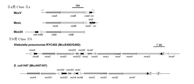
Ⅱ类微菌素在生物合成基因簇的结构和功能上有极大的相似性。例如,其生物合成基因簇一般由5个核心部分构成,分别是含有双甘氨酸信号序列的前体蛋白、免疫蛋白、带有C39肽酶结构域的吩嗪-1-羧酸转运蛋白(phenazine-1-carboxylic acid transporter,PCAT)[67]、膜融合蛋白(membrane fusion protein,MFP)和外膜因子蛋白TolC,目前已鉴定的Ⅱ类微菌素生物合成基因簇信息有7个。在Ⅱ类微菌素的生物合成过程中,前体蛋白与免疫蛋白的编码基因紧密关联,其中Ⅱa类微菌素的基因位于质粒上,而Ⅱb类微菌素的基因位于细菌染色体上,且Ⅱb类微菌素的成熟过程涉及C末端修饰[32]。如微菌素E492/G492需要通过其生物合成基因簇中的特定基因mceCmceI进行修饰,分别负责O-糖基化和C-糖基化过程[82]。通过这些修饰,微菌素E492/G492与肠杆菌素的衍生物N-(2,3-二羟基苯甲酰)-L-丝氨酸的三聚体结合,从而增强其抗菌活性。每种Ⅱ类微菌素都有其特定的免疫蛋白,对于微菌素分泌有重要作用[83]。Ⅱ类微菌素的生物合成基因簇见图8[84]
图8 Ⅱ类微菌素的生物合成基因簇

黑色部分为微菌素特定的免疫蛋白。Black part was specific immune protein of microcins.

Mcc:微菌素 microcin;Klebsiella pneumoniae:肺炎克雷伯菌;E. coli:大肠杆菌 Escherichia coli

Fig.8 Biosynthetic gene clusters of class Ⅱ microcins[84]

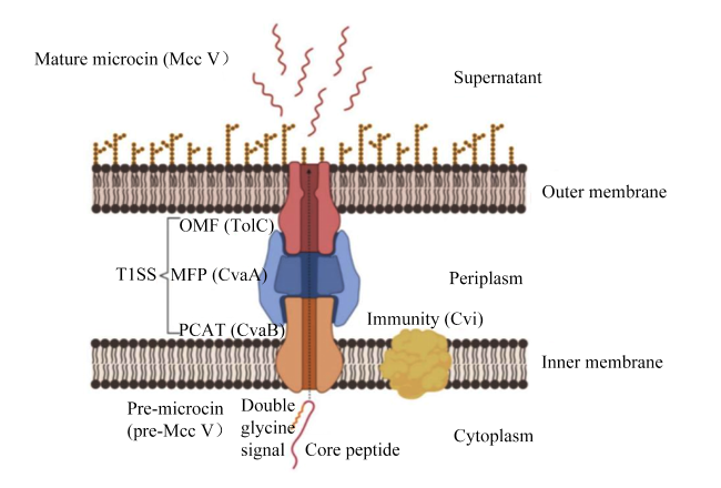
在分泌过程中,PCAT、MFP和TolC相互作用,形成一类非典型的Ⅰ型分泌系统(type Ⅰ secretion system,T1SS),直接将微菌素输送至外部环境,无需经过质粒中间体[85]。大肠杆菌产生的微菌素V是最早被发现采用这种复杂分泌系统的Ⅱ类微菌素(图9),其分泌机制涉及CvaC、Cvi、CvaB、CvaA和TolC等关键蛋白质[86]。具体而言,CvaC基因编码前体微菌素V,其携带1个N端信号肽。Cvi是一种免疫蛋白,与微菌素V同时表达,在宿主细胞内与微菌素V结合,防止其对自身细胞产生抗菌作用,提供自我保护机制。CvaB属于PCAT家族,具有C39肽酶结构域,能够识别前体微菌素V的信号肽,并在信号肽与成熟微菌素V之间进行精确切割,通过ATP水解,CvaB提供能量并引发构象变化,将成熟的微菌素V加载到其跨膜通道中。CvaA是一种MFP,位于内膜上,充当连接内膜转运蛋白CvaB和外膜通道TolC的桥梁。CvaA、CvaB和TolC共同组装形成分泌通道,协助微菌素V的跨膜转运,释放到细胞外环境。此外,跨膜通道的大小限制了可以结合的底物大小,因此改变Ⅱ类微菌素序列长度时需要考虑到T1SS输出微菌素可能具有尺寸限制[87]。目前,Ⅱ类微菌素在基因工程菌中的表达研究甚少,Kaur等[20]将带有谷胱甘肽-S-转移酶(GST)标签的重组微菌素N在大肠杆菌BL21中成功表达,但是可能由于肽的极端疏水性,表达的微菌素N无抑菌活性。
图9 微菌素V的Ⅰ型分泌系统

Mature microcin (Mcc V):成熟微菌素V mature microcin V;Supernatant:上清液;Outer membrane:外膜;T1SS:Ⅰ型分泌系统 type Ⅰ secretion system;OMF:外膜因子 outer membrane factor;MFP:膜融合蛋白 membrane fusion protein;PCAT:吩嗪-1-羧酸转运蛋白 phenazine-1-carboxylic acid transporter;Periplasm:胞间质;Immunity:免疫;Inner membrane:内膜;Cytoplasm:细胞质;Double glycine signal:双甘氨酸信号;Core peptide:核心肽;Pre-microcin (pre-Mcc V):前体微菌素V precursor microcin V。

Fig.9 T1SS of microcin V[61]

4 微菌素在畜禽生产中的应用

4.1 改善畜禽肠道健康和生长性能

微菌素在畜牧业中具有替代传统抗生素的潜力,能够促进动物健康和生产效率[88]。饲粮中添加微菌素能显著改善肉鸡和仔猪的生长性能,影响肠道微生物群的组成[89],改善肠道健康[34,90]。Dai等[91]研究发现,肉鸡饲粮中添加6 mg/kg的微菌素C(纯度大于99.95%)能显著改善平均日增重和料重比,肉鸡的体重提高了6.19%,且与添加45 mg/kg的金霉素效果相当。Shang等[92]研究发现,仔猪饲粮中添加500 mg/kg的微菌素C(纯度为1.19%)能够降低仔猪腹泻指数,提高营养物质表观消化率。饲粮中添加1.0 mg/kg的微菌素J25(纯度大于99.95%)可以改善断奶仔猪生长性能,并有效减少断奶后腹泻的发生,效果与添加20 mg/kg的硫酸黏菌素相似;微菌素J25还能够显著提高肠道绒毛高度,降低隐窝深度,维持肠道良好结构[93]。此外,微菌素还能提升回肠和盲肠中乳酸盐和短链脂肪酸含量,减少炎症,改善肠道屏障功能[92,94]

4.2 提高免疫力

肠道上皮细胞能感知细菌入侵,并通过Toll样受体(Toll-like receptor,TLR)激活免疫系统[95]。研究发现,微菌素C能通过抑制肉鸡空肠TLR2、TLR4及其下游髓样分化因子88(MyD88)、促炎细胞因子[肿瘤坏死因子-α(TNF-α)和干扰素-γ(IFN-γ)]以及趋化因子[白细胞介素-8(IL-8)]的mRNA表达,提高血清免疫球蛋白G(IgG)和免疫球蛋白M(IgM)含量,提高回肠分泌型免疫球蛋白A(sIgA)含量[91];增强断奶仔猪和肉鸡体液免疫反应和吞噬细胞功能,从而增强肠道黏膜的宿主免疫力,减轻肠道炎症和帮助调节免疫系统[10]。此外,Wang等[96]研究发现,饲粮中添加1.0 mg/kg的微菌素J25(纯度大于99.95%)可以显著下调大肠杆菌或沙门氏菌攻毒肉鸡血清中促炎因子TNF-α、白细胞介素-1β(IL-1β)和白细胞介素-6(IL-6)含量。Yu等[93]研究发现,饲粮中添加1.0~2.0 mg/kg的微菌素J25(纯度大于99.95%)能够显著降低断奶仔猪血清中细胞因子IL-1β、IL-6和TNF-α含量,提高抗炎细胞因子白细胞介素-10(IL-10)含量。以上研究表明,微菌素能够提高免疫力并维持免疫平衡。

5 小结和展望

随着禁止饲用抗生素在畜禽生产中的应用,养殖业面临着发病率和死亡率增加以及养殖效益下降的严峻挑战。研发绿色、天然和安全的饲用抗生素替代品是目前的研究热点。微菌素作为一类由革兰氏阴性菌产生的天然肽类小分子,仅对同类或亲缘较近的菌种有抑菌效果,能够精准抑制肠道病原体,降低病原菌抗生素耐药性传播的风险。微菌素具有套索结构、线性多杂环芳香环结构以及N末端“双甘氨酸”信号序列等多样且复杂结构,保障了其较高的稳定性和抑菌活性,也为设计新型抗菌药物提供了依据。此外,微菌素在畜禽生产中还能改善肠道健康、促进生长和增强免疫功能,在畜禽生产中具有广阔应用前景。
不过,在微菌素的研究中,仍面临若干挑战。首先,微菌素的生物合成成本高昂,化学合成工艺复杂,限制了其大规模应用。其次,目前微菌素研究的种类较为有限,研究集中于少数几种,尚未覆盖更广泛的微菌素家族。最后,动物试验虽然对微菌素的效果有所研究,但缺乏对其代谢途径及作用机制的深入探讨,并且针对不同动物和生长阶段的添加剂量尚无明确的规范指导。因此,未来可以从以下几个方向开展进一步研究:1)通过基因工程和合成生物学技术优化微生物的代谢途径,提升微菌素的产量,并降低其生产成本;2)扩大对不同细菌种类及其生态环境中微菌素的探索,特别是结合基因组筛选和生物信息学分析,识别出更多潜在的微菌素基因簇,并通过功能验证丰富微菌素的库;3)加强药代动力学研究,明确微菌素在动物体内的代谢途径和作用机制,并根据不同动物和生长阶段,制定合理的剂量指南,以确保其应用的安全性和有效性,为其在畜牧业中的应用提供更为全面的理论基础。
[1]
YANG W Q, LI J, YAO Z L, et al. A review on the alternatives to antibiotics and the treatment of antibiotic pollution:current development and future prospects[J]. The Science of the Total Environment, 2024, 926:171757.

[2]
ADEGBEYE M J, ADETUYI B O, IGIRIGI A I, et al. Comprehensive insights into antibiotic residues in livestock products:distribution,factors,challenges,opportunities,and implications for food safety and public health[J]. Food Control, 2024, 163:110545.

[3]
MEHDI Y, LÉTOURNEAU-MONTMINY M P, GAUCHER M L, et al. Use of antibiotics in broiler production:global impacts and alternatives[J]. Animal Nutrition, 2018, 4(2):170-178.

[4]
WANG J, DENG L F, CHEN M X, et al. Phytogenic feed additives as natural antibiotic alternatives in animal health and production:a review of the literature of the last decade[J]. Animal Nutrition, 2024, 17:244-264.

[5]
WICKRAMASURIYA S S, AULT J, RITCHIE S, et al. Alternatives to antibiotic growth promoters for poultry:a bibliometric analysis of the research journals[J]. Poultry Science, 2024, 103(9):103987.

[6]
SASSONE-CORSI M, NUCCIO S P, LIU H, et al. Microcins mediate competition among Enterobacteriaceae in the inflamed gut[J]. Nature, 2016, 540(7632):280-283.

[7]
TELHIG S, BEN SAID L, ZIRAH S, et al. Bacteriocins to thwart bacterial resistance in gram negative bacteria[J]. Frontiers in Microbiology, 2020, 11:586433.

[8]
TELHIG S, BEN SAID L, TORRES C, et al. Evaluating the potential and synergetic effects of microcins against multidrug-resistant Enterobacteriaceae[J]. Microbiology Spectrum, 2022, 10(3):e0275221.

[9]
YU H T, SHANG L J, YANG G X, et al. Biosynthetic microcin J25 exerts strong antibacterial,anti-inflammatory activities,low cytotoxicity without increasing drug-resistance to bacteria target[J]. Frontiers in Immunology, 2022, 13:811378.

[10]
DAI Z Q, SHANG L J, WEI Y S, et al. Immunomodulatory effects of microcin C7 in cyclophosphamide-induced immunosuppressed mice[J]. Journal of Agricultural and Food Chemistry, 2023, 71(34):12700-12714.

[11]
BAQUERO F, LANZA V F, BAQUERO M R, et al. Microcins in Enterobacteriaceae:peptide antimicrobials in the eco-active intestinal chemosphere[J]. Frontiers in Microbiology, 2019, 10:2261.

[12]
ASENSIO C, PÉREZ-DÍAZ J C, MARTÍNEZ M C, et al. A new family of low molecular weight antibiotics from enterobacteria[J]. Biochemical and Biophysical Research Communications, 1976, 69(1):7-14.

PMID

[13]
GUIJARRO J I, GONZÁLEZ-PASTOR J E, BALEUX F, et al. Chemical structure and translation inhibition studies of the antibiotic microcin C7[J]. The Journal of Biological Chemistry, 1995, 270(40):23520-23532.

[14]
GARCÍA-BUSTOS J F, PEZZI N, ASENSIO C. Microcin 7:purification and properties[J]. Biochemical and Biophysical Research Communications, 1984, 119(2):779-785.

[15]
SALOMÓN R A, FARÍAS R N. Microcin 25,a novel antimicrobial peptide produced by Escherichia coli[J]. Journal of Bacteriology, 1992, 174(22):7428-7435.

[16]
MARTÍNEZ J L, PÉREZ-DÍAZ J C. Isolation, characterization,and mode of action on Escherichia coli strains of microcin D93[J]. Antimicrobial Agents and Chemotherapy, 1986, 29(3):456-460.

[17]
LI Y, HAN Y, ZENG Z W, et al. Discovery and bioactivity of the novel lasso peptide microcin Y[J]. Journal of Agricultural and Food Chemistry, 2021, 69(31):8758-8767.

DOI PMID

[18]
GAILLARD-GENDRON S, VIGNON D, COTTENCEAU G, et al. Isolation, purification and partial amino acid sequence of a highly hydrophobic new microcin named microcin L produced by Escherichia coli[J]. FEMS Microbiology Letters, 2000, 193(1):95-98.

[19]
GILSON L, MAHANTY H K, KOLTER R. Four plasmid genes are required for colicin V synthesis,export,and immunity[J]. Journal of Bacteriology, 1987, 169(6):2466-2470.

[20]
KAUR K, TARASSOVA O, DANGETI R V, et al. Characterization of a highly potent antimicrobial peptide microcin N from uropathogenic Escherichia coli[J]. FEMS Microbiology Letters, 2016, 363(11):fnw095.

[21]
ZSCHÜTTIG A, ZIMMERMANN K, BLOM J, et al. Identification and characterization of microcin S,a new antibacterial peptide produced by probiotic Escherichia coli G3/10[J]. PLoS One, 2012, 7(3):e33351.

[22]
EBERHART L J, DERINGER J R, BRAYTON K A, et al. Characterization of a novel microcin that kills enterohemorrhagic Escherichia coli O157:H7 and O26[J]. Applied and Environmental Microbiology, 2012, 78(18):6592-6599.

[23]
VASSILIADIS G, DESTOUMIEUX-GARZÓN D, LOMBARD C, et al. Isolation and characterization of two members of the siderophore-microcin family,microcins M and H47[J]. Antimicrobial Agents and Chemotherapy, 2010, 54(1):288-297.

[24]
PATZER S I, BAQUERO M R, BRAVO D, et al. The colicin G,H and X determinants encode microcins M and H47,which might utilize the catecholate siderophore receptors FepA,Cir,Fiu and IroN[J]. Microbiology (Reading, England), 2003, 149(Pt 9):2557-2570.

[25]
LAVIÑA M, GAGGERO C, MORENO F. Microcin H47,a chromosome-encoded microcin antibiotic of Escherichia coli[J]. Journal of Bacteriology, 1990, 172(11):6585-6588.

[26]
MORTZFELD B M, BHATTARAI S K, BUCCI V. Expanding the toolbox:novel class Ⅱb microcins show activity against gram-negative ESKAPE and plant pathogens[J/OL]. bioRxiv, 2024.[2024-04-22]. https://pubmed.ncbi.nlm.nih.gov/39253482/.

[27]
TODOROV S D, DE MELO FRANCO B D G, TAGG J R. Bacteriocins of Gram-positive bacteria having activity spectra extending beyond closely-related species[J]. Beneficial Microbes, 2019, 10(3):315-328.

DOI PMID

[28]
PONS A M, DELALANDE F, DUARTE M, et al. Genetic analysis and complete primary structure of microcin L[J]. Antimicrobial Agents and Chemotherapy, 2004, 48(2):505-513.

[29]
WOOLEY R E, RITCHIE B W, CURRIN M F, et al. In vitro inhibition of Salmonella organisms isolated from reptiles by an inactivated culture of microcin-producing Escherichia coli[J]. American Journal of Veterinary Research, 2001, 62(9):1399-1401.

[30]
PALMER J D, MORTZFELD B M, PIATTELLI E, et al. Microcin H47:a class Ⅱb microcin with potent activity against multidrug resistant Enterobacteriaceae[J]. ACS Infectious Diseases, 2020, 6(4):672-679.

[31]
YU H T, LI N, ZENG X F, et al. A comprehensive antimicrobial activity evaluation of the recombinant microcin J25 against the foodborne pathogens Salmonella and E.coli O157:H7 by using a matrix of conditions[J]. Frontiers in Microbiology, 2019, 10:1954.

[32]
THOMAS X, DESTOUMIEUX-GARZÓN D, PEDUZZI J, et al. Siderophore peptide,a new type of post-translationally modified antibacterial peptide with potent activity[J]. Journal of Biological Chemistry, 2004, 279(27):28233-28242.

[33]
LOPEZ F E, VINCENT P A, ZENOFF A M, et al. Efficacy of microcin J25 in biomatrices and in a mouse model of Salmonella infection[J]. The Journal of Antimicrobial Chemotherapy, 2007, 59(4):676-680.

[34]
NAIMI S, ZIRAH S, TAHER M B, et al. Microcin J25 exhibits inhibitory activity against Salmonella Newport in continuous fermentation model mimicking swine colonic conditions[J]. Frontiers in Microbiology, 2020, 11:988.

[35]
POEY M E, AZPIROZ M F, LAVIÑA M. Comparative analysis of chromosome-encoded microcins[J]. Antimicrobial Agents and Chemotherapy, 2006, 50(4):1411-1418.

PMID

[36]
DESTOUMIEUX-GARZÓN D, THOMAS X, SANTAMARIA M, et al. Microcin E492 antibacterial activity:evidence for a TonB-dependent inner membrane permeabilization on Escherichia coli[J]. Molecular Microbiology, 2003, 49(4):1031-1041.

[37]
KAZAKOV T, VONDENHOFF G H, DATSENKO K A, et al. Escherichia coli peptidase A,B,or N can process translation inhibitor microcin C[J]. Journal of Bacteriology, 2008, 190(7):2607-2610.

[38]
NOVIKOVA M, METLITSKAYA A, DATSENKO K, et al. The Escherichia coli Yej transporter is required for the uptake of translation inhibitor microcin C[J]. Journal of Bacteriology, 2007, 189(22):8361-8365.

[39]
SEREBRYAKOVA M, TSIBULSKAYA D, MOKINA O, et al. A Trojan-horse peptide-carboxymethyl-cytidine antibiotic from Bacillus amyloliquefaciens[J]. Journal of the American Chemical Society, 2016, 138(48):15690-15698.

[40]
METLITSKAYA A, KAZAKOV T, KOMMER A, et al. Aspartyl-tRNA synthetase is the target of peptide nucleotide antibiotic microcin C[J]. Journal of Biological Chemistry, 2006, 281(26):18033-18042.

DOI PMID

[41]
KAZAKOV T, METLITSKAYA A, SEVERINOV K. Amino acid residues required for maturation,cell uptake,and processing of translation inhibitor microcin C[J]. Journal of Bacteriology, 2007, 189(5):2114-2118.

[42]
YANG G X, SHANG L J, LIU L, et al. Engineering and purification of microcin C7 variants resistant to trypsin and analysis of their biological activity[J]. Antibiotics-Basel, 2023, 12(9):1346.

[43]
ZUKHER I, PAVLOV M, TSIBULSKAYA D, et al. Reiterative synthesis by the ribosome and recognition of the N-terminal formyl group by biosynthetic machinery contribute to evolutionary conservation of the length of antibiotic microcin C peptide precursor[J]. mBio, 2019, 10(2):e00768-19.

[44]
DESTOUMIEUX-GARZÓN D, PEDUZZI J, REBUFFAT S. Focus on modified microcins:structural features and mechanisms of action[J]. Biochimie, 2002, 84(5/6):511-519.

[45]
DA HORA G C A, OH M, MIFFLIN M C, et al. Lasso peptides:exploring the folding landscape of nature’s smallest interlocked motifs[J]. Journal of the American Chemical Society, 2024, 146(7):4444-4454.

[46]
BAQUERO F, BEIS K, CRAIK D J, et al. The pearl jubilee of microcin J25:thirty years of research on an exceptional lasso peptide[J]. Natural Product Reports, 2024, 41(3):469-511.

[47]
MOHRI K, NHAT K P H, ZOUDA M, et al. Lasso peptide microcin J25 variant containing RGD motif as a PET probe for integrin avβ3 in tumor imaging[J]. European Journal of Pharmaceutical Sciences, 2023, 180:106339.

[48]
MATHAVAN I, ZIRAH S, MEHMOOD S, et al. Structural basis for hijacking siderophore receptors by antimicrobial lasso peptides[J]. Nature Chemical Biology, 2014, 10(5):340-342.

DOI PMID

[49]
LAI P K, KAZNESSIS Y N. Free energy calculations of microcin J25 variants binding to the FhuA receptor[J]. Journal of Chemical Theory and Computation, 2017, 13(7):3413-3423.

[50]
DE CRISTÓBAL R E, SOLBIATI J O, ZENOFF A M, et al. Microcin J25 uptake:His5 of the MccJ25 lariat ring is involved in interaction with the inner membrane MccJ25 transporter protein SbmA[J]. Journal of Bacteriology, 2006, 188(9):3324-3328.

[51]
BRAFFMAN N R, PISCOTTA F J, HAUVER J, et al. Structural mechanism of transcription inhibition by lasso peptides microcin J25 and capistruin[J]. Proceedings of the National Academy of Sciences of the United States of America, 2019, 116(4):1273-1278.

DOI PMID

[52]
PAVLOVA O, MUKHOPADHYAY J, SINEVA E, et al. Systematic structure-activity analysis of microcin J25[J]. The Journal of Biological Chemistry, 2008, 283(37):25589-25595.

[53]
VINCENT P A, MORERO R D. The structure and biological aspects of peptide antibiotic microcin J25[J]. Current Medicinal Chemistry, 2009, 16(5):538-549.

PMID

[54]
VIZÁN J L, HERNÁNDEZ-CHICO C, DEL CASTILLO I, et al. The peptide antibiotic microcin B17 induces double-strand cleavage of DNA mediated by E.coli DNA gyrase[J]. The EMBO Journal, 1991, 10(2):467-476.

[55]
BAYER A, FREUND S, JUNG G. Post-translational heterocyclic backbone modifications in the 43-peptide antibiotic microcin B17:structure elucidation and NMR study of a 13C,15N-labelled gyrase inhibitor[J]. European Journal of Biochemistry, 1995, 234(2):414-426.

[56]
HEDDLE J G, BLANCE S J, ZAMBLE D B, et al. The antibiotic microcin B17 is a DNA gyrase poison:characterisation of the mode of inhibition[J]. Journal of Molecular Biology, 2001, 307(5):1223-1234.

[57]
PIERRAT AND O A, MAXWELL A. Evidence for the role of DNA Strand passage in the mechanism of action of microcin B17 on DNA gyrase[J]. Biochemistry, 2005, 44(11):4204-4215.

DOI PMID

[58]
PIERRAT O A, MAXWELL A. The action of the bacterial toxin microcin B17:insight into the cleavage-religation reaction of DNA gyrase[J]. Journal of Biological Chemistry, 2003, 278(37):35016-35023.

[59]
COLLIN F, MAXWELL A. The microbial toxin microcin B17:prospects for the development of new antibacterial agents[J]. Journal of Molecular Biology, 2019, 431(18):3400-3426.

[60]
HÅVARSTEIN L S, HOLO H, NES I F. The leader peptide of colicin V shares consensus sequences with leader peptides that are common among peptide bacteriocins produced by Gram-positive bacteria[J]. Microbiology, 1994, 140:2383-2389.

[61]
PARKER J K, DAVIES B W. Microcins reveal natural mechanisms of bacterial manipulation to inform therapeutic development[J]. Microbiology, 2022, 168(4):001175.

[62]
RÍOS COLOMBO N S, CHALÓN M C, NAVARRO S A, et al. Pediocin-like bacteriocins:new perspectives on mechanism of action and immunity[J]. Current Genetics, 2018, 64:345-351.

[63]
RÍOS COLOMBO N S, CHALON M C, DUPUY F G, et al. The case for class Ⅱ bacteriocins:a biophysical approach using “suicide probes” in receptor-free hosts to study their mechanism of action[J]. Biochimie, 2019, 165:183-195.

[64]
TRUJILLO M, RODRÍGUEZ E, LAVIÑA M. ATP synthase is necessary for microcin H47 antibiotic action[J]. Antimicrobial Agents and Chemotherapy, 2001, 45(11):3128-3131.

PMID

[65]
BIÉLER S, SILVA F, BELIN D. The polypeptide core of microcin E492 stably associates with the mannose permease and interferes with mannose metabolism[J]. Research in Microbiology, 2010, 161(8):706-710.

DOI PMID

[66]
MORISSET D, BERJEAUD J M, MARION D, et al. Mutational analysis of mesentericin y105,an anti-listeria bacteriocin,for determination of impact on bactericidal activity,in vitro secondary structure,and membrane interaction[J]. Applied and Environmental Microbiology, 2004, 70(8):4672-4680.

[67]
BEIS K, REBUFFAT S. Multifaceted ABC transporters associated to microcin and bacteriocin export[J]. Research in Microbiology, 2019, 170(8):399-406.

DOI PMID

[68]
ASSRIR N, PAVELKOVA A, DAZZONI R, et al. Initial molecular recognition steps of McjA precursor during microcin J25 lasso peptide maturation[J]. Chembiochem:a European Journal of Chemical Biology, 2016, 17(19):1851-1858.

[69]
YAN K P, LI Y Y, ZIRAH S, et al. Dissecting the maturation steps of the lasso peptide microcin J25 in vitro[J]. Chembiochem:a European Journal of Chemical Biology, 2012, 13(7):1046-1052.

[70]
BOUNTRA K, HAGELUKEN G, CHOUDHURY H G, et al. Structural basis for antibacterial peptide self-immunity by the bacterial ABC transporter McjD[J]. The EMBO Journal, 2017, 36:3062-3079.

[71]
ROY R S, KIM S, BALEJA J D, et al. Role of the microcin B17 propeptide in substrate recognition:solution structure and mutational analysis of McbA1-26[J]. Chemistry & Biology, 1998, 5(4):217-228.

[72]
CHEUNG W L, PAN S J, LINK A J. Much of the microcin J25 leader peptide is dispensable[J]. Journal of the American Chemical Society, 2010, 132(8):2514-2515.

DOI PMID

[73]
YORGEY P, DAVAGNINO J, KOLTER R. The maturation pathway of microcin B17,a peptide inhibitor of DNA gyrase[J]. Molecular Microbiology, 1993, 9(4):897-905.

[74]
DONG S H, KULIKOVSKY A, ZUKHER I, et al. Biosynthesis of the RiPP Trojan horse nucleotide antibiotic microcin C is directed by the N-formyl of the peptide precursor[J]. Chemical Science, 2019, 10(8):2391-2395.

[75]
SMITS S H J, SCHMITT L, BEIS K. Self-immunity to antibacterial peptides by ABC transporters[J]. FEBS Letters, 2020, 594(23):3920-3942.

DOI PMID

[76]
SOLBIATI J O, CIACCIO M, FARÍAS R N, et al. Sequence analysis of the four plasmid genes required to produce the circular peptide antibiotic microcin J25[J]. Journal of Bacteriology, 1999, 181(8):2659-2662.

PMID

[77]
DELGADO M A, SOLBIATI J O, CHIUCHIOLO M J, et al. Escherichia coli outer membrane protein TolC is involved in production of the peptide antibiotic microcin J25[J]. Journal of Bacteriology, 1999, 181(6):1968-1970.

[78]
GARRIDO M C, HERRERO M, KOLTER R, et al. The export of the DNA replication inhibitor microcin B17 provides immunity for the host cell[J]. The EMBO Journal, 1988, 7(6):1853-1862.

[79]
TIKHONOV A, KAZAKOV T, SEMENOVA E, et al. The mechanism of microcin C resistance provided by the MccF peptidase[J]. The Journal of Biological Chemistry, 2010, 285(49):37944-37952.

[80]
DELGADO M A, VINCENT P A, FARÍAS R N, et al. YojI of Escherichia coli functions as a microcin J25 efflux pump[J]. Journal of Bacteriology, 2005, 187(10):3465-3470.

[81]
ZHANG G W, LIN M, QIN M M, et al. Establishing heterologous production of microcins J25 and Y in Bacillus subtilis[J]. Journal of Agricultural and Food Chemistry, 2023, 71(14):5600-5613.

[82]
NOLAN E M, WALSH C T. Investigations of the MceIJ-catalyzed posttranslational modification of the microcin E492 C-terminus:linkage of ribosomal and nonribosomal peptides to form “Trojan horse” antibiotics[J]. Biochemistry, 2008, 47(35):9289-9299.

[83]
GÉRARD F, PRADEL N, WU L F. Bactericidal activity of colicin V is mediated by an inner membrane protein,SdaC,of Escherichia coli[J]. Journal of Bacteriology, 2005, 187(6):1945-1950.

[84]
VASSILIADIS G, DESTOUMIEUX-GARZÓN D, PEDUZZI J. Class Ⅱ microcins[M]. New York: Springer New York, 2011:309-332.

[85]
HWANG J, ZHONG X, TAI P C. Interactions of dedicated export membrane proteins of the colicin V secretion system:CvaA,a member of the membrane fusion protein family,interacts with CvaB and TolC[J]. Journal of Bacteriology, 1997, 179(20):6264-6270.

[86]
KIM S Y, PARKER J K, GONZALEZ-MAGALDI M, et al. Export of diverse and bioactive peptides through a type Ⅰ secretion system[J/OL]. bioRxiv:the Preprint Server for Biology, 2023.[2024-04-25]. https://pubmed.ncbi.nlm.nih.gov/36747863/.

[87]
LIN D Y W, HUANG S, CHEN J. Crystal structures of a polypeptide processing and secretion transporter[J]. Nature, 2015, 523(7561):425-430.

[88]
ABD E H M E, EL S M T, SHAFI M E, et al. Probiotics in poultry feed:a comprehensive review[J]. Journal of Animal Physiology and Animal Nutrition, 2020, 104(6):1835-1850.

[89]
YU H T, WANG Y M, ZENG X F, et al. Therapeutic administration of the recombinant antimicrobial peptide microcin J25 effectively enhances host defenses against gut inflammation and epithelial barrier injury induced by enterotoxigenic Escherichia coli infection[J]. The FASEB Journal, 2020, 34(1):1018-1037.

[90]
BEN SAID L, EMOND-RHEAULT J G, SOLTANI S, et al. Phenomic and genomic approaches to studying the inhibition of multiresistant Salmonella enterica by microcin J25[J]. Environmental Microbiology, 2020, 22(7):2907-2920.

[91]
DAI Z Q, SHANG L J, WANG F M, et al. Effects of antimicrobial peptide microcin C7 on growth performance,immune and intestinal barrier functions,and cecal microbiota of broilers[J]. Frontiers in Veterinary Science, 2021, 8:813629.

[92]
SHANG L J, ZHOU J Y, TU J Y, et al. Evaluation of effectiveness and safety of microcin C7 in weaned piglets[J]. Animals, 2022, 12(23):3267.

[93]
YU H T, DING X L, LI N, et al. Dietary supplemented antimicrobial peptide microcin J25 improves the growth performance,apparent total tract digestibility,fecal microbiota,and intestinal barrier function of weaned pigs[J]. Journal of Animal Science, 2017, 95(11):5064-5076.

[94]
SHANG L J, YU H T, LIU H B, et al. Recombinant antimicrobial peptide microcin J25 alleviates DSS-induced colitis via regulating intestinal barrier function and modifying gut microbiota[J]. Biomedicine & Pharmacotherapy, 2021, 139:111127.

[95]
LAVELLE E C, MURPHY C, O’NEILL L A J, et al. The role of TLRs,NLRs,and RLRs in mucosal innate immunity and homeostasis[J]. Mucosal Immunology, 2010, 3(1):17-28.

[96]
WANG G, SONG Q L, HUANG S, et al. Effect of antimicrobial peptide microcin J25 on growth performance,immune regulation,and intestinal microbiota in broiler chickens challenged with Escherichia coli and Salmonella[J]. Animals, 2020, 10(2):345.

Outlines

/