REVIEW

Composition of Intestinal Fungi in Swine and Its Potential Health and Nutritional Significance

  • HUANG Shuangmei ,
  • LUO Yuheng , *
Expand
  • Animal Disease-Resistance Nutrition Biotechnology Engineering Research Center of the Ministry of Education, Key Laboratory of Animal Nutrition and Feed Engineering in Sichuan Province, Key Laboratory of Animal Disease-Resistance of Ministry of Education and Sichuan Province, Key Laboratory of Animal Disease-Resistance Nutrition and Feed of Ministry of Agriculture and Rural Affairs, Animal Nutrition Institute, Sichuan Agricultural University, Chengdu 611130, China
* professor, E-mail:

Received date: 2024-09-05

  Online published: 2025-04-15

Abstract

The gut microbes of monogastric animals such as pigs are composed of bacteria, fungi, archaea and viruses, and the proportion of fungi in gut microbes is less than 1%, which has been overlooked in previous studies. However, recent studies have shown that intestinal fungi are closely related to the maintenance of host intestinal barrier function, intestinal immune response, and post-intestinal microbial fermentation. Moreover, the volume of a single fungal cell is about 100 times that of a single bacterial cell, and can encode a large number of functional enzymes, which can affect host nutrient metabolism and body health to a certain extent. Therefore, fungi are an important member of gut microbiome of monogastric animals that cannot be ignored. Yet, to date, there is still a lack of research on intestinal fungi in monogastric animals such as pigs, which limits our understanding of the overall function of the gut microbiome. In this paper, the composition, influencing factors, and effects of intestinal fungi in swine gut on host health and nutrient digestion and absorption were comprehensively reviewed, providing ideas for further understanding its biological functions.

Cite this article

HUANG Shuangmei , LUO Yuheng . Composition of Intestinal Fungi in Swine and Its Potential Health and Nutritional Significance[J]. Chinese Journal of Animal Nutrition, 2025 , 37(4) : 2134 -2141 . DOI: 10.12418/CJAN2025.181

肠道是营养物质消化吸收的主要场所,也是机体的第1道免疫防线。猪肠道中存在着数量庞大而组成复杂的微生物群,包括细菌、古菌、真菌、病毒等,其细胞数量是宿主本身细胞总数的10倍之多(1013~1014个)[1]。这些微生物与宿主的共生关系在很大程度上影响肠道的生物学功能。肠道微生物全面参与宿主营养物质的消化吸收[2]、肠道屏障功能的形成和维持、宿主免疫系统的发生发育和免疫应答的不同途径[3]。尽管真菌数量仅占肠道微生物组的0.1%左右[4],但有限的研究表明其在肠道微生态的平衡和维持中扮演着极为重要的角色[5-6]。目前关于单胃动物肠道真菌的研究主要集中于其对疾病发生、发展的影响,但越来越多的研究表明,单胃动物肠道真菌不仅与肠道健康息息相关,还可能参与宿主能量代谢过程[7-8]。本文检索了近年来猪肠道真菌的相关文献报道,结合部分人、小鼠和反刍动物消化道真菌的研究,较为全面地综述了猪肠道真菌区系组成及其对宿主营养和健康的影响,为进一步研究猪肠道真菌组的生物学功能提供参考。

1 猪肠道真菌的组成

整体而言,担子菌门(Basidiomycota)和子囊菌门(Ascomycota)是猪肠道中的主要真菌门类[9-10],但不同品种猪肠道真菌门类存在一定差异。Wang等[11]研究了饲粮和遗传背景对猪肠道真菌组成和多样性的影响,发现本地品种桃源黑猪和引进品种杜洛克猪结肠真菌均以子囊菌门和微孢子门(Microsporidia)为主,并以哈萨克斯坦酵母属(Kazachstania)、毛霉菌属(Mucor)、厌氧鞭菌属(Anaeromyces)、瘤胃壶菌属(Piromyces)和新美鞭菌属(Neocallimastix)为代表属,但2个品种间肠道真菌区系多样性存在显著差异;而湘村黑猪作为二者的杂交品种,其肠道真菌多样性虽然更接近桃源黑猪,但也呈现出二者的混合特征;这与Moeller等[12]在小鼠上的研究结果一致,提示单胃动物肠道真菌群落可能通过杂合遗传垂直传播。Li等[10]对成华猪、约克夏猪和藏猪粪便样本中的真菌群落进行ITS高通量测序并测定了短链脂肪酸(short-chain fatty acids,SCFA)的含量,发现不同品种猪具有独特的肠道真菌群落组成,其中Loreleia是成华猪和约克夏猪肠道中相对丰度最高的真菌属,而藏猪肠道中相对丰度最高的真菌属则是Russula;相关性分析表明,绒毛菌属(Tomentella)相对丰度与样本中的乙酸和丁酸含量均呈显著正相关,Loreleia相对丰度与丙酸含量呈正相关,而NephromaTaiwanofungus相对丰度则与乙酸和丙酸含量呈明显负相关。Hu等[13]研究了7个品种的断奶仔猪和育肥猪肠道真菌群落,发现Kazachstania slooffiae是杜洛克×长白×约克(DLY)猪、藏猪、莱芜猪、沙子岭猪、从江小型猪、环江小香猪和宁乡猪断奶仔猪肠道中的优势真菌物种;热带念珠菌(Candida tropicalis)、季也蒙迈耶氏酵母(Meyerozyma guilliermondii)和芽枝状枝孢霉(Cladosporium cladosporioides)是成年小型藏猪肠道中特有的优势真菌物种;藤仓赤霉(Gibberella fujikuroi)和限制性马拉色菌(Malassezia restricta)是成年莱芜猪肠道中的特有优势真菌物种;成年从江小型猪育肥猪肠道内的特有优势真菌物种则为黄曲霉菌(Aspergillus flavus)和季也蒙迈耶氏酵母。
针对不同肠段食糜的研究提示了猪肠道真菌组成在不同消化道部位的差异。对断奶仔猪盲肠和结肠食糜的研究表明,虽然担子菌门和子囊菌门是这两段肠道中的主要真菌门类,但真菌组成特征在两段肠道之间存在明显差异,即盲肠真菌多样性明显高于结肠;与结肠相比,盲肠中的复膜孢酵母属(Saccharomycopsis)、节担菌属(Wallemia)和木拉克属(Mrakia)相对丰度更高,而树干毕赤酵母属(Scheffersomyces)、曲霉属(Aspergillus)、青霉属(Penicillium)和毛霉菌属相对丰度则相反[14]。对育肥猪胃、十二指肠、空肠、回肠、盲肠、结肠内容物的研究表明,担子菌门和毛霉菌门(Mucoromycota)的相对丰度按胃—小肠—大肠的顺序逐渐增加[15]

2 猪肠道真菌组成的影响因素

消化道微生物群落结构受年龄[16]、饲粮[17]、健康状态[18]、宿主遗传[11]和环境等因素影响,除遗传因素外,饲粮结构和成分对消化道微生物的影响研究的最多。反刍动物瘤胃厌氧真菌和部分环境真菌的相关研究表明,由于真菌基因组编码大量碳水化合物活性酶(carbohydrate-active enzymes,CAZymes),能有效降解饲粮或自然环境中的木质纤维素[19-20],但猪肠道真菌是否影响宿主后肠纤维发酵尚缺乏直接证据。与低纤维饲粮(粗纤维占2%~3%)相比,摄入高纤维饲粮(粗纤维占6%~7%)的杜洛克猪结肠中微孢子门相对丰度降低,同时毛霉菌属、刺盘孢属(Colletotrichum)、Hortaea和节丝皮菌属(Arthroderma)等菌属相对丰度也明显下降,但新美鞭菌属、副球孢子菌属(Paracoccidioides)、梨孢属(Pyricularia)、节担菌属和Neonectria等菌属相对丰度升高[11]。Luo等[21]采用L9(34)正交设计研究了仔猪后肠真菌群落对饲粮碳水化合物组合模式(DCHO)的响应规律,其中涉及的DCHO包括直链淀粉与支链淀粉的比例(AM/AP,2∶1、1∶1、1∶2)、非淀粉多糖(NSP,1%、2%、3%)和甘露寡糖(MOS,400、800、1 200 mg/kg BW),研究结果表明,仔猪结肠真菌群落对DCHO的响应顺序为MOS>AM/AP>NSP;以复膜孢酵母属、木拉克属、节担菌属、Cantharellus、散囊菌属(Eurotium)、Solicocozyma和青霉菌属为代表的低丰度真菌属大部分与DCHO相关,也与葡萄糖和果糖的浓度以及结肠消化液中β-D-葡萄糖苷酶活性有关,说明这些真菌可能与猪结肠碳水化合物的降解密切相关。上述结果为猪结肠真菌群落受饲粮碳水化合物来源的影响提供了直接证据。
早期针对无菌动物和人类的研究发现,消化道微生物群落可能通过母体-胎儿垂直传播[22],这一规律或许同样适用于猪的肠道真菌。一项对猪和野猪生产周期内各阶段肠道真菌的研究发现,母体Kazachstania slooffiae的定植水平对后代生命早期(出生第11天)和后期(断奶后第119天)的肠道真菌区系有塑造作用;相对家猪而言,野猪具有更复杂而独特的肠道真菌组成,推测可能与其摄入饲草且生存环境中含有大量不同种类的真菌有关[23]。仔猪肠道真菌的组成存在明显的窝间差异,母体真菌组对于仔猪早期真菌群的形成至关重要[24]。仔猪出生后其粪便中的真菌数量较少且表现出高度的可变性。断奶前,母乳喂养的仔猪肠道中亚罗酵母属(Yarrowia)和葡萄孢盘菌属(Botryotinia)真菌的相对丰度较高,而配方奶喂养的仔猪肠道中则以曲霉属为主,且马拉色菌属(Malassezia)的相对丰度降低[25];断奶后,仔猪肠道真菌负荷显著增加且组成趋于稳定,优势菌属为Kazachstania slooffiae[16,26-28]。此外,腹泻仔猪可能具有相对独特的肠道真菌群落。例如,与健康断奶藏仔猪相比,腹泻藏仔猪肠道中罗兹菌门(Rozellomycota)、丝膜菌属(Cortinarius)和哈萨克斯坦酵母属等真菌类群的相对丰度在腹泻组中增加,而DerxomycesLecanicillium的相对丰度下降[29]
大多数肠道微生物相关研究均忽略了采样时间或批次对微生物组成的影响。据Li等[15]报道,不同采样批次可能影响育肥猪肠道真菌测序结果的代表性。该研究表明,虽然担子菌门和子囊菌门是育肥猪全肠道真菌的优势门类,但第1批次样品中前者相对丰度明显较高,第2批次样品则后者相对丰度更高;此外,2批样本中的优势真菌属完全不同,分别为Naganishia、红酵母属(Rhodotorula)、镰刀菌属(Fusarium)、被孢霉属(Mortierella)和念珠菌属(Candida),以及哈萨克斯坦酵母属、毛霉菌、丝孢酵母属(Trichosporon)、Nothophoma和镰刀菌属,由此衍生的核心真菌群落也截然不同[15]。这一发现提示,即使在相同的饲养条件下,猪肠道真菌的组成也会随采样批次和时间发生极大变化,这与同一物种具有相对稳定的细菌组成不同。
人肠道真菌物种大部分被怀疑是食源性的[30]。对此,本实验室曾采用SourceTracker软件进行溯源分析,发现仔猪饲粮样品中最丰富的真菌属是浪梗霉属(Polythrincium,24.45%)、镰刀菌属(3.22%)、Issagenkia(2.97%)、曲霉属(2.26%)和间座壳属(Diaporthe,1.50%),与其结肠食糜中检测到的真菌种属组成完全不同[21]。这一结果也说明,猪肠道真菌并非来自饲粮或外部环境,其系统发生过程尚需进化生态学验证。

3 猪肠道真菌对宿主健康和养分消化吸收的可能影响

真菌属于真核生物,具有细胞壁,典型真菌细胞体积约为细菌的100倍,携带大量遗传信息[31],可推测其对宿主健康和代谢的影响方式多种多样。

3.1 猪肠道真菌对宿主健康的可能影响

真菌的细胞壁由(1-3)β-D-葡聚糖和几丁质等多种大分子多糖组成,并且其结构多样[32]。现有研究表明,肠道真菌可以通过抑制或促进局部炎症反应来影响宿主整体免疫反应[33-34]。使用抗真菌药物诱导小鼠肠道真菌失调可加重急性或慢性结肠炎病症[34]。限制马拉色菌是一种皮肤常驻真菌,克罗恩病患者肠道中的限制性马拉色菌可被抗酿酒酵母抗体(anti-Saccharomyces cerevisiae antibodies,ASCA)识别,并通过胱天蛋白酶富集域家族成员9(caspase recruitment domain-containing protein 9,CARD9)相关信号通路引起先天性免疫反应,从而加剧小鼠结肠炎[35]。白色念珠菌(Candida albicans)作为一种机会致病菌,可定植在肠道、阴道、口腔黏膜和皮肤等部位[36],当白色念珠菌与宿主稳态被破坏时,白色念珠菌可通过辅助性T细胞17(Th17)介导肠道炎症[37-38],还可以分泌肽毒素(念珠菌溶血素)侵入宿主组织,引起细胞损伤[39-41]。鉴于上述真菌物种均是猪肠道内的优势真菌种群,上述研究结果提示,猪肠道真菌可能在特定疾病状态下影响宿主健康。除条件致病性外,某些真菌物种也可能对宿主健康具有积极作用。如前所述,Kazachstania slooffiae是断奶仔猪肠道内的常驻真菌,现有证据表明其可能具有缓解断奶应激的作用[28,42-43],并且可以通过激活sirtuin 5(SIRT5)基因表达来抑制赖氨酸琥珀酰化,从而促进肠上皮糖酵解,介导蛋白质的翻译后修饰,参与细胞代谢[13]
作为肠道微生物组成员之一,真菌可以通过平衡与细菌之间的互作关系影响宿主各项生理功能。例如,在抗生素暴露导致细菌菌群失调的情况下,布拉氏酵母菌(Saccharomyces boulardii)或酿酒酵母(Saccharomyces cerevisiae)等共生真菌通过细胞壁甘露聚糖保护黏膜组织免受损伤并积极校准免疫细胞的活性,在功能上替代肠道细菌[44]。布拉氏酵母菌是一种潜在益生菌,可治疗多种疾病,包括改善肠道屏障和肠道免疫反应等[45-46];在炎症情况下刺激肠道细胞的生长和分化[47-48];刺激蛋白质和能量的产生,恢复结肠上皮细胞的代谢活动[49-51];恢复结肠SCFA的正常水平[52-53];稳定胃肠道屏障功能,加强肠细胞紧密连接等[50-51,54]。进一步研究发现,布拉氏酵母菌可通过分泌胞外酶(如磷酸酶和蛋白酶等)使艰难梭菌(Clostridium difficile)和大肠杆菌(Escherichia coli)产生的毒素失活[55-56],而艰难梭菌是造成医院感染的主要菌种之一[57]。对猪肠道真菌组和细菌组的分析表明,某些肠道真菌属,如哈萨克斯坦酵母属与Alloprevotella、乳杆菌属(Lactobacillus)、普雷沃氏菌属(Prevotella)、罕见小球菌属(Subdoligranulum)等细菌属的相对丰度呈显著正相关,而曲霉属则与产SCFA的细菌属[丁酸球菌属(Butyricicoccus)、Subdoligranulum和梭形杆菌属(Fusicatenibacter)]的相对丰度呈负相关[27]。对健康人类肠道真菌的研究则表明,限制性马拉色菌与多糖利用细菌拟杆菌属的相对丰度呈正相关,曲霉属则与瘤胃球菌科(Ruminococcaceae)的相对丰度呈正相关[58]。这些研究结果说明肠道真菌可以直接或间接影响细菌的组成和代谢,实现对微生物组功能的调控。

3.2 肠道真菌对宿主养分消化吸收的可能影响

对多糖的利用和相关代谢物的产生是肠道真菌发挥生理学功能的重要途径。碳水化合物是动物饲粮中提供能量的主要物质。多糖是一类结构复杂的大分子碳水化合物聚合物,也是饲粮纤维的主要成分,主要依靠后肠微生物分泌的CAZymes进行发酵。CAZymes是指与碳水化合物的合成、修饰和分解相关的酶的总称[59],负责糖缀合物、低聚糖和多糖的合成和分解,其编码基因通常占微生物基因组的1%~5%[60]
动物消化道内的真菌绝大多数为厌氧真菌,编码功能强大、种类复杂的CAZymes[61],反刍动物和白蚁肠道真菌已被证实具有分解多种植物多糖的能力[62-63]。据Lange等[61]报道,芽枝霉门(Blastocladiomycota)、壶菌门(Chytridiomycota)、隐真菌门(Cryptomycota)和新美鞭菌门(Neocallimastigomycota)等真菌成员可以分泌纤维素酶、半纤维素酶、裂解多糖单加氧酶、辅助活性酶等主要的CAZymes,具有较强的纤维素分解能力。部分真菌物种,如Caulochytrium protosteloidesGonapodya prolifera,其基因组编码极为丰富的CAZymes[64-65],后者编码靶向纤维素和半纤维素的酶组合,还具有多样化的果胶活性酶谱[61,66-67]。针对Neocallimastix californiaePiromyces finisAnaeromyces robustus等3种瘤胃真菌CAZymes的研究显示,它们可以编码多达312个非催化Dockerin结构域和多种CAZymes[62,66-67]。另一项关于12种肠道真菌的研究发现,几乎所有真菌物种中均存在3种CAZymes:GH31(α-葡萄糖苷酶,EC 3.2.1.20)、GH37(海藻糖酶,EC3.2.1.28)和GH47(α-1,2-甘露糖苷酶,EC 3.2.1.113)[61],这些CAZymes针对的多糖恰好是植物性饲料中饲粮纤维的主要成分。
目前尚无直接证据表明猪肠道真菌也能通过分泌CAZymes降解植物多糖。但Li等[14]报道,断奶仔猪结肠和盲肠中的真菌区系与相应食糜样本中的乙酸、丙酸、异丁酸和异戊酸含量之间存在显著相关性。另外,不同结构淀粉(AM/AP)的消化与猪肠道中低丰度真菌的比例有关[21]。这些结果说明猪肠道真菌很可能与细菌发酵效率密切相关,从而间接影响饲粮碳水化合物在猪后肠的发酵利用。然而,猪肠道中究竟哪些真菌物种编码CAZymes,以及这些真菌物种的CAZymes表达谱和表达调控机制仍需深入研究。

4 小结

猪肠道真菌在肠道微生物组中扮演着不可忽视的角色。真菌可直接参与或通过其次生代谢物间接影响宿主免疫和健康,同时也影响细菌、古菌等其他肠道微生物的组成和功能。肠道真菌可编码多种功能酶,借鉴其他物种(人类、小鼠、反刍动物)肠道真菌的研究结果,可推测其在很大程度上影响猪后肠微生物发酵,且猪肠道内的低丰度真菌物种可能具有较强的多糖利用能力,其对宿主健康的潜在作用机制如图1,这些生物学过程的发生机制亟待深入研究。以上研究或可为饲料预消化领域研究提供新思路。
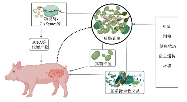
图1 猪后肠真菌对宿主健康的潜在作用机制

CAZymes:碳水化合物活性酶 carbohydrate-active enzymes;SCFA:短链脂肪酸 short-chain fatty acids。

Fig.1 Potential mechanisms of hindgut fungi on host health in swine

[1]
FLINT H J, SCOTT K P, LOUIS P, et al. The role of the gut microbiota in nutrition and health[J]. Nature Reviews Gastroenterology & Hepatology, 2012, 9(10):577-589.

[2]
TREMAROLI V, BÄCKHED F. Functional interactions between the gut microbiota and host metabolism[J]. Nature, 2012, 489(7415):242-249.

[3]
CERF-BENSUSSAN N, GABORIAU-ROUTHIAU V. The immune system and the gut microbiota:friends or foes?[J]. Nature Reviews Immunology, 2010, 10(10):735-744.

[4]
LI J Y, CHEN D W, YU B, et al. Fungi in gastrointestinal tracts of human and mice:from community to functions[J]. Microbial Ecology, 2018, 75(4):821-829.

[5]
ILIEV I D, CADWELL K. Effects of intestinal fungi and viruses on immune responses and inflammatory bowel diseases[J]. Gastroenterology, 2021, 160(4):1050-1066.

DOI PMID

[6]
ILIEV I D, LEONARDI I. Fungal dysbiosis:immunity and interactions at mucosal barriers[J]. Nature Reviews Immunology, 2017, 17(10):635-646.

[7]
SUN S S, SUN L, WANG K, et al. The gut commensal fungus,candida parapsilosis,promotes high fat-diet induced obesity in mice[J]. Communications Biology, 2021, 4(1):1220.

[8]
ZHOU X Y, ZHANG X, YU J. Gut mycobiome in metabolic diseases:mechanisms and clinical implication[J]. Biomedical Journal, 2024, 47(3):100625.

[9]
HU J, NIE Y F, CHEN J W, et al. Gradual changes of gut microbiota in weaned miniature piglets[J]. Frontiers in Microbiology, 2016,7:1727.

[10]
LI J Y, CHEN D W, YU B, et al. The fungal community and its interaction with the concentration of short-chain fatty acids in the faeces of Chenghua,Yorkshire and Tibetan pigs[J]. Microbial Biotechnology, 2020, 13(2):509-521.

[11]
WANG T, LIU J H, LUO Y H, et al. Combined effects of host genetics and diet on porcine intestinal fungi and their pathogenic genes[J]. Frontiers in Microbiology, 2023,14:1192288.

[12]
MOELLER A H, SUZUKI T A, PHIFER-RIXEY M, et al. Transmission modes of the mammalian gut microbiota[J]. Science, 2018, 362(6413):453-457.

DOI PMID

[13]
HU J, CHEN J W, HOU Q L, et al. Core-predominant gut fungus Kazachstania slooffiae promotes intestinal epithelial glycolysis via lysine desuccinylation in pigs[J]. Microbiome, 2023, 11(1):31.

[14]
LI J Y, LUO Y H, CHEN D W, et al. The fungal community and its interaction with the concentration of short-chain fatty acids in the caecum and colon of weaned piglets[J]. Journal of Animal Physiology and Animal Nutrition, 2020, 104(2):616-628.

DOI PMID

[15]
LI J Y, CHEN D W, YU B, et al. Batch and sampling time exert a larger influence on the fungal community than gastrointestinal location in model animals:a meaningful case study[J]. Frontiers in Nutrition, 2022,9:1021215.

[16]
ARFKEN A M, FREY J F, SUMMERS K L. Temporal dynamics of the gut bacteriome and mycobiome in the weanling pig[J]. Microorganisms, 2020, 8(6):868.

[17]
HEISEL T, MONTASSIER E, JOHNSON A, et al. High-fat diet changes fungal microbiomes and interkingdom relationships in the murine gut[J]. mSphere, 2017, 2(5):e00351-17.

[18]
RAY K. Dysbiosis in fungal microbiota in IBD[J]. Nature Reviews Gastroenterology & Hepatology, 2016, 13(4):188.

[19]
BHAGAT N R, KUMAR S, KUMARI R, et al. A review on rumen anaerobic fungi:current understanding on carbohydrate fermentation and roughages digestion in ruminants[J]. Applied Biochemistry and Microbiology, 2023, 59(3):231-249.

[20]
XIE C L, GONG W B, ZHU Z H, et al. Comparative secretome of white-rot fungi reveals co-regulated carbohydrate-active enzymes associated with selective ligninolysis of ramie stalks[J]. Microbial Biotechnology, 2021, 14(3):911-922.

[21]
LUO Y H, LI J Y, ZHOU H, et al. The nutritional significance of intestinal fungi:alteration of dietary carbohydrate composition triggers colonic fungal community shifts in a pig model[J]. Applied and Environmental Microbiology, 2021, 87(10):e00038-21.

[22]
MURPHY K M, LE S M, WILSON A E, et al. The microbiome as a maternal effect:a systematic review on vertical transmission of microbiota[J]. Integrative and Comparative Biology, 2023, 63(3):597-609.

[23]
PRISNEE T L, RAHMAN R, FOUHSE J M, et al. Tracking the fecal mycobiome through the lifespan of production pigs and a comparison to the feral pig[J]. Applied and Environmental Microbiology, 2023, 89(11):e0097723.

[24]
PRISNEE T L, FOUHSE J M, DIETHER N E, et al. Maternal mycobiome,but not antibiotics,alters fungal community structure in neonatal piglets[J]. Applied and Environmental Microbiology, 2022, 88(24):e0159322.

[25]
ELOLIMY A, ROSA F, TRIPP P, et al. Bacterial and fungal adaptations in cecum and distal colon of piglets fed with dairy-based milk formula in comparison with human milk[J]. Frontiers in Microbiology, 2022,13:801854.

[26]
SUMMERS K L, FREY J F, RAMSAY T G, et al. The piglet mycobiome during the weaning transition:a pilot study[J]. Journal of Animal Science, 2019, 97(7):2889-2900.

[27]
ARFKEN A M, FREY J F, RAMSAY T G, et al. Yeasts of burden: exploring the mycobiome-bacteriome of the piglet GI tract[J]. Frontiers in Microbiology, 2019,10:2286.

[28]
FEEHAN B, RAN Q H, DORMAN V, et al. Stability and volatility shape the gut bacteriome and Kazachstania slooffiae dynamics in preweaning,nursery and adult pigs[J]. Scientific Reports, 2022, 12(1):15080.

[29]
KONG Q H, LIU S Z, LI A Y, et al. Characterization of fungal microbial diversity in healthy and diarrheal Tibetan piglets[J]. BMC Microbiology, 2021, 21(1):204.

DOI PMID

[30]
AUCHTUNG T A, FOFANOVA T Y, STEWART C J, et al. Investigating colonization of the healthy adult gastrointestinal tract by fungi[J]. mSphere, 2018, 3(2):e00092-18.

[31]
HOEKSTRA R F. Population genetics of filamentous fungi[J]. Antonie van Leeuwenhoek, 1994, 65(3):199-204.

[32]
CORTÉS J C G, CURTO M Á, CARVALHO V S D, et al. The fungal cell wall as a target for the development of new antifungal therapies[J]. Biotechnology Advances, 2019, 37(6):107352.

[33]
ILIEV I D, FUNARI V A, TAYLOR K D, et al. Interactions between commensal fungi and the C-type lectin receptor dectin-1 influence colitis[J]. Science, 2012, 336(6086):1314-1317.

DOI PMID

[34]
WHEELER M L, LIMON J J, BAR A S, et al. Immunological consequences of intestinal fungal dysbiosis[J]. Cell Host & Microbe, 2016, 19(6):865-873.

[35]
LIMON J J, TANG J, LI D L, et al. Malassezia is associated with Crohn’s disease and exacerbates colitis in mouse models[J]. Cell Host & Microbe, 2019, 25(3):377-388.e6.

[36]
VAUTIER S, DRUMMOND R A, CHEN K, et al. Candida albicans colonization and dissemination from the murine gastrointestinal tract:the influence of morphology and th17 immunity[J]. Cellular Microbiology, 2015, 17(4):445-450.

[37]
ACOSTA-RODRIGUEZ E V, RIVINO L, GEGINAT J, et al. Surface phenotype and antigenic specificity of human interleukin 17-producing T helper memory cells[J]. Nature Immunology, 2007, 8(6):639-646.

[38]
BACHER P, HOHNSTEIN T, BEERBAUM E, et al. Human anti-fungal Th17 immunity and pathology rely on cross-reactivity against Candida albicans[J]. Cell, 2019, 176(6):1340-1355.e15.

[39]
MOYES D L, WILSON D, RICHARDSON J P, et al. Candidalysin is a fungal peptide toxin critical for mucosal infection[J]. Nature, 2016, 532(7597):64-68.

[40]
ALLERT S, FÖRSTER T M, SVENSSON C M, et al. Candida albicans -induced epithelial damage mediates translocation through intestinal barriers[J]. mBio, 2018, 9(3):e00915-e00918.

[41]
NAGLIK J R, GAFFEN S L, HUBE B. Candidalysin:discovery and function in Candida albicans infections[J]. Current Opinion in Microbiology, 2019,52:100-109.

[42]
URUBSCHUROV V, BÜSING K, FREYER G, et al. New insights into the role of the porcine intestinal yeast,Kazachstania slooffiae,in intestinal environment of weaned piglets[J]. FEMS Microbiology Ecology, 2017, 93(2):fiw245.

[43]
SUMMERS K L, FOSTER FREY J, ARFKEN A M. Characterization of Kazachstania slooffiae,a proposed commensal in the porcine gut[J]. Journal of Fungi, 2021, 7(2):146.

[44]
JIANG T T, SHAO T Y, ANG W X G, et al. Commensal fungi recapitulate the protective benefits of intestinal bacteria[J]. Cell Host & Microbe, 2017, 22(6):809-816.e4.

[45]
KELESIDIS T, POTHOULAKIS C. Efficacy and safety of the probiotic Saccharomyces boulardii for the prevention and therapy of gastrointestinal disorders[J]. Therapeutic Advances in Gastroenterology, 2012, 5(2):111-125.

[46]
SUN S Y, XU X X, LIANG L, et al. Lactic acid-producing probiotic Saccharomyces cerevisiae attenuates ulcerative colitis via suppressing macrophage pyroptosis and modulating gut microbiota[J]. Frontiers in Immunology, 2021,12:777665.

[47]
BARC M C, CHARRIN-SARNEL C, ROCHET V, et al. Molecular analysis of the digestive microbiota in a gnotobiotic mouse model during antibiotic treatment:influence of Saccharomyces boulardii[J]. Anaerobe, 2008, 14(4):229-233.

[48]
SWIDSINSKI A, LOENING-BAUCKE V, VERSTRAELEN H, et al. Biostructure of fecal microbiota in healthy subjects and patients with chronic idiopathic diarrhea[J]. Gastroenterology, 2008, 135(2):568-579.

DOI PMID

[49]
CZERUCKA D, PICHE T, RAMPAL P. Review article:yeast as probiotics-Saccharomyces boulardii[J]. Alimentary Pharmacology & Therapeutics, 2007, 26(6):767-778.

[50]
SZAJEWSKA H, SKÓRKA A, DYLAG M. Meta-analysis:Saccharomyces boulardii for treating acute diarrhoea in children[J]. Alimentary Pharmacology & Therapeutics, 2007, 25(3):257-264.

[51]
ZANELLO G, MEURENS F, BERRI M, et al. Saccharomyces boulardii effects on gastrointestinal diseases[J]. Current Issues in Molecular Biology, 2009, 11(1):47-58.

[52]
SEZER A, USTA U, CICIN I. The effect of Saccharomyces boulardii on reducing irinotecan-induced intestinal mucositis and diarrhea[J]. Medical Oncology, 2009, 26(3):350-357.

[53]
BREVES G, FAUL K, SCHRÖDER B, et al. Application of the colon-simulation technique for studying the effects of Saccharomyces boulardii on basic parameters of porcine cecal microbial metabolism disturbed by clindamycin[J]. Digestion, 2000, 61(3):193-200.

[54]
WU X, VALLANCE B A, BOYER L, et al. Saccharomyces boulardii ameliorates Citrobacter rodentium-induced colitis through actions on bacterial virulence factors[J]. American Journal of Physiology:Gastrointestinal and Liver Physiology, 2008, 294(1):G295-G306.

[55]
CASTAGLIUOLO I, RIEGLER M F, VALENICK L, et al. Saccharomyces boulardii protease inhibits the effects of Clostridium difficile toxins A and B in human colonic mucosa[J]. Infection and Immunity, 1999, 67(1):302-307.

[56]
BUTS J P, DEKEYSER N, STILMANT C, et al. Saccharomyces boulardii produces in rat small intestine a novel protein phosphatase that inhibits Escherichia coli endotoxin by dephosphorylation[J]. Pediatric Research, 2006, 60(1):24-29.

[57]
CZEPIEL J, DRÓŻDŻ M, PITUCH H, et al. Clostridium difficile infection:review[J]. European Journal of Clinical Microbiology & Infectious Diseases, 2019, 38(7):1211-1221.

[58]
MAAS E, PENDERS J, VENEMA K. Fungal-bacterial interactions in the human gut of healthy individuals[J]. Journal of Fungi, 2023, 9(2):139.

[59]
HUANG L, ZHANG H, WU P Z, et al. Dbcan-seq:a database of carbohydrate-active enzyme(CAZyme)sequence and annotation[J]. Nucleic Acids Research, 2017, 46(D1):D516-D521.

[60]
AMERI R, GARCÍA J L, DERENFED A B, et al. Genome sequence and carbohydrate active enzymes (CAZymes) repertoire of the thermophilic caldicoprobacter algeriensis TH7C1T[J]. Microbial Cell Factories, 2022, 21(1):91.

DOI PMID

[61]
LANGE L, BARRETT K, PILGAARD B, et al. Enzymes of early-diverging,zoosporic fungi[J]. Applied Microbiology and Biotechnology, 2019, 103(17):6885-6902.

[62]
SOLOMON K V, HAITJEMA C H, HENSKE J K, et al. Early-branching gut fungi possess a large,comprehensive array of biomass-degrading enzymes[J]. Science, 2016, 351(6278):1192-1195.

[63]
AHMAD F, YANG G Y, LIANG S Y, et al. Multipartite symbioses in fungus-growing termites (Blattodea:Termitidae,Macrotermitinae) for the degradation of lignocellulose[J]. Insect Science, 2021, 28(6):1512-1529.

[64]
AHRENDT S R, QUANDT C A, CIOBANU D, et al. Leveraging single-cell genomics to expand the fungal tree of life[J]. Nature Microbiology, 2018, 3(12):1417-1428.

DOI PMID

[65]
POWELL M J. Zoospore structure of the mycoparasitic chytrid caulochytrium protostelioides olive[J]. American Journal of Botany, 1981,68:1074-1089.

[66]
CHANG Y, WANG S S, SEKIMOTO S, et al. Phylogenomic analyses indicate that early fungi evolved digesting cell walls of algal ancestors of land plants[J]. Genome Biology and Evolution, 2015, 7(6):1590-1601.

DOI PMID

[67]
HAITJEMA C H, GILMORE S P, HENSKE J K, et al. A parts list for fungal cellulosomes revealed by comparative genomics[J]. Nature Microbiology, 2017,2:17087.

Outlines

/