REVIEW

Research Progress on Intestinal Pathogenesis and Nutritional Regulation of Porcine Epidemic Diarrhea Virus

  • YANG Jiebo , 1 ,
  • YAN Honglin 1 ,
  • WU Aimin 2 ,
  • ZHANG Yong , 1, 2, *
Expand
  • 1 College of Life Science and Engineering, Southwest University of Science and Technology, Mianyang 621010, China
  • 2 Key Laboratory of Animal Disease-Resistance Nutrition of Ministry of Education, Institute of Animal Nutrition, Sichuan Agricultural University, Chengdu 611130, China
* associate professor, E-mail:

Received date: 2024-08-15

  Online published: 2025-04-15

Abstract

Porcine epidemic diarrhea virus (PEDV) is a highly infectious enteric coronavirus, which can cause atrophic enteritis, watery diarrhea and dehydration in newborn piglets, and is the main cause of death in newborn piglets. The provision of nutrients is essential for sustaining animal growth and health, while also exerting direct or indirect effects on the virus by inhibiting PEDV replication and activity, as well as repairing intestinal damage caused by PEDV infection through enhancing piglet immune performance, antioxidant capacity, and anti-inflammatory ability to maintain intestinal health. Nutrients play an anti-PEDV role in alleviating the effects of PEDV on piglets mainly by enhancing immunity and inhibiting PEDV activity of piglets. Therefore, based on a review of the pathogenesis of PEDV in the intestine, this paper focuses on the research progress on the effects of PEDV on piglets and the nutritional regulation in alleviating PEDV, aiming to provide theoretical reference for further study of the mechanism of nutrition regulation on PEDV and development of antiviral nutrition regulation technology.

Cite this article

YANG Jiebo , YAN Honglin , WU Aimin , ZHANG Yong . Research Progress on Intestinal Pathogenesis and Nutritional Regulation of Porcine Epidemic Diarrhea Virus[J]. Chinese Journal of Animal Nutrition, 2025 , 37(4) : 2142 -2156 . DOI: 10.12418/CJAN2025.182

猪流行性腹泻是由猪流行性腹泻病毒(porcine epizootic diarrhea virus,PEDV)引起猪的一种高度传染性肠道疾病[1]。PEDV感染会导致新生仔猪死亡率极高,严重制约着养猪业的健康发展[2]。新生仔猪感染PEDV后,可引起急性水样腹泻、呕吐和脱水,发病后死亡率高达80%~100%[3]。PEDV于1971年在英国首次被发现[4],此后PEDV迅速传播到欧洲和中国、日本、韩国等亚洲国家[5]。2013年4月,在美国规模化养猪场中首次发现了高毒力PEDV毒株[6]。这种新变种PEDV毒株造成美国约800万头仔猪死亡,约占总猪群的10%,给美国养猪业造成了重大经济损失[7]。常新见等[8]调查了2017—2019年我国华东地区养猪场的病毒性腹泻,发现PEDV的阳性率为62.6%,由此可见PEDV是引起我国猪场病毒性腹泻的主要病原体。由于高毒力PEDV新变种的出现,使得现有的防控技术在防控当前PEDV上的效果较差,对我国养猪业造成严重影响[9]。营养物质是维持动物生长和生产过程中所必需的重要因素,可在病毒感染过程中,通过增强动物机体免疫力和调节肠道菌群来抑制病毒在肠道中的复制,从而维持动物的肠道健康[10]。本文重点介绍了PEDV的肠致病机制和对仔猪的影响以及防控PEDV的营养调控作用研究进展,旨在为生产中开发抗病毒营养调控技术以及为当前养猪生产中合理利用营养物质来有效防控PEDV提供理论参考。

1 PEDV的肠致病机制

PEDV是引起仔猪流行性腹泻的主要病原体,其是一种具有包膜的单股正链RNA病毒,在冠状病毒分类上属于套式病毒目、冠状病毒科和甲型冠状病毒属[11]。PEDV基因结构中的开放阅读框(open reading frame,ORF)2和ORF4~ORF6共编码4种结构蛋白,即刺突蛋白(spike protein,S蛋白,150~220 ku)、包膜蛋白(envelope protein,E蛋白,7 ku)、膜蛋白(membrane protein,M蛋白,20~30 ku)和核衣壳蛋白(nucleocapsid protein,N蛋白,58 ku)[12]。氨基肽酶N(aminopeptidase N,APN)是一种大小为150 ku的糖基化跨膜蛋白,PEDV通过与肠细胞上的受体APN相互作用,从而进入肠道并进行复制[13]。PEDV具有溶细胞作用,肠上皮细胞受到感染后会迅速发生坏死,从而导致小肠绒毛萎缩,破坏肠道屏障功能[14]。研究发现,PEDV抗原主要在猪的小肠(十二指肠、空肠和回肠)绒毛肠上皮细胞中被检测到[6,15]。在对哺乳仔猪的研究中,用PEDV感染3日龄仔猪12 h后,可在十二指肠和空肠近端检测到PEDV抗原,且PEDV复制后迅速扩散到整个空肠和回肠[16]。Jung等[2]研究指出,PEDV感染后肠上皮细胞表达APN,可通过细胞质膜(如内质网和高尔基体)出芽而快速组装。在PEDV潜伏期,整个小肠可见PEDV抗原阳性细胞,30%~50%的绒毛肠上皮细胞呈阳性[17],同时在急性感染期还可在空肠部位观察到空泡化的肠上皮细胞和大量细胞脱落[2]。PEDV感染引起的腹泻主要是由于受感染的肠细胞死亡、肠道功能紊乱以及与刷状缘膜结合的消化酶(乳糖酶、蔗糖酶和麦芽糖酶)减少,从而造成消化不良和吸收不良;研究还发现,PEDV感染仔猪30~120 h后,在仔猪的小肠中观察到闭锁小带蛋白(ZO)-1和E-钙黏蛋白(E-cadherin)呈无序、不规则分布,并且其表达水平显著降低[18]。在感染PEDV哺乳仔猪的小肠中,绒毛肠上皮细胞的紧密连接和黏附连接被破坏[19],而紧密连接的破坏会导致上皮跨膜电阻降低,使PEDV更容易在小肠中复制[20]。以上研究表明,肠道完整性受损和肠道先天免疫功能不全是导致哺乳仔猪更易感染PEDV的主要因素。
PEDV的一个经典毒株CV777主要感染仔猪的小肠部位,并在其中进行复制和感染[21]。PEDV主要通过诱导仔猪肠上皮细胞的凋亡和抑制细胞的增殖来增强其在肠道中的复制[22]。体外研究发现,PEDV通过调控磷脂酰肌醇3-激酶(PI3K)/丝氨酸-苏氨酸激酶(Akt)和哺乳动物雷帕霉素靶蛋白(mTOR)信号通路来降低猪小肠上皮细胞系-J2(IPEC-J2)的增殖活性,并抑制细胞周期停滞在S期,诱导上皮细胞的凋亡[23]。此外,PEDV还通过激活p38丝裂原活化蛋白激酶(MAPK)和c-Jun氨基末端激酶(JNK)信号通路来调控病毒在靶细胞中的复制,从而促进PEDV的感染[24]。在PEDV感染期间,PEDV还以蛋白酶依赖性方式降解信号转导和转录激活因子(STAT)1蛋白的表达,进而干扰Ⅰ型干扰素(IFN-Ⅰ)信号通路,调控PEDV的复制和感染[25]。同时,PEDV感染还会促进细胞间融合,引起细胞持续坏死,从而导致仔猪出现严重腹泻甚至死亡[19]。据报道,PEDV感染宿主细胞的机制主要是依靠丝氨酸蛋白酶水解和低pH,并通过内吞作用进入细胞[26]
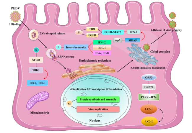
PEDV侵入宿主细胞主要依赖其结构蛋白与受体的相互作用,其感染宿主肠道上皮细胞的主要机制见图1。PEDV的S蛋白通过与转铁蛋白受体1(TfR1)的胞外结构域相互作用来增强TfR1的内化,从而促进病毒进入[27]。S蛋白的S1亚单位是识别和结合细胞受体的关键区域,可与受体相互作用,促进病毒进入细胞[28]。Yang等[29]研究发现,PEDV的S蛋白通过激活表皮生长因子受体(EGFR)和EGFR-STAT3信号通路来抑制IFN-Ⅰ的抗病毒活性,进而增强PEDV的复制。E蛋白和M蛋白分别通过与干扰素调节因子(interferon regulatory factor,IRF)3和IRF7的相互作用来抑制IFN-Ⅰ的免疫应答[30]。E蛋白可诱导内质网应激,在抑制宿主细胞凋亡和逃避宿主先天免疫反应中发挥重要作用[31]。此外,N蛋白同样在促进病毒复制和逃避宿主先天免疫反应中具有重要作用,可与核仁中的核磷脂共存并相互作用,促进病毒的复制并抑制细胞凋亡[32]。据报道,N蛋白可与核内磷酸化蛋白(p53)的相互作用来诱导细胞周期停滞在S期,以此促进病毒的复制[33]。同时,N蛋白还可通过阻断核因子-κB(NK-κB)的核易位来抑制干扰素(IFN)-λ的产生,并与TANK结合激酶1(TBK1)的相互作用来阻碍TBK1与IRF3的结合,从而抑制IRF3的激活和IFN-Ⅰ的生成[34]。辅助蛋白ORF3主要存在于内质网中,可引起内质网凋亡或自噬相关的应激反应,PEDV的ORF3通过调节葡萄糖调节蛋白78(GRP78)的表达水平和激活蛋白激酶R样内质网激酶(PERK)-真核起始因子2α(eIF2α)信号通路来诱导内质网应激,促进细胞自噬标志物微管关联蛋白轻链3-Ⅰ(LC3-Ⅰ)向微管关联蛋白轻链3-Ⅱ(LC3-Ⅱ)的转化来诱导细胞自噬[35]。在受感染细胞的表面,ORF3可与细胞质膜上的S蛋白相互作用来促进病毒的复制[36]。此外,ORF3还可通过抑制核酸结合蛋白(p65)的表达和干扰p65的核易位来抑制NF-κB的活化,减少促炎细胞因子白细胞介素-6(IL-6)和白细胞介素-8(IL-8)的产生[37]。PEDV的ORF3还通过抑制病毒感染引起的细胞凋亡来促进病毒的复制[38]。研究发现,PEDV的非结构蛋白(non-structural protein,nsp)4通过诱导促炎细胞因子[白细胞介素-1α(IL-1α)、白细胞介素-1β(IL-1β)和肿瘤坏死因子-α(TNF-α)]和趋化因子[趋化因子2(CCL2)、趋化因子5(CCL5)和趋化因子C-X-C基序配体(CXCL)]的过表达来促进PEDV的复制[39]。此外,PEDV nsp6还可通过调节PI3K/Akt/mTOR信号通路来诱导细胞自噬,增强PEDV在IPEC-J2细胞中的复制,诱导肠上皮细胞的损伤[40]。PEDV nsp16具有2'-O甲基转移酶(2'-O MTase)活性,可通过调节视黄酸诱导基因-Ⅰ(retinoic acid-inducible gene-Ⅰ,RIG-Ⅰ)和黑色素瘤分化相关基因5(melanoma differentiation related gene 5,MDA5)介导的先天免疫来促进病毒复制[41]。综上所述,PEDV主要通过在小肠细胞中感染和复制,从而诱导肠上皮细胞损伤,引起仔猪萎缩性肠炎、腹泻、呕吐和死亡,这对深入认识和研究PEDV的肠致病机制和开发抗PEDV营养调控技术具有重要意义。
图1 PEDV感染宿主肠上皮细胞的机制示意图

PEDV:猪流行性腹泻病毒 porcine epidemic diarrhea virus;Binding:附着;Viral capsid release:病毒衣壳释放;RNA release:RNA释放;Replication:复制;Transcription:转录;Translation:翻译;Furin-mediated maturation:弗林蛋白酶介导的成熟;Release of viral progeny:释放病毒原;Mitochondria:线粒体;Endoplasmic reticulum:内质网;Nucleus:细胞核;Golgi complex:高尔基复合体;S:刺突蛋白 spike protein;TfR1:转铁蛋白受体1 transferrin receptor 1;EGFR:表皮生长因子受体 epidermal growth factor receptor;STAT3:信号转导和转录激活因子3 signal transducer and activator of transcription 3;IFN-Ⅰ:Ⅰ型干扰素 type Ⅰ interferon;nsp3:非结构蛋白3 non-structural protein 3;MDA5:黑色素瘤分化相关基因5 melanoma differentiation related gene 5;E:包膜蛋白 envelope protein;Innate immunity:先天免疫;IFN-Ⅲ:Ⅲ型干扰素 type Ⅲ interferon;RIG-Ⅰ:视黄酸诱导基因-Ⅰ retinoic acid-inducible gene-Ⅰ;IL-6:白细胞介素-6 interleukin-6;IL-8:白细胞介素-8 interleukin-8;N:核衣壳蛋白 nucleocapsid protein;NK-κB:核因子-κB nuclear factor-κB;TBK1:TANK结合激酶1 TANK binding kinase 1;IFR3:干扰素调节因子3 interferon regulatory factor 3;ORF3:辅助蛋白3 auxiliary protein 3;GRP78:葡萄糖调节蛋白78 glucose-regulated protein 78;PERK:蛋白激酶R样内质网激酶 protein kinase R-like endoplasmic reticulum kinase;eIF2α:真核起始因子2α phosphorylation of eukaryotic initiation factor 2α;LC3-Ⅰ:微管关联蛋白轻链3-Ⅰ microtubule-associated protein light chain 3-Ⅰ;LC3-Ⅱ:微管关联蛋白轻链3-Ⅱ microtubule-associated protein light chain 3-Ⅱ;M:膜蛋白 membrane protein;Protein synthesis:蛋白质合成;Viral replication:病毒复制。

红色实线表示抑制,蓝色实线表示激活。Solid red line represented inhibition, and solid blue line represented activation.

Fig.1 Mechanism diagram of PEDV infecting host intestinal epithelial cells

2 PEDV对仔猪的影响

2.1 PEDV对仔猪生长性能的影响

PEDV是一种具有高度传染性的肠道冠状病毒,可导致仔猪发生严重水样腹泻、呕吐和脱水,严重时造成死亡[3]。此外,PEDV可感染所有年龄段的猪,其中对哺乳仔猪的影响最大,其主要临床特征为引起仔猪萎缩性肠炎、腹泻、呕吐和脱水[42]。研究发现,PEDV对1周龄以下的哺乳仔猪影响最为严重,感染后其发病率为100%,死亡率高达80%~100%[6]。PEDV感染肠道上皮细胞后,主要引起肠绒毛萎缩、坏死和脱落,进而影响营养物质的吸收,导致仔猪生长性能降低[43]。5日龄仔猪在接种rPEDVGFP-S-OKN1和rPEDVGFP-S1-OKN1菌株2~4 d后,受感染仔猪出现水样腹泻、厌食、嗜睡、严重脱水、体重减轻和生长迟缓;而在攻毒7 d后,所有受感染仔猪均死亡,并在其粪便中检测到脱落的PEDV抗原[44]。在PEDV感染仔猪的研究中,对试验仔猪攻毒12 h后,可观察到受感染仔猪的粪便呈现水样并伴有腥味,同时并出现呕吐和呼吸困难等临床症状;而在感染后24~96 h内,所有PEDV感染仔猪死亡[45]。Zhang等[46]研究表明,PEDV攻毒哺乳仔猪后,可显著降低其平均日增重,同时伴有腹泻和呕吐症状。PEDV在小肠上皮细胞中的复制会抑制钠离子(Na+)-钾离子(K+)-ATP酶和钙离子(Ca2+)-镁离子(Mg2+)-ATP酶的活性,而Na+、K+和Ca2+含量的显著变化与这2种酶的活性下调有关,这表明PEDV感染可能会导致水和电解质失衡,从而引起仔猪水样腹泻和脱水,降低其生长性能[47]。此外,哺乳仔猪在感染PEDV后,还会通过降低其氨基酸转运蛋白的表达,改变血浆和空肠中的氨基酸谱,进而影响小肠黏膜中脂质和葡萄糖的吸收和代谢,导致仔猪生长迟缓并引起腹泻[48]

2.2 PEDV对仔猪免疫性能的影响

病毒感染会诱导宿主的先天抗病毒免疫反应来抑制病毒在宿主细胞中的复制,并激活启动机体的适应性免疫反应,防止病毒感染。宿主的先天免疫系统是抑制病毒感染的第一道防线,其与INF、炎性细胞因子、细胞凋亡和自噬等有关[49]。肠道黏膜中的杯状细胞、潘氏细胞和微褶皱细胞(M细胞),有助于维持肠道稳态的免疫反应。研究发现,用PEDV攻毒断奶仔猪后,会显著提高其空肠中白细胞介素-1(IL-1)和TNF-α的表达,同时并通过降低潘氏细胞和M细胞的活性以及聚合免疫球蛋白受体(pIgR)的表达来影响断奶仔猪的先天黏膜免疫[9]。而哺乳仔猪在感染PEDV后,其血液和回肠中产生IFN-γ的自然杀伤(NK)细胞的数量和活性显著降低,表明NK细胞先天免疫功能的缺陷可能是导致哺乳仔猪更易感染PEDV的原因[50]。Zhang等[51]报道,PEDV感染7日龄仔猪,会显著降低其脾脏中超氧化物歧化酶和谷胱甘肽过氧化物酶的活性以及IL-1β、白细胞介素-10(IL-10)和TNF-α的mRNA表达水平。由此表明,PEDV感染会显著降低仔猪的抗氧化水平、抗炎能力和抗病毒免疫反应。IFN-α/β对控制病毒感染和启动适应性免疫反应具有重要作用[52]。研究发现,PEDV可通过抑制肠上皮细胞IFN-λ的生成来影响仔猪的抗病毒免疫反应[53]

2.3 PEDV对仔猪肠道健康的影响

2.3.1 PEDV对肠道屏障的影响

PEDV是引起仔猪肠道疾病的主要病原,其主要病理变化包括感染猪肠上皮细胞、急性黏膜损伤、肠绒毛变短以及肠壁变薄,最终导致肠壁细胞细胞质空泡化和脱落,从而破坏肠道屏障的完整性[2,6]。肠道屏障的完整性可以保护肠道免受外来物质的影响,并允许营养物质穿过上皮[22]。PEDV主要在十二指肠、空肠和回肠上皮细胞中复制和感染,并诱导上皮细胞的凋亡和坏死[54]。在PEDV感染期间,空肠和回肠中负责分泌黏蛋白和维持肠完整性的杯状细胞数量显著减少,导致黏液层受损,引起继发感染[55]。PEDV可急性感染空肠绒毛上皮细胞,引起急性、萎缩性肠炎,同时并伴有病毒血症,使哺乳仔猪出现严重腹泻和脱水,这是造成哺乳仔猪死亡的主要原因[18]。研究发现,感染PEDV仔猪的肠道绒毛高度、绒毛宽度和杯状细胞数量均显著低于未感染仔猪,并且其血清中D-乳酸含量和二胺氧化酶活性提高,表明感染PEDV会显著影响肠道屏障的完整性和正常功能[56]。此外,PEDV具有高度肠道致病性,可破坏猪肠道屏障的完整性和隐窝干细胞增殖[57],降低紧密连接蛋白的表达,进而抑制猪的生长[19]。肠上皮细胞作为肠黏膜屏障最重要的组成部分,其通过紧密连接蛋白发挥调节作用,不仅能维持肠道屏障的功能,还能阻碍病原菌和病毒的侵入,是抵御肠道感染的第一道防线[58]。体外研究发现,PEDV感染IPEC-J2细胞后,破坏了肠道上皮屏障的完整性,并降低了IPEC-J2细胞中紧密连接蛋白ZO-1、ZO-2、闭合蛋白(occludin)和封闭蛋白1(claudin 1,CLDN1)的表达[59]。在感染PEDV的仔猪中,其小肠中的ZO-1和E-cadherin分布异常且表达减少,并伴有紧密连接和黏附连接结构和功能的缺陷[19]。综上所述,紧密连接参与了PEDV感染过程,而紧密连接的损伤会促进PEDV在肠道内的复制;紧密连接蛋白作为肠上皮细胞的重要连接方式,在维持仔猪肠黏膜屏障的完整性和抵抗PEDV感染方面发挥着重要作用,但还需要进一步研究来阐明紧密连接蛋白在PEDV感染中的作用以及分子机制。

2.3.2 PEDV对肠道菌群的影响

肠道菌群是整个肠道内稳定的微环境,在肠道黏膜免疫系统的发育中起着至关重要的作用,其主要与其他病原菌竞争营养物质和结合位点,抑制其侵入肠道黏膜[60]。当肠道菌群的稳态被破坏时,会导致肠道内致病菌的数量增加,有益菌的数量减少,从而引发炎症或腹泻[61]。在PEDV感染期间,与肠道健康相关的菌群,如拟杆菌属(Bacteroides)、丁酸梭菌(Clostridium butyricum)和嗜冷杆菌属(Psychrobacter)的丰度显著降低,但肠球菌属(Enterococcus)、梭菌属(Fusobacterium)、埃希氏菌属(Escherichia)和脱硫弧菌属(Desulfovibrio)等致病菌的丰度却显著提高[62]。在对PEDV感染仔猪的研究中发现,受感染仔猪小肠中包括梭菌属和志贺氏菌属(Shigella)在内的病原微生物丰度显著提高,然而,肠道中有益菌嗜冷杆菌属和普雷沃氏菌属(Prevotella)的丰度却显著降低,PEDV感染破坏了肠道内环境的稳态,引起仔猪腹泻[63]。同样,也有研究报道,PEDV感染后,仔猪肠道中的有益菌群厚壁菌门(Firmicutes)、拟杆菌门(Bacteroidetes)和变形菌门(Proteobacteria)丰度显著降低,致病菌丰度显著提高[64]。肠道微生物可以通过刺激先天免疫系统和产生抗菌化合物来阻止病原菌入侵[54]。PEDV会导致仔猪和母猪肠道菌群失调,PEDV诱导的肠道内环境稳态的失调提高了志贺氏菌属、肠球菌属、梭菌属和韦荣球菌属(Veillonella)的丰度,使肠道中致病菌数量增加[65]。Dong等[66]在长白×约克夏仔猪的研究中发现,PEDV感染仔猪肠道中埃希氏菌属和志贺氏菌属的丰度均高于未感染仔猪,但PEDV感染显著降低了其肠道中唾液乳酸杆菌(Lactobacillus salivarius)的丰度。综上所述,仔猪感染PEDV会破坏其肠道菌群的稳态,使肠道中益生菌的数量减少,而使致病菌的数量增加,从而导致仔猪发生腹泻和脱水等症状。目前,营养调控对肠道微生物的作用机制还需要进一步研究,从而为PEDV引起的肠道损伤修复和作用机制提供理论参考。

3 缓解PEDV感染的营养调控措施

营养物质是维持动物生长和健康的重要因素,可通过增强动物机体的免疫性能来提高动物的抗病力;同时可在病毒感染过程中,抑制病毒的复制和活性。氨基酸、维生素、微量元素、益生菌和植物提取物等物质已被证实可抑制PEDV在肠道中的复制和活性,并可缓解PEDV感染所引起的肠道损伤。

3.1 营养性添加剂

3.1.1 氨基酸

氨基酸作为猪生长所必需的营养物质,具有维持动物肠道健康、抗炎、抗氧化应激和抑制细胞凋亡等多种生理功能,对提高猪的免疫性能和维持肠道黏膜的完整性具有重要作用[67]。据报道,精氨酸可通过调节免疫反应、紧密连接和肠道代谢来改善肠道炎症[68]。饲粮中添加精氨酸可提高断奶仔猪的免疫性能和生长性能,改善小肠形态,并通过调节NF-κB信号通路激活肠道先天免疫反应[69]。蛋氨酸能够调节动物的新陈代谢和先天免疫反应,并通过产生谷胱甘肽增强机体的抗氧化能力,对维持肠道完整性和增强肠道抗氧化能力发挥着重要作用[70]。此外,支链氨基酸(亮氨酸、缬氨酸和异亮氨酸)也能通过改善肠道形态的完整性和促进免疫球蛋白的生成来增强肠道的免疫防御系统,从而预防肠道炎症[71]。研究表明,半胱氨酸可通过抑制NF-κB通路和激活核因子E2相关因子2(Nrf2)信号通路,在肠道屏障中发挥抗炎和抗氧化等保护作用[72]。N-乙酰半胱氨酸是由L-半胱氨酸衍生而来的一种氨基酸,是动物机体内重要的生物活性物质。Zhang等[51]在感染PEDV哺乳仔猪的研究中发现,给PEDV感染仔猪饲喂N-乙酰半胱氨酸不仅能减轻PEDV感染引起的肠道损伤,还能改善PEDV感染仔猪的脾脏、淋巴结和腓肠肌中的抗氧化和炎症水平。同时,饲粮中添加N-乙酰半胱氨酸还可缓解PEDV引起的腹泻,改善仔猪的肠道功能[73]。N-乙酰半胱氨酸主要通过抑制炎症反应和改善物质转运来缓解PEDV诱导的肠道损伤,并通过提高空肠和回肠中益生菌的丰度来改善小肠的吸收功能[74-75]。综上所述,氨基酸不仅能缓解PEDV感染引起的肠道损伤,还能通过改善仔猪的肠道屏障来预防PEDV。

3.1.2 维生素

维生素是一类维持动物正常生理功能的必需营养素,能够清除体内的自由基,提高机体的抗氧化能力并减少细胞损伤,同时还能增强机体的免疫功能,抑制病原菌对机体的侵袭[76]。研究发现,对感染PEDV的妊娠母猪饲喂维生素A,能够增强妊娠母猪的肠道-乳腺-分泌型免疫球蛋白A(sIgA)轴,从而提高其血液、乳汁和回肠中的抗PEDV免疫球蛋白A(IgA)水平,表明饲喂维生素A可提高母猪妊娠期间的肠道免疫力和对哺乳仔猪的泌乳免疫保护能力[77]。维生素D能够抑制冠状病毒在肠道细胞中的复制,并通过提高仔猪的免疫功能,缓解其感染症状[78]。饲粮中添加25-羟基维生素D3(155.5 μg),可降低PEDV感染仔猪肠道中炎症细胞因子和IFN信号通路相关基因的表达,并通过提高PEDV感染小肠中紧密连接蛋白的表达,缓解PEDV感染引起的肠道损伤,并维持肠道屏障的完整性[79]。体外研究发现,1,25-二羟基维生素D3可通过调节NF-κB和Janus激酶(JAK)/STAT信号通路,抑制IPEC-J2细胞中PEDV诱导的促炎细胞因子表达,有效缓解PEDV感染引起的肠道炎症和肠道损伤[80]。Li等[81]报道,烟酰胺通过激活MAPK和细胞外信号调节激酶(ERK)信号通路来下调转录因子的表达,抑制PEDV在Vero细胞中的复制,由此表明烟酰胺可抑制PEDV在仔猪肠道中的复制和感染。

3.1.3 微量元素

微量元素是维持动物生长和代谢过程中所必需的营养素,具有调节动物生长发育、促进新陈代谢以及增强机体免疫性能和抗应激能力等多种生理功能。锌是动物机体所必需的微量元素,具有抗菌和抗炎特性,对肠道发育和免疫功能具有重要作用[82-83]。饲粮中添加氧化锌可改善PEDV感染仔猪的生长性能、肠道氧化还原状态以及肠道形态和功能,缓解PEDV感染引起的腹泻和临床症状;此外,氧化锌还可通过调节中性粒细胞活性,对感染PEDV仔猪发挥抗病毒、抗氧化和抗炎作用,对修复肠道损伤和维持屏障正常功能具有重要作用[84]。单宁酸螯合锌是一种新型的有机锌螯合物,可提高PEDV感染仔猪的抗氧化能力,改善PEDV诱导的肠道黏膜损伤和吸收功能,以此来促进仔猪的生长,缓解PEDV导致的生长迟缓[46]。另外,Li等[85]体外研究表明,氯化锂也能有效抑制PEDV在Vero细胞中的进入和复制,主要通过抑制PEDV RNA及其蛋白在Vero细胞中的表达,以及抑制PEDV诱导的早期和晚期细胞凋亡来发挥抗病毒作用。综上所述,微量元素同样具有抗PEDV的重要作用,可通过抑制PEDV在肠道中的复制来缓解PEDV诱导的肠道损伤,这为微量元素在预防PEDV生产中的应用提供了一定指导。

3.2 非营养性添加剂

3.2.1 益生菌

益生菌是指对动物宿主健康有益的一类微生物,具有调节肠道菌群、提高免疫力、调控肠道屏障功能以及抑制病原菌生长和繁殖等多种生物学功能[86],是肠道先天性和适应性免疫系统的关键激活剂,可以发出抗菌和炎症反应信号来预防仔猪腹泻[87]。此外,益生菌还可通过改善肠道菌群的稳态和刺激肠道杯状细胞以及上皮细胞分泌黏蛋白、抗菌肽和防御素等抗菌物质来形成免疫屏障,抑制病原菌的侵入[88]。其机制主要是益生菌通过产生的抗菌化合物、次级代谢物和细菌素在肠道黏膜上竞争共同的黏附受体来抑制病原菌,并通过刺激肠黏膜中的先天性和适应性免疫防御机制,激活NF-κB信号通路来诱导肠细胞中防御素和潘氏细胞中抗菌因子的表达,从而与病原菌竞争营养物质和共同位点来抑制病原菌的定植和感染[89]。同时,益生菌还可通过提高肠道紧密连接蛋白的表达来缓解冠状病毒对肠道的损伤[90]。据报道,枯草芽孢杆菌(Bacillus subtilis)及其表面活性蛋白可以调节猪肠道上皮细胞的先天抗病毒免疫反应,抑制传染性胃肠炎病毒进入肠上皮细胞[91]。采用地衣芽孢杆菌(Bacillus licheniformis)饲喂PEDV感染仔猪,可抑制PEDV在肠道中的复制和感染活性,并缓解其腹泻和呕吐等症状;同时,还在仔猪粪便中观察到PEDV RNA脱落减少,表明地衣芽孢杆菌对PEDV具有显著的抗病毒作用[92]。据报道,通过给PEDV感染小鼠口服干酪乳杆菌(Lactobacillus casei),可诱导其黏膜免疫应答和血清抗体的生成,同时显著提高感染小鼠病毒中和抗体、免疫球蛋白G(IgG)和sIgA水平[93],表明干酪乳杆菌可能具有保护仔猪免受PEDV感染的作用。乳酸菌可抑制PEDV在细胞中的复制,在用乳酸菌上清液处理Vero细胞后,采用泰国PEDV流行毒株进行攻毒发现,乳酸菌可降低泰国PEDV流行毒株对Vero细胞的感染性和在Vero细胞中的复制[94]。Huang等[95]通过体内和体外研究表明,植物乳杆菌(Lactobacillus plantarum)能够抑制PEDV的复制,且具有抗病毒活性,能够抑制PEDV对细胞的吸附;同时,植物乳杆菌能减轻炎症反应,并诱导受损细胞的凋亡。此外,在仔猪饲粮中添加鼠李糖乳杆菌GG(Lactobacillus rhamnosus GG),同样可改善PEDV感染仔猪的肠道形态,并通过增强其肠道中的抗氧化和抗炎能力来缓解PEDV感染引起的空肠黏膜炎症和脂质代谢紊乱[96]。综上所述,益生菌不仅能抑制病原菌的生长,还能缓解PEDV引起的肠道损伤,从而为深入研究益生菌抗PEDV的调控作用机制提供了新的思路。

3.2.2 植物提取物

植物提取物是指从中草药中提取出来的多糖、单宁、皂苷、类黄酮、生物碱和多酚等生物活性成分,这些物质能够改善动物的肠道健康,提高机体的免疫力和抗氧化水平,同时具有抗菌、抗炎和解毒活性等多种生物学功能,能够抑制病毒对宿主的感染[97]。给PEDV感染仔猪饲喂葛根素,可显著提高其小肠中有益菌(乳酸菌)的丰度,并通过增强受感染仔猪的抗炎和抗氧化能力来降低仔猪的发病率,同时还可显著改善PEDV感染引起的肠道黏膜损伤[98]。厚朴酚是一种天然多酚化合物,具有抗菌、抗炎和抗氧化等多种生物学功能[99]。体外研究表明,厚朴酚可通过干扰病毒复制过程来抑制PEDV在Vero细胞中的复制,在体外表现出较强的抗PEDV活性[100]。月桂酸甘油酯是一种存在于棕榈油和椰子油中的天然化合物,具有促进营养物质吸收,提高动物生长性能,减少炎性细胞因子产生,以及增强动物机体免疫力的作用[101]。饲粮中添加月桂酸甘油酯可有效抑制PEDV的复制和缓解PEDV感染引起的临床症状,并通过改善肠道屏障的功能和提高机体抗氧化能力及免疫性能,促进仔猪从PEDV感染中恢复[102]。芦荟提取物同样具有抗PEDV作用,在体外研究中发现,其可显著抑制PEDV在Vero和IPEC-J2细胞中的增殖;而在体内研究中,其主要通过降低仔猪肠道中的病毒载量和病理变化来保护新生仔猪免受高致病性PEDV毒株的感染,由此表明,芦荟提取物可抑制PEDV在肠道中的复制和感染[103]。Rao等[104]在金丝桃提取物对PEDV体内外抑制作用的研究中发现,金丝桃提取物在体内和体外均具有抗PEDV病毒感染的活性,并可通过调节仔猪肠道微生物群和降低病毒滴度来抑制PEDV的复制。槲皮素是一种从芦丁中提取出来的天然多羟基类黄酮,具有免疫调节、抗炎和抗病毒等多种生理功能[105]。在PEDV感染过程中,给仔猪饲喂槲皮素可有效缓解PEDV感染引起的腹泻和肠道损伤,并抑制PEDV在肠道中的复制[106]。此外,甘草提取物也被证明具有抗PEDV的作用,主要以剂量依赖性方式抑制PEDV的复制和缓解PEDV感染引起的肠道损伤[107]。金丝桃苷是一种从山楂中提取出来的黄酮醇糖苷,主要通过降低仔猪肠道中PEDV的病毒载量来抑制其在细胞中的复制,从而缓解PEDV感染引起的肠道损伤[108]。鞣花酸是一种多酚二内酯,具有抗病毒和抗菌活性[109]。鞣花酸在体外可抑制PEDV在猪小肠上皮细胞系-1(IPEC-1)中的复制,而在体内主要通过提高PEDV感染仔猪血清和肠道中的抗炎和抗氧化能力来缓解PEDV感染引起的肠道损伤[110]。综上所述,植物提取物能够通过增强动物机体的肠道屏障功能和免疫性能来抑制PEDV的复制,这为深入认识营养的抗病毒作用机制以及为生产中营养调控和干预PEDV提供了借鉴。

3.2.3 其他

在饲粮中添加有机酸、中链脂肪酸、酵母多糖和黑水虻提取物等饲料添加剂可有效抑制PEDV的复制和活性。单宁酸是一类具有生物活性的多酚广泛存在于植物中,作为一种天然植物来源的抗生素,单宁酸具有抗氧化、抗菌、抗腹泻、抗病毒、抗癌和调节细胞凋亡等多种生物学功能[111]。研究发现,单宁酸可有效抑制PEDV在Vero细胞中的吸附和侵入,并通过抑制PEDV nsp5(3C样蛋白酶)的活性,抑制PEDV在细胞中的复制和感染[112]。苯甲酸是一种无味的芳香族羧酸,是一种天然代谢产物有机酸,具有抗菌、防腐、促进营养物质吸收和改善肠道屏障等多种生理功能[113]。苯甲酸作为一种有机酸饲料添加剂,可显著提高仔猪空肠和回肠中黏蛋白2的表达,并通过介导Wnt/Notch/MAPK信号通路来促进肠道杯状细胞的分化,从而增强肠道黏液屏障的完整性,保护仔猪免受PEDV的侵害[114]。中链脂肪酸主要存在于棕榈油、红棕榈油、棕榈仁油、椰子油和乳制品中,主要包括己酸(C6∶0)、辛酸(C8∶0)、癸酸(C10∶0)和月桂酸(C12∶0),具有抗菌、抗病毒、维持动物肠道健康和促进营养物质的消化吸收等多种生物学功能[115-116]。饲粮中添加0.5%~1.0%按1∶1∶1混合的中链脂肪酸(己酸、辛酸和癸酸)混合物可显著降低猪饲粮中的PEDV RNA水平以及活性,表明其具有抗PEDV病毒活性[117]。酵母多糖是一种生物活性化合物,主要由β葡聚糖和甘露聚糖组成,其具有免疫调节、抗氧化和抗炎特性[118]。给PEDV感染仔猪饲喂酵母多糖,可显著提高其血清和小肠中的抗氧化能力,并通过调节小肠中炎症基因和脂质代谢基因的mRNA水平,抑制PEDV在小肠中的复制,进而缓解PEDV引起的肠道损伤[119]。黑水虻是一种腐生水虱,其幼虫富含蛋白质、氨基酸、月桂酸和矿物质等营养物质,是一种优质的蛋白质原料,具有抑菌、抗炎、抗氧化和维持肠道健康等多种生理功能[120]。研究发现,黑水虻提取物可改善PEDV感染仔猪的肠道组织形态,并通过提高回肠中抗病毒相关基因的mRNA表达水平和降低PEDV感染过程中产生的氧化应激来有效缓解PEDV诱导的应激和肠道损伤[121]
部分营养物质抑制PEDV活性和修复肠道损伤的作用机制见表1
表1 缓解PEDV感染的营养调控作用机制

Table 1 Mechanism of nutritional regulation in alleviating PEDV infection

项目
Items
研究对象
Study object
作用机制
Mechanism of action
参考文献
References
N-乙酰半胱氨酸
N-acetylcysteine
7日龄仔猪 增强机体免疫和抗氧化
能力,抑制炎症反应
[51,74-75]
维生素A
Vitamin A
妊娠母猪 通过肠道-乳腺-sIgA轴提高母体IgA
水平和泌乳免疫保护能力
[77]
25-羟基维生素D3
25-hydroxyvitamin D3
24日龄仔猪,
IPEC-J2细胞
抑制NF-κB和JAK/STAT信号通路,下调
炎症细胞因子和IFN信号通路基因表达
[79-80]
烟酰胺
Niacinamide
Vero细胞 激活ERK1/2/MAPK通路,
抑制PEDV蛋白合成和复制
[81]
氧化锌
ZnO
7日龄仔猪 调节中性粒细胞,提高
抗病毒、抗氧化和抗炎能力
[84]
氯化锂LiCl Vero细胞 抑制病毒RNA和蛋白表达及细胞凋亡 [85]
干酪乳杆菌
Lactobacillus casei
小鼠 诱导黏膜免疫应答和血清抗体生成,
提高病毒中和抗体、IgG和sIgA水平
[93]
植物乳杆菌
Lactobacillus plantarum
Vero细胞,
7日龄仔猪
提高免疫相关蛋白和抗病毒因子的
转录水平,诱导受损细胞早期凋亡
[95]
鼠李糖乳杆菌
Lactobacillus rhamnosus
7日龄仔猪 介导TNF、PPAR和脂肪消化
吸收通路,调控肠道黏膜炎症
[96]
葛根素Puerarin 10日龄仔猪 增强肠黏膜屏障,抑制PEDV吸附 [98]
厚朴酚Magnolol Vero细胞 干扰PEDV复制过程,抑制病毒活性 [100]
月桂酸甘油酯
Monolaurin
7日龄仔猪 调节IFN途径,抑制
PEDV复制和促炎因子表达
[102]
槲皮素
Quercetin
Vero细胞
新生仔猪
下调NF-κB信号通路,
抑制促炎因子的表达
[106]
鞣花酸
Ellagic acid
7日龄仔猪 调节与JAK2/STAT3信号激活相关的
干扰素信号通路,促进肠道稳态
[110]
单宁酸Tannic acid Vero细胞 抑制PEDV 3Clpro活性和吸附入侵过程 [112]
苯甲酸
Benzoic acid
新生仔猪 介导Wnt/Notch/MAPK通路促进肠道
杯状细胞分化,增强黏液屏障
[114]

PEDV:猪流行性腹泻病毒 porcine epidemic diarrhea virus;sIgA:分泌型免疫球蛋白A secretory immunoglobulin A;IgA:免疫球蛋白A immunoglobulin A;IPEC-J2:猪小肠上皮细胞系-J2 intestinal porcine epithelial cell line-J2;NF-κB:核因子-κB nuclear factor-κB;JAK:Janus激酶 Janus kinase;STAT:信号转导和转录激活因子 signal transducer and activator of transcription;IFN:干扰素 interferon;ERK1/2:细胞外调节蛋白激酶1/2 extracellular regulated protein kinases 1/2;MAPK:丝裂原活化蛋白激酶 mitogen-activated protein kinase;IgG:免疫球蛋白G immunoglobulin G;TNF:肿瘤坏死因子 tumor necrosis factor;PPAR:过氧化物酶体增殖物激活受体 peroxisome proliferators-activated receptor;3Clpro:3C样蛋白酶 3C-like protease。

4 小结

综上所述,PEDV主要通过在仔猪小肠感染和复制来破坏肠道屏障的完整性,从而引起仔猪出现急性水样腹泻、呕吐和脱水,是造成新生仔猪死亡的主要病原。由于当前PEDV毒株的持续变异,使得现有的防控技术在防控当前高毒力PEDV上的效果较差,致使仔猪的发病率和死亡率极高,给我国养猪业造成严重影响。近年来,许多研究已证实了氨基酸、维生素、微量元素、益生菌和植物提取物等物质可抑制PEDV在肠道中的感染和复制,并通过提高仔猪的免疫性能、抗氧化能力和抗炎能力来缓解PEDV引起的肠道损伤,从而维持仔猪的肠道健康。虽然可通过营养调节动物机体的免疫系统、肠道菌群和物质代谢来修复病毒感染引起的组织损伤,但其与PEDV的相互作用机制还尚不清楚。因此,当前需要对营养物质、益生菌和植物提取物抗PEDV的调控作用机制进行深入研究,从而为当前生产中开发抗PEDV营养调控技术和合理使用营养调控来增强动物的抗病毒和抗病能力提供有价值的理论依据。
[1]
KOCHERHANS R, BRIDGEN A, ACKERMANN M, et al. Completion of the porcine epidemic diarrhoea coronavirus (PEDV) genome sequence[J]. Virus Genes, 2001, 23(2):137-144.

DOI PMID

[2]
JUNG K, SAIF L J. Porcine epidemic diarrhea virus infection:etiology,epidemiology,pathogenesis and immunoprophylaxis[J]. Veterinary Journal (London,England:1997), 2015, 204(2):134-143.

[3]
LI J, MURTAUGH M P. Dissociation of porcine reproductive and respiratory syndrome virus neutralization from antibodies specific to major envelope protein surface epitopes[J]. Virology, 2012, 433(2):367-376.

DOI PMID

[4]
WOOD E N. An apparently new syndrome of porcine epidemic diarrhoea[J]. Veterinary Record, 1977, 100(12):243-244.

PMID

[5]
SONG D, PARK B. Porcine epidemic diarrhoea virus:a comprehensive review of molecular epidemiology,diagnosis,and vaccines[J]. Virus Genes, 2012, 44(2):167-175.

[6]
STEVENSON G W, HOANG H, SCHWARTZ K J, et al. Emergence of porcine epidemic diarrhea virus in the United States:clinical signs,lesions,and viral genomic sequences[J]. Journal of Veterinary Diagnostic Investigation, 2013, 25(5):649-654.

[7]
ZHANG H, ZOU C C, PENG O Y, et al. Global dynamics of porcine enteric coronavirus PEDV epidemiology,evolution,and transmission[J]. Molecular Biology and Evolution, 2023, 40(3):msad052.

[8]
常新见, 周金柱, 殷杰, 等. 2017-2019年华东地区猪场主要病毒性腹泻病原调查[J]. 畜牧兽医学报, 2020, 51(12):3141-3150.

DOI

CHANG X J, ZHOU J Z, YIN J, et al. Investigation on pathogens of major viral diarrhea in pig farms in East China from 2017 to 2019[J]. Acta Veterinaria et Zootechnica Sinica, 2020, 51(12):3141-3150. (in Chinese)

DOI

[9]
CHEN Y M, HELM E T, GABLER N, et al. Alterations in intestinal innate mucosal immunity of weaned pigs during porcine epidemic diarrhea virus infection[J]. Veterinary Pathology, 2020, 57(5):642-652.

[10]
陈代文, 伍爱民, 余冰. 猪肠道病毒感染的致病机理与营养调控研究进展[J]. 动物营养学报, 2024, 36(4):2041-2056.

DOI

CHEN D W, WU A M, YU B. Pathogenic mechanisms and advances in nutritional regulation in porcine enteric virus infections[J]. Chinese Journal of Animal Nutrition, 2024, 36(4):2041-2056. (in Chinese)

DOI

[11]
WOO P C Y, LAU S K P, LAM C S F, et al. Discovery of seven novel mammalian and avian coronaviruses in the genus deltacoronavirus supports bat coronaviruses as the gene source of alphacoronavirus and betacoronavirus and avian coronaviruses as the gene source of gammacoronavirus and deltacoronavirus[J]. Journal of Virology, 2012, 86(7):3995-4008.

DOI PMID

[12]
SHEN Z, YANG Y L, YANG S Q, et al. Structural and biological basis of alphacoronavirus nsp1 associated with host proliferation and immune evasion[J]. Viruses, 2020, 12(8):812.

[13]
李宝贤, 马广鹏, 葛俊伟, 等. 猪流行性腹泻病毒功能性受体的鉴定[J]. 病毒学报, 2009, 25(3):220-225.

LI B X, MA G P, GE J W, et al. Porcine aminopeptidase N is a functional receptor for the PEDV coronavirus[J]. Chinese Journal of Virology, 2009, 25(3):220-225. (in Chinese)

[14]
JUNG K, WANG Q H, SCHEUER K A, et al. Pathology of US porcine epidemic diarrhea virus strain PC21A in gnotobiotic pigs[J]. Emerging Infectious Diseases, 2014, 20(4):662-665.

PMID

[15]
MADSON D M, MAGSTADT D R, ARRUDA P H E, et al. Pathogenesis of porcine epidemic diarrhea virus isolate (US/Iowa/18984/2013) in 3-week-old weaned pigs[J]. Veterinary Microbiology,2014, 174(1/2):60-68.

[16]
WANG Y H, GAO X J, YAO Y L, et al. The dynamics of Chinese variant porcine epidemic diarrhea virus production in Vero cells and intestines of 2-day old piglets[J]. Virus Research, 2015,208:82-88.

[17]
DEBOUCK P, PENSAERT M, COUSSEMENT W. The pathogenesis of an enteric infection in pigs,experimentally induced by the coronavirus-like agent,CV 777[J]. Veterinary Microbiology, 1981, 6(2):157-165.

[18]
JUNG K, AHN K, CHAE C. Decreased activity of brush border membrane-bound digestive enzymes in small intestines from pigs experimentally infected with porcine epidemic diarrhea virus[J]. Research in Veterinary Science, 2006, 81(3):310-315.

PMID

[19]
JUNG K, EYERLY B, ANNAMALAI T, et al. Structural alteration of tight and adherens junctions in villous and crypt epithelium of the small and large intestine of conventional nursing piglets infected with porcine epidemic diarrhea virus[J]. Veterinary Microbiology, 2015, 177(3/4):373-378.

[20]
ZONG Q F, HUANG Y J, WU L S, et al. Effects of porcine epidemic diarrhea virus infection on tight junction protein gene expression and morphology of the intestinal mucosa in pigs[J]. Polish Journal of Veterinary Sciences, 2019, 22(2):345-353.

DOI PMID

[21]
DUCATELLE R, COUSSEMENT W, DEBOUCK P, et al. Pathology of experimental CV777 coronavirus enteritis in piglets.Ⅱ. Electron microscopic study[J]. Veterinary Pathology, 1982, 19(1):57-66.

[22]
CURRY S M, SCHWARTZ K J, YOON K J, et al. Effects of porcine epidemic diarrhea virus infection on nursery pig intestinal function and barrier integrity[J]. Veterinary Microbiology, 2017,211:58-66.

[23]
SHEN X H, YIN L, PAN X C, et al. Porcine epidemic diarrhea virus infection blocks cell cycle and induces apoptosis in pig intestinal epithelial cells[J]. Microbial Pathogenesis, 2020,147:104378.

[24]
LEE C, KIM Y, JEON J H. JNK and p38 mitogen-activated protein kinase pathways contribute to porcine epidemic diarrhea virus infection[J]. Virus Research, 2016,222:1-12.

[25]
GUO L J, LUO X L, LI R, et al. Porcine epidemic diarrhea virus infection inhibits interferon signaling by targeted degradation of STAT1[J]. Journal of Virology, 2016, 90(18):8281-8292.

DOI PMID

[26]
PARK J E, CRUZ D J M, SHIN H J. Clathrin- and serine proteases-dependent uptake of porcine epidemic diarrhea virus into Vero cells[J]. Virus Research, 2014,191:21-29.

[27]
ZHANG S, CAO Y N, YANG Q. Transferrin receptor 1 levels at the cell surface influence the susceptibility of newborn piglets to PEDV infection[J]. PLoS Pathogens, 2020, 16(7):e1008682.

[28]
XIA S, YAN L, XU W, et al. A pan-coronavirus fusion inhibitor targeting the HR1 domain of human coronavirus spike[J]. Science Advances, 2019, 5(4):eaav4580.

[29]
YANG L J, XU J Y, GUO L J, et al. Porcine epidemic diarrhea virus-induced epidermal growth factor receptor activation impairs the antiviral activity of type I interferon[J]. Journal of Virology, 2018, 92(8):e02095-17.

[30]
LI S S, ZHU Z X, YANG F, et al. Porcine epidemic diarrhea virus membrane protein interacted with IRF7 to inhibit type Ⅰ IFN production during viral infection[J]. Journal of Immunology(Baltimore,Md.: 1950),2021, 206(12):2909-2923.

[31]
XU X G, ZHANG H L, ZHANG Q, et al. Porcine epidemic diarrhea virus E protein causes endoplasmic reticulum stress and up-regulates interleukin-8 expression[J]. Virology Journal, 2013,10:26.

[32]
SHI D, SHI H Y, SUN D B, et al. Nucleocapsid interacts with NPM1 and protects it from proteolytic cleavage,enhancing cell survival,and is involved in PEDV growth[J]. Scientific Reports, 2017,7:39700.

[33]
SU M J, SHI D, XING X X, et al. Coronavirus porcine epidemic diarrhea virus nucleocapsid protein interacts with p53 to induce cell cycle arrest in S-phase and promotes viral replication[J]. Journal of Virology, 2021, 95(16):e0018721.

[34]
DING Z, FANG L R, JING H Y, et al. Porcine epidemic diarrhea virus nucleocapsid protein antagonizes beta interferon production by sequestering the interaction between IRF3 and TBK1[J]. Journal of Virology, 2014, 88(16):8936-8945.

DOI PMID

[35]
ZOU D H, XU J X, DUAN X L, et al. Porcine epidemic diarrhea virus ORF3 protein causes endoplasmic reticulum stress to facilitate autophagy[J]. Veterinary Microbiology, 2019,235:209-219.

[36]
KAEWBORISUTH C, HE Q G, JONGKAEWWATTANA A. The accessory protein ORF3 contributes to porcine epidemic diarrhea virus replication by direct binding to the spike protein[J]. Viruses, 2018, 10(8):399.

[37]
WU Z J, CHENG L X, XU J X, et al. The accessory protein ORF3 of porcine epidemic diarrhea virus inhibits cellular interleukin-6 and interleukin-8 productions by blocking the nuclear factor-κB p65 activation[J]. Veterinary Microbiology, 2020,251:108892.

[38]
SI F S, HU X X, WANG C Y, et al. Porcine epidemic diarrhea virus (PEDV) ORF3 enhances viral proliferation by inhibiting apoptosis of infected cells[J]. Viruses, 2020, 12(2):214.

[39]
YU L Y, DONG J G, WANG Y W, et al. Porcine epidemic diarrhea virus nsp4 induces pro-inflammatory cytokine and chemokine expression inhibiting viral replication in vitro[J]. Archives of Virology, 2019, 164(4):1147-1157.

[40]
LIN H X, LI B, LIU M X, et al. Nonstructural protein 6 of porcine epidemic diarrhea virus induces autophagy to promote viral replication via the PI3K/Akt/mTOR axis[J]. Veterinary Microbiology, 2020,244:108684.

[41]
SHI P D, SU Y X, LI R Q, et al. PEDV nsp16 negatively regulates innate immunity to promote viral proliferation[J]. Virus Research, 2019,265:57-66.

[42]
LIN C N, CHUNG W B, CHANG S W, et al. US-like strain of porcine epidemic diarrhea virus outbreaks in Taiwan,2013-2014[J]. Journal of Veterinary Medical Science, 2014, 76(9):1297-1299.

[43]
WU Y, LI W, ZHOU Q F, et al. Characterization and pathogenicity of Vero cell-attenuated porcine epidemic diarrhea virus CT strain[J]. Virology Journal, 2019, 16(1):121.

DOI PMID

[44]
SUZUKI T, TERADA Y, ENJUANES L, et al. S1 subunit of spike protein from a current highly virulent porcine epidemic diarrhea virus is an important determinant of virulence in piglets[J]. Viruses, 2018, 10(9):467.

[45]
LI Z L, ZHANG W D, SU L J, et al. Difference analysis of intestinal microbiota and metabolites in piglets of different breeds exposed to porcine epidemic diarrhea virus infection[J]. Frontiers in Microbiology, 2022,13:990642.

[46]
ZHANG Z F, WANG S T, ZHENG L Y, et al. Tannic acid-chelated zinc supplementation alleviates intestinal injury in piglets challenged by porcine epidemic diarrhea virus[J]. Frontiers in Veterinary Science, 2022,9:1033022.

[47]
MANOHARAN P, GAYAM S, ARTHUR S, et al. Chronic and selective inhibition of basolateral membrane Na-K-ATPase uniquely regulates brush border membrane Na absorption in intestinal epithelial cells[J]. American Journal of Physiology Cell Physiology, 2015, 308(8):C650-C656.

[48]
ZHANG J M, ZHAO D, YI D, et al. Microarray analysis reveals the inhibition of intestinal expression of nutrient transporters in piglets infected with porcine epidemic diarrhea virus[J]. Scientific Reports, 2019, 9(1):19798.

DOI PMID

[49]
LI S S, YANG J P, ZHU Z X, et al. Porcine epidemic diarrhea virus and the host innate immune response[J]. Pathogens, 2020, 9(5):367.

[50]
LV C C, XIAO Y, LI X D, et al. Porcine epidemic diarrhea virus:current insights[J]. Virus Adaptation and Treatment, 2016,8:1-12.

[51]
ZHANG Y Y, TIAN J J, WANG C, et al. N-acetylcysteine administration improves the redox and functional gene expression levels in spleen,mesenteric lymph node and gastrocnemius muscle in piglets infected with porcine epidemic diarrhea virus[J]. Animals, 2023, 13(2):262.

[52]
LE BON A, TOUGH D F. Links between innate and adaptive immunity via type Ⅰ interferon[J]. Current Opinion in Immunology, 2002, 14(4):432-436.

[53]
SHAN Y, LIU Z Q, LI G W, et al. Nucleocapsid protein from porcine epidemic diarrhea virus isolates can antagonize interferon-λ production by blocking the nuclear factor-κB nuclear translocation[J]. Journal of Zhejiang University.Science.B, 2018, 19(7):570-580.

[54]
KIM Y, LEE C. Porcine epidemic diarrhea virus induces caspase-independent apoptosis through activation of mitochondrial apoptosis-inducing factor[J]. Virology, 2014,460/461:180-193.

[55]
JUNG K, SAIF L J. Goblet cell depletion in small intestinal villous and crypt epithelium of conventional nursing and weaned pigs infected with porcine epidemic diarrhea virus[J]. Research in Veterinary Science, 2017,110:12-15.

[56]
QIN W Y, QI X Y, XIE Y X, et al. LncRNA446 regulates tight junctions by inhibiting the ubiquitinated degradation of Alix after porcine epidemic diarrhea virus infection[J]. Journal of Virology, 2023, 97(3):e0188422.

[57]
JUNG K, ANNAMALAI T, LU Z Y, et al. Comparative pathogenesis of US porcine epidemic diarrhea virus (PEDV) strain PC21A in conventional 9-day-old nursing piglets vs. 26-day-old weaned pigs[J]. Veterinary Microbiology, 2015, 178(1/2):31-40.

[58]
LAUKOETTER M G, BRUEWER M, NUSRAT A. Regulation of the intestinal epithelial barrier by the apical junctional complex[J]. Current Opinion in Gastroenterology, 2006, 22(2):85-89.

DOI PMID

[59]
ZHAO S S, GAO J K, ZHU L Q, et al. Transmissible gastroenteritis virus and porcine epidemic diarrhoea virus infection induces dramatic changes in the tight junctions and microfilaments of polarized IPEC-J2 cells[J]. Virus Research, 2014,192:34-45.

[60]
GOULET O. Potential role of the intestinal microbiota in programming health and disease[J]. Nutrition Reviews, 2015,73 (Suppl.1):32-40.

[61]
YANG S S, LI Y, WANG B, et al. Acute porcine epidemic diarrhea virus infection reshapes the intestinal microbiota[J]. Virology, 2020,548:200-212.

[62]
YANG S S, LIU G L, SAVELKOUL H F J, et al. Mini-review:microbiota have potential to prevent PEDV infection by improved intestinal barrier[J]. Frontiers in Immunology, 2023,14:1230937.

[63]
KOH H W, KIM M S, LEE J S, et al. Changes in the swine gut microbiota in response to porcine epidemic diarrhea infection[J]. Microbes and Environments, 2015, 30(3):284-287.

[64]
LIU S Y, ZHAO L L, ZHAI Z X, et al. Porcine epidemic diarrhea virus infection induced the unbalance of gut microbiota in piglets[J]. Current Microbiology, 2015, 71(6):643-649.

DOI PMID

[65]
HUANG A N, CAI R J, WANG Q, et al. Dynamic change of gut microbiota during porcine epidemic diarrhea virus infection in suckling piglets[J]. Frontiers in Microbiology, 2019,10:322.

[66]
DONG W T, DING N, ZHANG Y, et al. Alterations of suckling piglet jejunal microbiota due to infection with porcine epidemic diarrhea virus and protection against infection by Lactobacillus salivarius[J]. Frontiers in Veterinary Science, 2021,8:771411.

[67]
HE F, WU C L, LI P, et al. Functions and signaling pathways of amino acids in intestinal inflammation[J]. BioMed Research International, 2018,2018:9171905.

[68]
YANG Z Y, LIAO S F. Physiological effects of dietary amino acids on gut health and functions of swine[J]. Frontiers in Veterinary Science, 2019,6:169.

[69]
TAN B, LI X G, KONG X F, et al. Dietary L-arginine supplementation enhances the immune status in early-weaned piglets[J]. Amino Acids, 2009, 37(2):323-331.

[70]
MARTÍNEZ Y, LI X, LIU G, et al. The role of methionine on metabolism,oxidative stress,and diseases[J]. Amino Acids, 2017, 49(12):2091-2098.

[71]
REN M, ZHANG S H, ZENG X F, et al. Branched-chain amino acids are beneficial to maintain growth performance and intestinal immune-related function in weaned piglets fed protein restricted diet[J]. Asian-Australasian Journal of Animal Sciences, 2015, 28(12):1742-1750.

DOI PMID

[72]
SONG Z H, TONG G, XIAO K, et al. L-cysteine protects intestinal integrity,attenuates intestinal inflammation and oxidant stress,and modulates NF-κB and Nrf2 pathways in weaned piglets after LPS challenge[J]. Innate Immunity, 2016, 22(3):152-161.

[73]
WANG L, ZHOU J, HOU Y Q, et al. N-acetylcysteine supplementation alleviates intestinal injury in piglets infected by porcine epidemic diarrhea virus[J]. Amino Acids, 2017, 49(12):1931-1943.

DOI PMID

[74]
ZHANG Q, LI P, LI H B, et al. Multifaceted effects and mechanisms of N-acetylcysteine on intestinal injury in a porcine epidemic diarrhea virus-infected porcine model[J]. Molecular Nutrition & Food Research, 2022, 66(24):2200369.

[75]
WU T, LYU Y, LI X N, et al. Impact of N-acetylcysteine on the gut microbiota in the piglets infected with porcine epidemic diarrhea virus[J]. Frontiers in Veterinary Science, 2021,7:582338.

[76]
孔维溧, 芦鑫荣, 侯琳琳, 等. 维生素与免疫系统健康[J]. 四川大学学报(医学版), 2023, 54(1):7-13.

KONG W L, LU X R, HOU L L, et al. Vitamins and immune system health[J]. Journal of Sichuan University (Medical Sciences), 2023, 54(1):7-13. (in Chinese)

[77]
LANGEL S N, PAIM F C, ALHAMO M A, et al. Oral vitamin A supplementation of porcine epidemic diarrhea virus infected gilts enhances IgA and lactogenic immune protection of nursing piglets[J]. Veterinary Research, 2019, 50(1):101.

DOI PMID

[78]
TIAN G, LIANG X F, CHEN D W, et al. Vitamin D3 supplementation alleviates rotavirus infection in pigs and IPEC-J2 cells via regulating the autophagy signaling pathway[J]. The Journal of Steroid Biochemistry and Molecular Biology, 2016,163:157-163.

[79]
YANG J W, TIAN G, CHEN D W, et al. Dietary 25-hydroxyvitamin D3 supplementation alleviates porcine epidemic diarrhea virus infection by improving intestinal structure and immune response in weaned pigs[J]. Animals, 2019, 9(9):627.

[80]
YANG J W, CHEN D W, TIAN G, et al. 1,25-dihydroxyvitamin D3 negatively regulates the inflammatory response to porcine epidemic diarrhea virus infection by inhibiting NF-κB and JAK/STAT signaling pathway in IPEC-J2 porcine epithelial cells[J]. International Journal of Molecular Sciences, 2022, 23(18):10603.

[81]
LI M X, ZHANG L P, PAN L, et al. Nicotinamide efficiently suppresses porcine epidemic diarrhea virus and porcine deltacoronavirus replication[J]. Viruses, 2023, 15(7):1591.

[82]
WESSELS I, MAYWALD M, RINK L. Zinc as a gatekeeper of immune function[J]. Nutrients, 2017, 9(12):1286.

[83]
ROSELLI M, FINAMORE A, GARAGUSO I, et al. Zinc oxide protects cultured enterocytes from the damage induced by Escherichia coli[J]. The Journal of Nutrition, 2003, 133(12):4077-4082.

[84]
ZHANG Q, WU T, LI S Y, et al. Protective effect of zinc oxide and its association with neutrophil degranulation in piglets infected with porcine epidemic diarrhea virus[J]. Oxidative Medicine and Cellular Longevity, 2021,2021:3055810.

[85]
LI H J, GAO D S, LI Y T, et al. Antiviral effect of lithium chloride on porcine epidemic diarrhea virus in vitro[J]. Research in Veterinary Science, 2018,118:288-294.

[86]
ZHANG Y, ZHANG Y Y, LIU F, et al. Mechanisms and applications of probiotics in prevention and treatment of swine diseases[J]. Porcine Health Management, 2023, 9(1):5.

DOI PMID

[87]
KAWASHIMA T D I, IKARI N, KOUCHI T, et al. The molecular mechanism for activating IgA production by Pediococcus acidilactici K15 and the clinical impact in a randomized trial[J]. Scientific Reports, 2018, 8(1):5065.

[88]
DO CARMO M S, SANTOS C I D, ARAÚJO M C, et al. Probiotics,mechanisms of action,and clinical perspectives for diarrhea management in children[J]. Food & Function, 2018, 9(10):5074-5095.

[89]
BRON P A, KLEEREBEZEM M, BRUMMER R J, et al. Can probiotics modulate human disease by impacting intestinal barrier function?[J]. British Journal of Nutrition, 2017, 117(1):93-107.

[90]
PAPARO L, TRIPODI L, BRUNO C, et al. Protective action of Bacillus clausii probiotic strains in an in vitro model of rotavirus infection[J]. Scientific Reports, 2020, 10(1):12636.

[91]
WANG X Q, HU W W, ZHU L Q, et al. Bacillus subtilis and surfactin inhibit the transmissible gastroenteritis virus from entering the intestinal epithelial cells[J]. Bioscience Reports, 2017, 37(2):BSR20170082.

[92]
PENG J Y, HORNG Y B, WU C H, et al. Evaluation of antiviral activity of Bacillus licheniformis-fermented products against porcine epidemic diarrhea virus[J]. AMB Express, 2019, 9(1):191.

[93]
YU M L, WANG L, MA S T, et al. Immunogenicity of eGFP-marked recombinant Lactobacillus casei against transmissible gastroenteritis virus and porcine epidemic diarrhea virus[J]. Viruses, 2017, 9(10):274.

[94]
SIRICHOKCHATCHAWAN W, TEMEEYASEN G, NILUBOL D, et al. Protective effects of cell-free supernatant and live lactic acid bacteria isolated from Thai pigs against a pandemic strain of porcine epidemic diarrhea virus[J]. Probiotics and Antimicrobial Proteins, 2018, 10(2):383-390.

DOI PMID

[95]
HUANG S L, YU Q H, XIE L Y, et al. Inhibitory effects of Lactobacillus plantarum metabolites on porcine epidemic diarrhea virus replication[J]. Research in Veterinary Science, 2021,139:32-42.

[96]
XU Z Y, ZHANG Q, WU M J, et al. Lactobacillus rhamnosus GG powder supplementation alleviates intestinal injury in piglets challenged by porcine epidemic diarrhea virus[J]. Frontiers in Cellular and Infection Microbiology, 2024,14:1371916.

[97]
MOHANTY S S, SAHOO C R, PAIDESETTY S K, et al. Role of phytocompounds as the potential anti-viral agent:an overview[J]. Naunyn-Schmiedeberg’s Archives of Pharmacology, 2023, 396(10):2311-2329.

[98]
WU M J, YI D, ZHANG Q, et al. Puerarin enhances intestinal function in piglets infected with porcine epidemic diarrhea virus[J]. Scientific Reports, 2021, 11(1):6552.

DOI PMID

[99]
KHATOON F, ALI S, KUMAR V, et al. Pharmacological features,health benefits and clinical implications of honokiol[J]. Journal of Biomolecular Structure and Dynamics, 2023, 41(15):7511-7533.

[100]
WANG X T, CHEN B Q, YU R S, et al. Magnolol,a neolignan-like drug,inhibits porcine epidemic diarrhea virus replication in cultured cells[J]. Pathogens, 2023, 12(2):263.

[101]
SCHLIEVERT P M, PETERSON M L. Glycerol monolaurate antibacterial activity in broth and biofilm cultures[J]. PLoS One, 2012, 7(7):e40350.

[102]
ZHANG Q, YI D, JI C Z, et al. Monolaurin confers a protective effect against porcine epidemic diarrhea virus infection in piglets by regulating the interferon pathway[J]. Frontiers in Immunology, 2021,12:797476.

[103]
XU Z C, LIU Y, PENG P, et al. Aloe extract inhibits porcine epidemic diarrhea virus in vitro and in vivo[J]. Veterinary Microbiology, 2020,249:108849.

[104]
RAO H Y, SU W W, ZHANG X L, et al. Hypericum japonicum extract inhibited porcine epidemic diarrhea virus in vitro and in vivo[J]. Frontiers in Pharmacology, 2023,14:1112610.

[105]
RAJESH R U, DHANARAJ S. A critical review on quercetin bioflavonoid and its derivatives:scope,synthesis,and biological applications with future prospects[J]. Arabian Journal of Chemistry, 2023, 16(8):104881.

[106]
GONG T, WU D D, FENG Y Z, et al. Inhibitory effects of quercetin on porcine epidemic diarrhea virus in vitro and in vivo[J]. Virology, 2024,589:109923.

[107]
BAI W F, ZHU Q H, WANG J, et al. Licorice extract inhibits porcine epidemic diarrhea virus in vitro and in vivo[J]. Journal of General Virology, 2024, 105(3):001964.

[108]
WANG J, SUN H B, SU M J, et al. Natural hyperoside extracted from hawthorn exhibits antiviral activity against porcine epidemic diarrhea virus in vitro and in vivo[J]. Virology, 2024,594:110037.

[109]
ALFEI S, TURRINI F, CATENA S, et al. Ellagic acid a multi-target bioactive compound for drug discovery in CNS? A narrative review[J]. European Journal of Medicinal Chemistry, 2019,183:111724.

[110]
SONG Z, DENG C F, CHEN Q Y, et al. Protective effects and mechanisms of ellagic acid on intestinal injury in piglets infected with porcine epidemic diarrhea virus[J]. Frontiers in Immunology, 2024,15:1323866.

[111]
WANG M H, XU X X, SHENG M X, et al. Tannic acid protects against colitis by regulating the IL17-NFκB and microbiota-methylation pathways[J]. International Journal of Biological Macromoleculesa, 2024, 274(Pt.1):133334.

[112]
曾一桐, 朱靓芸, 童庆芳, 等. 单宁酸对猪流行性腹泻病毒增殖的体外抑制作用[J]. 中国畜牧兽医, 2023, 50(10):4150-4159.

DOI

ZENG Y T, ZHU L Y, TONG Q F, et al. Inhibition of tannic acid on proliferation of porcine epidemic diarrhea virus in vitro[J]. China Animal Husbandry & Veterinary Medicine, 2023, 50(10):4150-4159. (in Chinese)

[113]
CUI C, WEI Y L, WANG Y B, et al. Dietary supplementation of benzoic acid and essential oils combination enhances intestinal resilience against LPS stimulation in weaned piglets[J]. Journal of Animal Science and Biotechnology, 2024, 15(1):4.

DOI PMID

[114]
LIU R L, TANG R F, LI Y C, et al. A novel function of benzoic acid to enhance intestinal barrier defense against PEDV infection in piglets[J]. Veterinary Microbiology, 2024,295:110152.

[115]
SZABÓ R T, KOVÁCS-WEBER M, ZIMBORÁN Á, et al. Effects of short- and medium-chain fatty acids on production, meat quality, and microbial attributes-a review[J]. Molecules, 2023, 28(13):4956.

[116]
JADHAV H B, ANNAPURE U S. Triglycerides of medium-chain fatty acids:a concise review[J]. Journal of Food Science and Technology, 2023, 60(8):2143-2152.

[117]
LERNER A B, COCHRANE R A, GEBHARDT J T, et al. Effects of medium chain fatty acids as a mitigation or prevention strategy against porcine epidemic diarrhea virus in swine feed[J]. Journal of Animal Science, 2020, 98(6):skaa159.

[118]
TAO Z K, YUAN H B, LIU M, et al. Yeast extract:characteristics,production,applications and future perspectives[J]. Journal of Microbiology and Biotechnology, 2023, 33(2):151-166.

[119]
LI H X, WU M J, LI Z H, et al. Effect of supplementation with yeast polysaccharides on intestinal function in piglets infected with porcine epidemic diarrhea virus[J]. Frontiers in Microbiology, 2024,15:1378070.

[120]
RIOLO K, ROTONDO A, LA TORRE G L, et al. Cytoprotective and antioxidant effects of hydrolysates from black soldier fly (Hermetia illucens)[J]. Antioxidants(Basel,Switzerland), 2023, 12(2):519.

[121]
YU C M, WU M J, SUN L Y, et al. Effect of supplementation with black soldier fly extract on intestinal function in piglets infected with porcine epidemic diarrhea virus[J]. Animals:an Open Access Journal from MDPI, 2024, 14(10):1512.

Outlines

/