RESEARCH PAPER

Comparison of Gastrointestinal Tissue Development between Hu Sheep and Karakul Sheep during Growth Period

  • WANG Jishu , 1 ,
  • E Guangxu 1 ,
  • ZHANG Yanlong 1 ,
  • BAI Tiantian 2 ,
  • ZHU Zhenyu 1 ,
  • DENG Shuyang 1 ,
  • YANG Cheng 1 ,
  • WANG Minggen 1 ,
  • PU Xuanxuan 1, 3, 4 ,
  • GUO Xuefeng , 1, 3, 4, *
Expand
  • 1 College of Animal Science and Technology, Tarim University, Alar 843300, China
  • 2 College of Life Science and Technology, Tarim University, Alar 843300, China
  • 3 Key Laboratory of Tarim Animal Husbandry Science and Technology of Xinjiang Production and Construction Group, Alar 843300, China
  • 4 Key Laboratory of Livestock and Forage Resources Utilization around Tarim, Ministry of Agriculture and Rural Affairs, Alar 843300, China
* professor, E-mail:

Received date: 2024-10-28

  Online published: 2025-06-12

Abstract

The purpose of this study was to compare the differences in gastrointestinal tissue development between Hu sheep and Karakul sheep during growth period under the same feeding mode. Twenty-four 15-day-old weaned lamb rams with similar body weight (8.5 to 9.0 kg) were selected, including 12 Hu sheep and 12 Karakul sheep. They were raised under the same mode for a total of 135 d. At 90 and 150 days of age, six lambs of each breed were selected for slaughter and sampling. The results showed as follows: 1) at 90 days of age, the rumen papilla height, abomasum mucosa layer thickness, duodenal villus height, jejunal villus height, jejunal crypt depth, ileal villus height and ileal villus height/crypt depth ratio of Hu sheep were extremely significantly lower than those of Karakul sheep (P<0.01), and the duodenal crypt depth of Hu sheep was significantly lower than that of Karakul sheep (P<0.05), while the other indicators of gastrointestinal tissue were not significantly different between Hu sheep and Karakul sheep (P>0.05). At 150 days of age, the omasum central muscle layer thickness of Hu sheep was extremely significantly lower than that of Karakul sheep (P<0.01), and the abomasum mucosa layer thickness of Hu sheep was significantly lower than that of Karakul sheep (P<0.05), while the other indicators of gastrointestinal tissue were not significantly different (P>0.05). 2) Age had a certain influence on the development of gastrointestinal tissue in sheep. With the increase of age, the abomasum mucosa layer thickness decreased significantly (P<0.05). In addition, the villus height, crypt depth and villus height/crypt depth ratio of each segment of small intestine were not significantly different at different ages (P>0.05). 3) The interaction between breed and age had significant or extremely significant effects on the rumen papilla height (P<0.05), rumen muscle layer thickness (P<0.01), omasum central muscle layer thickness (P<0.05), abomasum muscle layer thickness (P<0.01), abomasum mucosa layer thickness (P<0.01), jejunal villus height (P<0.01), jejunal crypt depth (P<0.01) and ileal villus height to crypt depth ratio (P<0.05). In conclusion, under the same feeding conditions, there are significant differences in the physiological structure of rumen, abomasum, duodenum, jejunum and ileum between Hu sheep and Karakul sheep at 90 days of age, but there is still difference in the physiological structure of abomasum at 150 days of age; the interaction between breed and age has significant or extremely significant effects on rumen papilla length, rumen muscular layer thickness, omasum central muscular layer thickness, abomasum muscular layer thickness, abomasum mucosal layer thickness, jejunal villus height, jejunal crypt depth and ileum villus height/crypt depth ratio.

Cite this article

WANG Jishu , E Guangxu , ZHANG Yanlong , BAI Tiantian , ZHU Zhenyu , DENG Shuyang , YANG Cheng , WANG Minggen , PU Xuanxuan , GUO Xuefeng . Comparison of Gastrointestinal Tissue Development between Hu Sheep and Karakul Sheep during Growth Period[J]. Chinese Journal of Animal Nutrition, 2025 , 37(6) : 3933 -3944 . DOI: 10.12418/CJAN2025.322

动物的胃肠道(gastrointestinal tract,GIT)是一条起始于口腔,终止于肛门的连续性管道系统,由5个主要解剖结构构成,即食管、胃、小肠(包括十二指肠、 空肠和回肠)、大肠(包括盲肠、阑尾、结肠和直肠)[1],主要功能是营养物质的消化和吸收。反刍动物拥有独特的消化系统,主要由瘤胃、网胃、瓣胃和皱胃及肠道构成,这使反刍动物能够有效地消化高纤维饲料,幼龄反刍动物胃肠道组织发育程度直接影响成年后的采食量和消化能力[2]。小肠是参与营养物质消化和吸收的重要部分,十二指肠从胃接收食糜(胃酸和食物的混合物),是营养物质吸收开始的地方,并通过酶分解食糜,能够在食糜到达空肠之前中和胃酸;此外,来自肝脏的胆汁也通过肝胰壶腹进入十二指肠,也参与消化过程;而空肠主要通过绒毛吸收碳水化合物、氨基酸和脂肪酸,空肠皱襞和回肠皱襞环状物增加表面积有利于增强营养物质吸收;回肠主要吸收在前2个小肠段未吸收的营养物质,如维生素B12和胆汁酸[3-4]。因此,反刍动物胃肠道组织的结构和功能对于营养物质的消化吸收起着关键作用。卡拉库尔羊是新疆南疆地区本地品种羊,有着优良的环境适应性和耐粗饲等特点[5],是当地饲养的主要品种,但多数处于传统散养模式[6]。湖羊适应于亚热带湿润气候,饲养环境通常为舍饲或半舍饲,需要精细化饲养[7],因其高繁殖性能[8],被新疆南疆地区大量引入。阐明卡拉库尔羊和湖羊对环境适应性差异和由此导致的饲养模式不同的原因,可为精细化饲养这2个品种绵羊提供理论依据。本研究旨在通过比较相同饲养模式下湖羊与卡拉库尔羊在胃肠道组织发育方面的差异,以深入了解湖羊和卡拉库尔羊生长发育过程中胃肠道组织的形态演变,分析不同发育阶段胃肠道组织的形态学特征,为制定科学的饲料配方提供理论依据,并进一步为湖羊和卡拉库尔羊的集约化养殖提供学指导。

1 材料与方法

1.1 试验设计

选取体重相近(8.5~9.0 kg)的15日龄断奶羔羊公羊,湖羊(n=12)和卡拉库尔羊(n=12)各12只,羔羊选择为同一品种内相同父系的后代。为消除外界环境、个体和饮食因素的影响,所有羔羊均分栏饲养,并接受统一模式饲养管理。试验期共135 d。

1.2 饲养管理及饲粮组成

试验于2023年8—12月在塔里木大学动物科学与技术学院动物试验站进行,试验获得塔里木大学科技伦理委员会的批准,批准编号为2024053。整个试验期间,羔羊均自由采食和饮水,每日分2次(08:00和18:00)饲喂。在试验期间所有羔羊在相同模式下饲养,试验开始后所有羔羊统一饲喂由羔羊颗粒饲料(羔羊颗粒饲料为商品饲料,原料主要包括玉米、豆粕、乳清粉、石粉、氯化钠、维生素、氨基酸)与青干草配制的基础饲粮;当羔羊饲养至31日龄时开始使用棉籽壳饲粮代替基础饲粮进行饲喂,首日替代量为当日总量的6%,随后每日的替代量均按照当日总量的6%增加,在羔羊45日龄时完成棉籽壳饲粮对基础饲粮的全部替换;试验羔羊饲养至150日龄时试验结束。试验全期羔羊自由采食,自由饮水。试验中所使用的棉籽壳饲粮是根据《肉羊营养需要量》(NY/T 816—2021)[9]中绵羊饲养标准并结合新疆南疆地区农户通用的地方品种绵羊的饲喂模式,以棉籽壳作为基础饲粮的粗饲料来源进行配制。基础饲粮和棉籽壳饲粮组成及营养水平见表1
表1 基础饲粮和棉籽壳饲粮组成及营养水平(风干基础)

Table 1 Composition and nutrient levels of basal diet and cottonseed hull diet (air-dry basis) %

项目
Items
基础饲粮
Basal diet
棉籽壳饲粮
Cottonseed
hull diet
原料Ingredients
羔羊颗粒饲料Lamb pellet feed 60.00
青干草Green hay 40.00
棉籽壳Cottonseed hulls 65.00
玉米Corn 20.00
豆粕Soybean meal 8.70
麦麸Wheat bran 4.00
氯化钠NaCl 0.80
碳酸钙CaCO3 0.50
预混料Premix1) 1.00
合计Total 100.00 100.00
营养水平Nutrient levels2)
干物质DM 91.52 91.37
代谢能ME/(MJ/kg) 9.73 9.84
粗蛋白质CP 13.00 9.12
粗灰分Ash 3.31 3.67
粗脂肪EE 13.18 7.79
中性洗涤纤维NDF 40.03 61.88
酸性洗涤纤维ADF 27.23 52.36

1)预混料为每千克饲粮提供 Premix provided the following per kilogram of the diet:VA 1 800 IU,VD 600 IU,VE 30 mg,Fe 65 mg,Se 0.15 mg,I 0.6 mg,Cu 10 mg,Mn 28 mg,Zn 45 mg。
2)代谢能为计算值,其余营养水平均为实测值。干物质、粗灰分、粗蛋白质、粗脂肪、酸性洗涤纤维和中性洗涤纤维含量的测定分别参考GB/T 6435—2014[10]、GB/T 6438—2007[11]、GB/T 6432—2018[12]、GB/T 6433—2006[13]、NY/T 1459—2022[14]和GB/T 20806—2022[15]。ME was a calculated value, while the others were measured values. The contents of DM, Ash, CP, EE, NDF and ADF were measured according to GB/T 6435—2014[10], GB/T 6438—2007[11], GB/T 6432—2018[12], GB/T 6433—2006[13], NY/T 1459—2022[14] and GB/T 20806—2022[15], respectively.

1.3 胃肠道组织样品采集

在90和150日龄时,每个品种分别选取6只试验羊,按照《实验动物 福利伦理审查指南》(GB/T 35892—2018)[16],先行麻醉(5 mg/kg丙泊酚推注)后进行屠宰[17]。采集瘤胃、网胃、瓣胃、皱胃、十二指肠、空肠和回肠组织样品,用生理盐水冲洗以去除内容物,并在4%多聚甲醛溶液中固定,用于制备石蜡切片[18];另采集瘤胃组织样品,用生理盐水冲洗以去除内容物,并在电镜专用固定液中固定,用于制备电镜切片[19]

1.4 组织学形态观测

将制作好的石蜡切片置于NIKON E200电子显微镜下观察,并用TL-507显微镜图像处理系统拍照取像,用Image J软件测量各指标的数值。参考Graham等[19]、Namei等[20]和Wilson等[21]的报道,本试验测定的胃肠道组织形态指标包括:瘤胃乳头高度、瘤胃乳头宽度、瘤胃肌层厚度、瘤胃黏膜层厚度、网胃乳头高度、网胃乳头宽度、瓣胃中央肌层厚度、瓣胃肌层厚度、皱胃黏膜层厚度、皱胃肌层厚度以及小肠(十二指肠、回肠、空肠)绒毛高度、隐窝深度和绒隐比。

1.5 统计分析

试验数据经Excel 2019整理后,使用SPSS 26.0软件进行独立样本t检验和双因素方差分析,以平均值±标准差表示结果,P<0.05表示差异显著,P<0.01表示差异极显著,P>0.05表示差异不显著。

2 结果与分析

2.1 光学显微镜下90和150日龄湖羊和卡拉库尔羊胃肠道组织形态比较
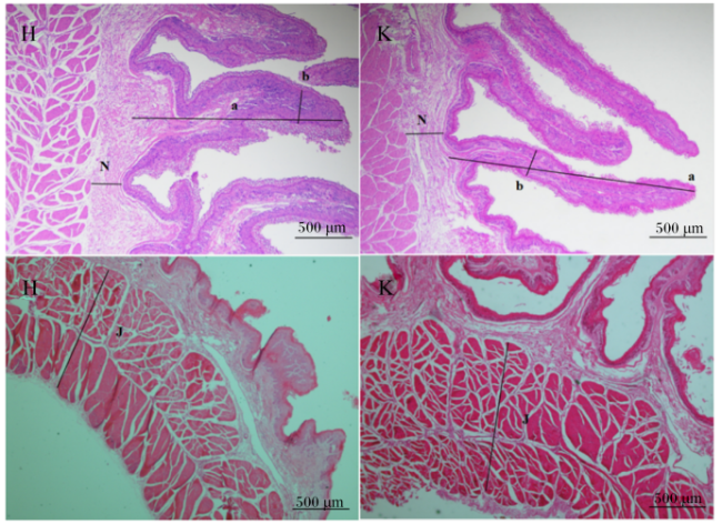
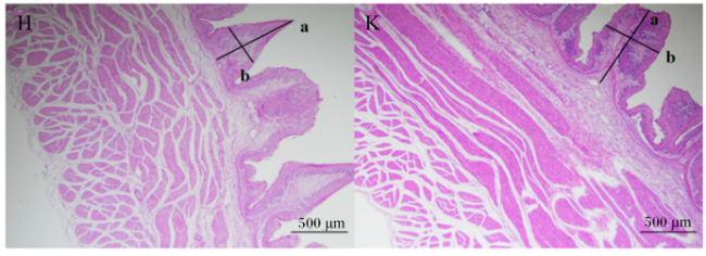
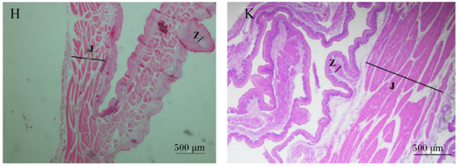
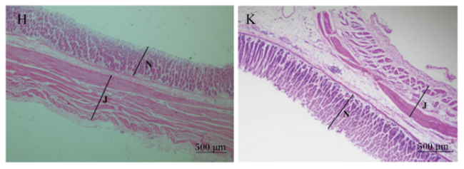
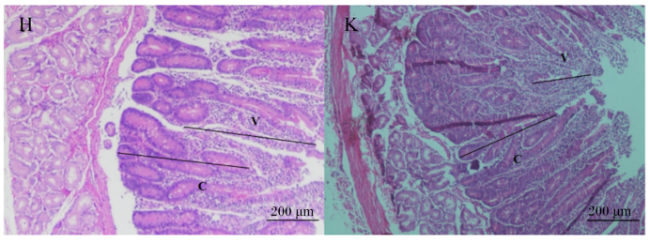
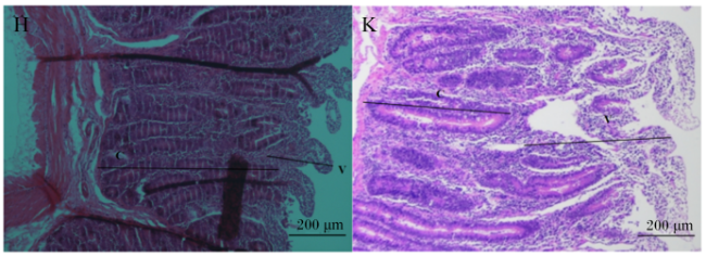
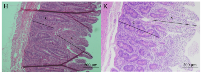
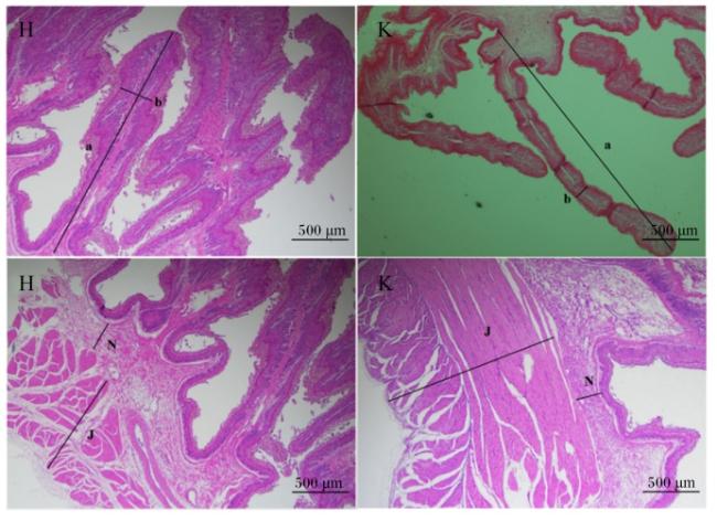
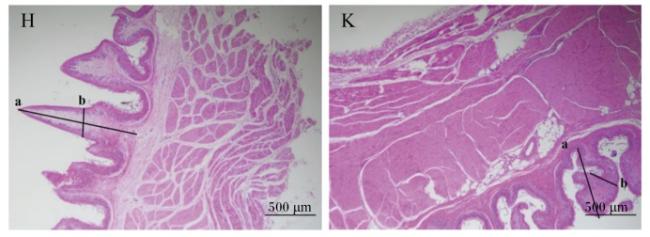
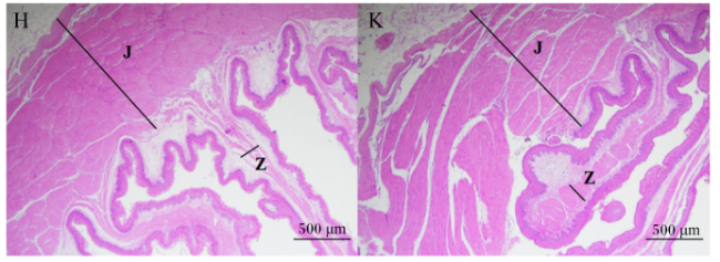
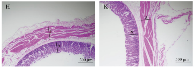
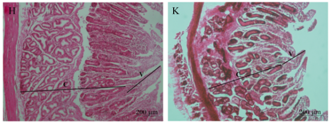
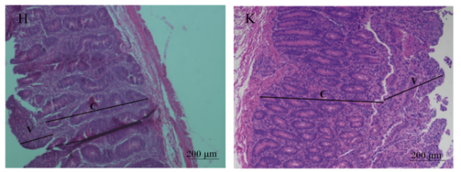
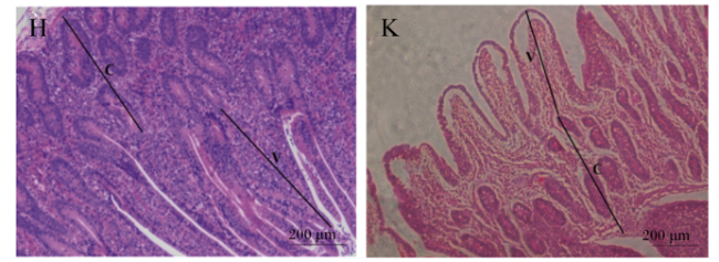
光学显微镜下90和150日龄湖羊和卡拉库尔羊胃肠道组织形态见图1图14
图1 90日龄湖羊和卡拉库尔羊瘤胃组织形态比较

H表示湖羊,K表示卡拉库尔羊;a表示乳头高度,b表示乳头宽度,J表示肌层厚度,Z表示中央肌层厚度,N表示黏膜层厚度,V表示绒毛高度,C表示隐窝深度。下图同。

Fig.1 Comparison of rumen tissue morphology between Hu sheep and Karakul sheep at 90 days of age (40×)

H represents Hu sheep, K represents Karakul sheep; a indicates papilla height, b indicates papilla width, J indicates muscle layer thickness, Z indicates central muscle layer thickness, N indicates mucosa layer thickness, V indicates villus height, and C indicates crypt depth. The same as below.

图2 90日龄湖羊和卡拉库尔羊网胃组织形态比较

Fig.2 Comparison of reticulum tissue morphology between Hu sheep and Karakul sheep at 90 days of age (40×)

图3 90日龄湖羊和卡拉库尔羊瓣胃组织形态比较

Fig.3 Comparison of omasum tissue morphology between Hu sheep and Karakul sheep at 90 days of age (40×)

图4 90日龄湖羊和卡拉库尔羊皱胃组织形态比较

Fig.4 Comparison of abomasum tissue morphology between Hu sheep and Karakul sheep at 90 days of age (40×)

图5 90日龄湖羊和卡拉库尔羊十二指肠组织形态比较

Fig.5 Comparison of duodenum tissue morphology between Hu sheep and Karakul sheep at 90 days of age (100×)

图6 90日龄湖羊和卡拉库尔羊空肠组织形态比较

Fig.6 Comparison of jejunum tissue morphology between Hu sheep and Karakul sheep at 90 days of age (100×)

图7 90日龄湖羊和卡拉库尔羊回肠组织形态比较

Fig.7 Comparison of ileum tissue morphology between Hu sheep and Karakul sheep at 90 days of age (100×)

图8 150日龄湖羊和卡拉库尔羊瘤胃组织形态比较

Fig.8 Comparison of rumen tissue morphology between Hu sheep and Karakul sheep at 150 days of age (40×)

图9 150日龄湖羊和卡拉库尔羊网胃组织形态比较

Fig.9 Comparison of reticulum tissue morphology between Hu sheep and Karakul sheep at 150 days of age (40×)

图10 150日龄湖羊和卡拉库尔羊瓣胃组织形态比较

Fig.10 Comparison of omasum tissue morphology between Hu sheep and Karakul sheep at 150 days of age (40×)

图11 150日龄湖羊和卡拉库尔羊皱胃组织形态比较

Fig.11 Comparison of abomasum tissue morphology between Hu sheep and Karakul sheep at 150 days of age (40×)

图12 150日龄湖羊和卡拉库尔羊十二指肠组织形态比较

Fig.12 Comparison of duodenum tissue morphology between Hu sheep and Karakul sheep at 150 days of age (100×)

图13 150日龄湖羊和卡拉库尔羊空肠组织形态比较(100×)

Fig.13 Comparison of jejunum tissue morphology between Hu sheep and Karakul sheep at 150 days of age (100×)

图14 150日龄湖羊和卡拉库尔羊回肠组织形态比较(100×)

Fig.14 Comparison of ileum tissue morphology between Hu sheep and Karakul sheep at 150 days of age (100×)

表2可知,在90日龄,湖羊的瘤胃乳头高度、皱胃黏膜层厚度、十二指肠绒毛高度、空肠绒毛高度、空肠隐窝深度、回肠绒毛高度和回肠绒隐比极显著低于卡拉库尔羊(P<0.01),湖羊的十二指肠隐窝深度显著低于卡拉库尔羊(P<0.05),其他指标2个品种羊之间均差异不显著(P>0.05)。在150日龄,湖羊瓣胃中央肌层厚度极显著低于卡拉库尔羊(P<0.01),湖羊皱胃黏膜层厚度显著低于卡拉库尔羊(P<0.05),其他指标2个品种羊之间均差异不显著(P>0.05)。
表2 90和150日湖羊和卡拉库尔羊胃肠道组织形态比较

Table 2 Comparison of gastrointestinal tissue morphology between Hu sheep and Karakul sheep at 90 and 150 days of age

项目
Items
90日龄90 days of age 150日龄150 days of age PP-value
湖羊
Hu sheep
卡拉库尔羊
Karakul sheep
湖羊
Hu sheep
卡拉库尔羊
Karakul sheep
品种
Breed
日龄
Day of
age
品种×日龄
Breed×
day of age
瘤胃乳头高度
Rumen papilla height/μm
1 400.62
±252.23B
2 363.93
±357.06A
2 257.85
±700.63
2 150.43
±89.64
<0.001 0.750 0.011
瘤胃乳头宽度
Rumen papilla width/μm
340.80
±43.40
328.74
±64.73
349.92
±66.42
304.73
±11.95
0.738 0.204 0.483
瘤胃肌层厚度
Rumen muscle layer thickness/μm
607.91
±162.14
1 103.92
±104.05
1 103.92
±214.26
1 183.65
±39.04
0.655 0.086 <0.001
瘤胃黏膜层厚度
Rumen mucosa layer thickness/μm
49.89
±6.64
58.63
±10.84
307.23
±80.79
299.00
±144.54
0.163 0.914 0.822
网胃乳头高度
Reticulum papilla height/μm
472.92
±165.16
392.77
±28.02
440.67
±165.82
480.61
±8.58
0.342 0.619 0.272
网胃乳头宽度
Reticulum papilla width/μm
281.05
±52.27
287.12
±80.57
287.12
±80.57
304.92
±5.56
0.355 0.648 0.340
瓣胃肌层厚度
Omasum muscle layer thickness/μm
304.92
±5.56
967.24
±241.66
967.24
±241.66
1 067.99
±46.61
0.183 0.408 0.708
瓣胃中央肌层厚度
Omasum central muscle layer
thickness/μm
318.47
±56.30
318.60
±34.99
165.15
±29.99B
244.01
±36.34A
0.997 0.101 0.046
皱胃肌层厚度
Abomasum muscle layer
thickness/μm
490.00
±223.27
376.24
±3.99
180.31
±125.46
351.47
±87.70
0.318 0.101 <0.001
皱胃黏膜层厚度
Abomasum mucosa layer
thickness/μm
379.80
±158.23B
890.37
±10.10A
179.23
±109.45b
339.55
±81.17a
<0.001 0.030 <0.001
十二指肠绒毛高度
Duodenum villus height/μm
525.82
±20.76B
582.94
±17.30A
619.00
±199.10
652.28
±63.08
<0.001 0.731 0.803
十二指肠隐窝深度
Duodenum crypt depth/μm
254.09
±28.96b
326.39
±51.09a
288.67
±68.16
297.62
±11.79
0.025 0.786 0.138
十二指肠绒隐比
Duodenum VH/CD
2.09
±0.24
1.83
±0.31
2.13
±0.34
2.20
±0.21
0.177 0.731 0.214
空肠绒毛高度
Jejunum villus height/μm
400.45
±44.77B
616.86
±4.13A
704.97
±48.53
722.57
±8.29
<0.001 0.466 <0.001
空肠隐窝深度
Jejunum crypt depth/μm
249.32
±30.17B
383.36
±9.43A
308.57
±20.77
331.02
±15.75
<0.001 0.090 <0.001
空肠绒隐比
Jejunum VH/CD
1.62
±0.17
1.62
±0.04
2.30
±0.02
2.19
±0.09
0.994 0.061 0.238
回肠绒毛高度
Ileum villus height/μm
417.64
±40.21B
558.12
±41.13A
658.21
±56.12
719.61
±62.63
<0.001 0.141 0.102
回肠隐窝深度
Ileum crypt depth/μm
235.19
±23.08
258.94
±8.02
314.78
±41.46
337.05
±36.10
0.062 0.392 0.956
回肠绒隐比
Ileum VH/CD
1.78
±0.12B
2.16
±0.11A
2.12
±0.31
2.14
±0.07
<0.001 0.888 0.044

同一日龄下,同行数据肩标不同小写字母表示差异显著(P<0.05),不同大写字母表示差异极显著(P<0.01),无字母表示差异不显著(P>0.05)。

Under the same age and in the same column, data shoulder with different lowercase letters mean significant difference (P<0.05), and with different uppercase letters mean extremely significant difference (P<0.01), while with no letter mean no significant difference (P>0.05).

表2可知,随着日龄增加,皱胃黏膜层厚度显著降低(P<0.05),其他指标在不同日龄间无显著差异(P>0.05)。品种与日龄的交互作用对瘤胃乳头高度(P<0.05)、瘤胃肌层厚度(P<0.01)、瓣胃中央肌层厚度(P<0.05)、皱胃肌层厚度(P<0.01)、皱胃黏膜层厚度(P<0.01)、空肠绒毛高度(P<0.01)、空肠隐窝深度(P<0.01)及回肠绒隐比(P<0.05)有显著或极显著影响,说明品种和日龄的交互作用对这些组织结构发育影响较大。

2.2 扫描电镜下90和150日龄湖羊和卡拉库尔羊瘤胃组织形态比较

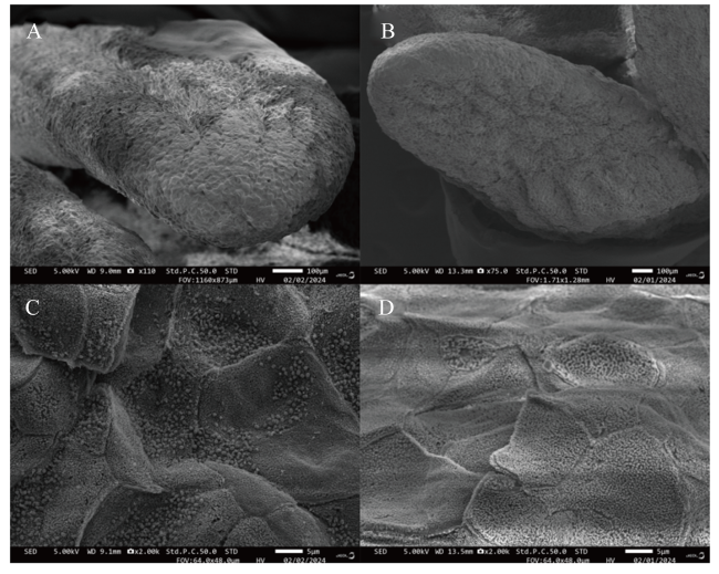
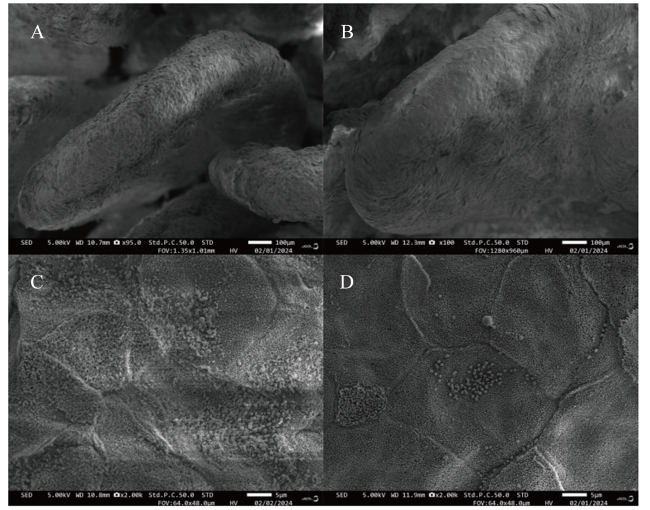
扫描电镜结果显示,90和150日龄湖羊和卡拉库尔羊的瘤胃乳头发育良好,呈现出舌形等特征,瘤胃乳头顶端并未出现脓肿现象,且角质层平整、呈规律的鳞片状(图15-A图15-B图16-A图16-B);同时,表面的细胞未出现角质化不全的现象(图15-C图15-D图16-C图16-D)。
图15 90日龄湖羊和卡拉库尔羊瘤胃乳头扫描电镜图比较

A、C:湖羊瘤胃组织形态;B、D:卡拉库尔羊瘤胃组织形态。下图同。

Fig.15 Comparison of scanning electron microscopy image of rumen papilla between Hu sheep and Karakul sheep at 90 days of age

A, C: rumen tissue morphology of Hu sheep; B, D: rumen tissue morphology of Karakul sheep. The same as below.

图16 150日龄湖羊和卡拉库尔羊瘤胃乳头扫描电镜图比较

Fig.16 Comparison of scanning electron microscopy image of rumen papilla between Hu sheep and Karakul sheep at 150 days of age

3 讨论

3.1 90和150日龄湖羊和卡拉库尔羊胃组织形态比较

反刍动物的瘤胃主要是通过微生物发酵和蠕动来消化吸收大量植物性饲料中的碳水化合物,其发育直接影响反刍动物的消化能力[2,22]。瘤胃乳头是存在于反刍动物瘤胃壁上的突起结构,主要分布在瘤胃内壁的前庭区域,其通过增加食糜与瘤胃的接触面积提升瘤胃对营养物质的吸收能力,因此瘤胃乳头的高度是评定瘤胃组织形态学发育最重要的指标之一[23]。本研究结果显示,90日龄湖羊的瘤胃乳头高度极显著低于卡拉库尔羊,这为解释湖羊较卡拉库尔羊对粗饲料的适应性差提供了可能的理由[24]。李峰鹏[25]开展了类似的研究,在相同条件饲养下,将6月龄西门塔尔杂交牛、宣汉黄牛和犏牛饲喂至30月龄,结果显示西门塔尔杂交牛瘤胃乳头高度显著高于宣汉黄牛和犏牛,宣汉黄牛和犏牛乳头高度无显著差异,而宣汉黄牛乳头宽度显著大于犏牛,说明在相同饲养条件下,不同品种牛存在瘤胃乳头发育的差异。考虑到瘤胃乳头处于生长发育过程中,而本试验中的2个品种绵羊限定在相同饲养管理模式下,因此认定品种为唯一影响因素,饲喂到150日龄时,2个品种绵羊的瘤胃乳头高度则差异不显著,这是否与动物的补偿生长机制[26-27]有关,有待进一步验证。
瓣胃的胃壁有许多叶状折层,这些折层有助于吸收饲料中的水分,同时进一步分解食糜。有研究发现,摄入粗饲料后物理刺激前胃消化,能够加剧瓣胃上皮的角质化以及黏膜、肌层的发育[28]。本研究发现,90日龄时,湖羊瓣胃中央肌层厚度与卡拉库尔羊没有显著差异,但150日龄时湖羊瓣胃中央肌层厚度极显著低于卡拉库尔羊,推测可能是卡拉库尔羊对粗饲料的适应性更强。在相同舍饲条件下,成年无角陶赛特羊瓣胃肌层厚度显著高于成年特克赛尔羊和萨福克羊[29],全年舍饲且饲养管理一致的成年德国美利奴羊瓣胃中央肌层的厚度显著高于蒙古羊、无角陶赛特羊、特克塞尔羊和特克塞尔羊[29]。上述结果表明,相同饲养条件下,不同品种绵羊瓣胃结构存在差异。但目前关于瓣胃肌层作用机制研究较少,缺乏相关文献报道,是否因此进一步影响了营养物质的消化吸收有待进一步研究。
皱胃又称真胃,是反刍动物唯一具有分泌功能的胃,皱胃分泌的胃酸和消化酶能够帮助动物消化蛋白质和其他营养成分[30]。皱胃在运动机制上与单胃动物的胃具有一定的相似性,但也有其独特的生理特征,皱胃内富含腺体细胞,其黏膜层结构包含胶原纤维、脂肪组织及黏膜下神经丛等组织成分,可以更好地消化如蛋白质等营养物质[31]。有报道显示,在相同舍饲条件下,18月龄无角陶赛特羊的皱胃肌层最为发达,其次是蒙古羊,而德国美利奴羊的肌层最薄,各品种间存在显著差异;萨福克羊皱胃黏膜肌层最厚,而蒙古羊最薄[32]。本研究发现,90日龄时湖羊皱胃黏膜层厚度极显著低于卡拉库尔羊,150日龄时湖羊皱胃黏膜层厚度和肌层厚度均显著低于卡拉库尔羊,说明不同品种绵羊的皱胃组织形态有差异,这可能是由于精料在经过前胃的消化后,到达皱胃时可消化物质减少,导致湖羊的皱胃黏膜层和肌层较薄。
反刍动物的胃黏膜表面特征性突起,如瘤胃乳头和网胃瓣叶等,能够显著增加饲料与胃上皮的接触面积,以增大吸收营养物质的表面积,有助于胃上皮发育和增强对营养物质的吸收效率,所以乳头高度和宽度是反刍动物胃组织形态学的重要参考指标[33]。据报道,饲喂绵羊较高水平精料的饲粮可以促进瘤胃乳头高度的增加和乳头宽度的降低[34]。另外,乔灵等[35]研究发现,瘤胃乳头的发育还与饲料的类型有关,瘤胃乳头高度变化为干草羊>青草羊>放牧羊,瘤胃乳头宽度变化为青草羊>放牧羊>干草羊。本研究发现,在150日龄时卡拉库尔羊的瘤胃乳头高度和宽度与湖羊的网胃乳头高度和宽度均低于90日龄,推测其原因可能与150日龄时饲粮中粗饲料比例显著提高有关。本研究发现,150日龄时卡拉库尔羊和湖羊的皱胃肌层厚度和黏膜层厚度均低于90日龄。王斯琴塔娜[36]发现,饲喂优质青干草和玉米秸秆对犊牛消化道组织的形态会产生不同影响,秸秆组犊牛皱胃肌层厚度较青干草组低。我们推测,在90日龄时,饲粮过渡已经完成,但饲粮中的纤维水平尚未达到预期标准;而至150日龄时,饲粮中的纤维水平已进入稳定状态,这可能是导致绵羊皱胃收缩频率下降的原因。

3.2 90和150日龄湖羊和卡拉库尔羊肠道组织形态比较

小肠在动物体内扮演着至关重要的角色,作为营养物质消化与吸收的核心场所,其功能性表现与生理架构紧密相连,因此,肠黏膜的组织结构是动物健康状况的关键指示器[37-38]。小肠绒毛显著提升了小肠的表面积,从而增强了小肠对营养物质的吸收能力,绒毛高度与其营养物质吸收能力呈正相关,绒毛高度增加,使营养物质的吸收效率提升,此外,较长的绒毛结构构成了一道物理屏障,有利于抵御有害病原体的侵袭,绒隐比越大,表明肠黏膜的吸收和消化功能更为强,因此小肠绒毛高度和隐窝深度是评估其消化与吸收能力的关键参数[39-41]。有研究发现,18月龄西门塔尔杂交牛十二指肠绒毛高度显著高于宣汉黄牛和犏牛,而宣汉黄牛空肠和回肠绒毛高度显著高于西门塔尔杂交牛,犏牛十二指肠、空肠和回肠的绒隐比均小于宣汉黄牛[42]。孙青松等[43]研究表明,成年西门塔尔牛空肠绒毛高度较草原红牛和利木赞牛更高,绒隐比也大于草原红牛和利木赞牛。本研究中,90日龄湖羊十二指肠和空肠绒毛高度、隐窝深度以及回肠绒毛高度和绒隐比均显著低于卡拉库尔羊,但在150日龄时湖羊和卡拉库尔羊的十二指肠、空肠和回肠的绒毛高度、隐窝深度及绒隐比均无显著差异,推测卡拉库尔羊在90日龄前具有更强的肠道发育优势,但在后期湖羊隐窝深度和绒隐比的代偿性升高可能暗示其独特的适应策略。
小肠绒毛内富含的毛细血管网络及淋巴组织为营养物质的吸收及免疫防御提供了结构基础,绒毛的高度与其消化吸收的表面积成正比,因此,绒毛高度增加,相应的消化吸收能力亦随之增强[44],隐窝深度的降低可作为肠上皮细胞更新速率的一个指标,随着隐窝深度的变浅,肠道上皮细胞的分泌功能得到提升,进而强化了化学消化能力[45]。本研究表明,在150日龄时卡拉库尔羊的十二指肠和空肠隐窝深度均低于90日龄,而在150日龄时湖羊十二指肠及空肠隐窝深度高于90日龄,说明卡拉库尔羊发育状态较好。

4 结论

综上所述,在相同饲养条件下,90日龄的湖羊与卡拉库尔羊在瘤胃、皱胃、十二指肠、空肠和回肠的生理结构上存在差异,而到150日龄时两者在皱胃的生理结构上仍存在差异。品种与日龄的交互作用对绵羊的瘤胃乳头高度、瘤胃肌层厚度、瓣胃中央肌层厚度、皱胃肌层厚度、皱胃黏膜层厚度、空肠绒毛高度、空肠隐窝深度及回肠绒隐比有显著或极显著影响。
[1]
PATEL B, GIZZI A, HASHEMI J, et al. Biomechanical constitutive modeling of the gastrointestinal tissues:a systematic review[J]. Materials & Design, 2022,217:110576.

[2]
何婷莉, 吴振岭, 张峰硕, 等. 不同蛋白质水平日粮对藏羊屠宰性能、脏器发育和体尺指标的影响[J]. 饲料工业, 2024, 45(8):109-114.

HE T L, WU Z L, ZHANG F S, et al. Effects of diets with different protein levels on slaughter performance,organ development and body size indexes in tibetan sheep[J]. Feed Industry, 2024, 45(8):109-114.(in Chinese)

[3]
COLLINS J T, NGUYEN A, BADIREDDY M. Anatomy,abdomen and pelvis,small intestine[M/OL]// Anon.StatPearls.Treasure Island: StatPearls Publishing, 2024:NBK459366[2024-10-01].https://pubmed.ncbi.nlm.nih.gov/29083773/

[4]
HUNDT M, WU C Y, YOUNG M. Anatomy,abdomen and pelvis:biliary ducts[M/OL]// Anon.StatPearls.Treasure Island: StatPearls Publishing, 2023:NBK459246[2024-10-01].https://pubmed.ncbi.nlm.nih.gov/29083810/.

[5]
韩亚儒. 中国绵羊地方品种的种质特性及其生态分布规律的研究[D]. 硕士学位论文. 泰安: 山东农业大学, 2016.

HAN Y R. The research on chinese indigenous sheep breeds germplasm characteristics and their ecological distributing law[D]. Master’s Thesis. Tai’an: Shandong Agricultural University, 2016.(in Chinese)

[6]
李莲瑞, 陈根元, 王晓斌, 等. 新疆卡拉库尔羊的研究现状及前景[J]. 中国草食动物, 2008, 28(2):63-64.

LI L R, CHEN G Y, WANG X B, et al. Research status and prospect of Xinjiang Karakul sheep[J]. China Herbivore Science, 2008, 28(2):63-64.(in Chinese)

[7]
SUN M X, CHEN M Y, LI S Y, et al. Study on structure and properties of Hu sheep wool[J]. Journal of Natural Fibers, 2023, 20(1):2160405.

[8]
LI R, YANG P, LI M, et al. A Hu sheep genome with the first ovine Y chromosome reveal introgression history after sheep domestication[J]. Science China Life Sciences, 2021, 64(7):1116-1130.

[9]
中华人民共和国农业农村部. 肉羊营养需要量标准:NY/T 816—2021[S]. 北京: 中国农业出版社, 2021.

Ministry of Agriculture and Rural Affairs of the People’s Republic of China. Nutrient requirements of meat-type sheep and goats:NY/T 816—2021[S]. Beijing: China Agriculture Press, 2021.(in Chinese)

[10]
中华人民共和国国家质量监督检验检疫总局, 中国国家标准化管理委员会. 饲料中水分的测定:GB/T 6435—2014[S]. 北京: 中国标准出版社, 2015.

General Administration of Quality Supervision,Inspection and Quarantine of the People’s Republic of China, Standardization Administration of the People’s Republic of China. Determination of moisture in feedstuffs:GB/T 6435—2014[S]. Beijing: Standards Press of China, 2015.(in Chinese)

[11]
中华人民共和国国家质量监督检验检疫总局, 中国国家标准化管理委员会. 饲料中粗灰分的测定:GB/T 6438—2007[S]. 北京: 中国标准出版社, 2007.

General Administration of Quality Supervision, Inspection and Quarantine of the People’s Republic of China, Standardization Administration of the People’s Republic of China. Animal feeding stuffs—determination of crude ash:GB/T 6438—2007[S]. Beijing: Standards Press of China, 2007.(in Chinese)

[12]
国家市场监督管理总局, 国家标准化管理委员会. 饲料中粗蛋白的测定凯氏定氮法:GB/T 6432—2018[S]. 北京: 中国标准出版社, 2018.

State Administration for Market Regulation, Standardization Administration of the People’s Republic of China. Determination of crude protein in feeds—Kjeldahl method:GB/T 6432—2018[S]. Beijing: Standards Press of China, 2018.(in Chinese)

[13]
全国饲料工业标准化技术委员会. 饲料中粗脂肪的测定:GB/T 6433—2006[S]. 北京: 中国标准出版社, 2006.

National Feed Industry Standardization Technical Committee. Determination of crude fat in feeds:GB/T 6433—2006[S]. Beijing: Standards Press of China, 2006.(in Chinese)

[14]
中华人民共和国农业农村部. 饲料中酸性洗涤纤维的测定:NY/T 1459—2022[S]. 北京: 中国农业出版社, 2022.

Ministry of Agriculture and Rural Affairs of the People’s Republic of China. Determination of acidic detergent fiber in feed:NY/T 1459—2022[S]. Beijing: China Agriculture Press, 2022.(in Chinese)

[15]
中华人民共和国国家质量监督检验检疫总局, 中国国家标准化管理委员会. 饲料中中性洗涤纤维(NDF)的测定:GB/T 20806—2022[S]. 北京: 中国标准出版社, 2022.

General Administration of Quality Supervision,Inspection and Quarantine of the People’s Republic of China, Standardization Administration of the People’s Republic of China. Determination of neutral detergent fiber (NDF) in feeds:GB/T 20806—2022[S]. Beijing: Standards Press of China, 2022.(in Chinese)

[16]
中华人民共和国国家质量监督检验检疫总局, 中国国家标准化管理委员会. 实验动物福利伦理审查指南:GB/T 35892—2018[S]. 北京: 中国标准出版社, 2018.

General Administration of Quality Supervision,Inspection and Quarantine of the People’s Republic of China, Standardization Administration of the People’s Republic of China. Laboratory animal—guideline for ethical review of animal welfare:GB/T 35892—2018[S]. Beijing: Standards Press of China, 2018.(in Chinese)

[17]
窦新雨, 刘宇, 刘啸, 等. 小尾寒羊颈椎前路椎间盘切除融合模型的建立及评估[J]. 中国实验动物学报, 2024, 32(2):139-150.

DOU X Y, LIU Y, LIU X, et al. Establishment and evaluation of anterior cervical discectomy fusion model in small-tailed Han sheep model[J]. Acta Laboratorium Animalis Scientia Sinica, 2024, 32(2):139-150.(in Chinese)

[18]
娜梅拉, 李科南, 娜仁花. 不同日龄下内蒙古绒山羊胃肠道组织形态学发育的研究[J]. 饲料研究, 2024, 47(6):6-11.

NA M L, LI K N, NA R H. Study on histomorphological development of gastrointestinal tract of cashmere goats in Inner Mongolia at different ages[J]. Feed Research, 2024, 47(6):6-11.(in Chinese)

[19]
GRAHAM C, SIMMONS N L. Functional organization of the bovine rumen epithelium[J]. American Journal of Physiology:Regulatory, Integrative and Comparative Physiology, 2005, 288(1):R173-R181.

[20]
NAMEI E, SUN W, PAN D, et al. The advanced paraffin-section preparation technique based on multiple cumulus-oocyte complexes rather than ovaries in ovine[J]. Reproductive Biology, 2021, 21(1):100473.

[21]
WILSON F D, CUMMINGS T S, BARBOSA T M, et al. Comparison of two methods for determination of intestinal villus to crypt ratios and documentation of early age-associated ratio changes in broiler chickens,[J]. Poultry Science, 2018, 97(5):1757-1761.

DOI PMID

[22]
MCSWEENEY C, MACKIE R. Improving rumen function[M]. Cambridge: Burleigh Dodds Science Publishing, 2020.

[23]
刘婷, 李发弟, 李冲, 等. 断奶时间对不同日龄湖羊羔羊瘤胃形态及表皮生长相关基因表达的影响[J]. 动物营养学报, 2016, 28(5):1384-1393.

DOI

LIU T, LI F D, LI C, et al. Effects of weaner time on rumen morphology and gene expressions involved in rumen epidermis growth of Hu lambs at different days of age[J]. Chinese Journal of Animal Nutrition, 2016, 28(5):1384-1393.

[24]
时乾. 湖羊部分繁殖性状及其羔羊生长发育研究[D]. 硕士学位论文. 南京: 南京农业大学, 2007.

SHI Q. A dissertation submitted to Nanjing agricultural university in partial fulfillment of the requirements for the master degree[D]. Master’s Thesis. Nanjing: Nanjing Agricultural University, 2007.(in Chinese)

[25]
李峰鹏. 不同品种(系)肉用牛胃肠道微生物组成及瘤胃上皮转录组差异性比较[D]. 硕士学位论文. 雅安: 四川农业大学, 2019.

LI F P. Comparison of microbial compositions in gastrointestinal tract and rumen epithelial transcriptome among different breeds of beef cattle[D]. Master’s Thesis. Ya’an: Sichuan Agricultural University, 2019.(in Chinese)

[26]
TURGEON O A J, BRINK D R, BARTLE S J, et al. Effects of growth rate and compensatory growth on body composition in lambs[J]. Journal of Animal Science, 1986, 63(3):770-780.

PMID

[27]
MISZURA A A, FERRAZ M V C, CARDOSO R C, et al. Implications of growth rates and compensatory growth on puberty attainment in Nellore heifers[J]. Domestic Animal Endocrinology, 2021,74:106526.

[28]
ARTHINGTON J D, KALMBACHER R S. Effect of early weaning on the performance of three-year-old,first-calf beef heifers and calves reared in the subtropics[J]. Journal of Animal Science, 2003, 81(5):1136-1141.

[29]
阚向东. 不同品种肉用绵羊前胃黏膜组织学结构分析[J]. 中国畜禽种业, 2016, 12(11):76-77.

KAN X D. Analysis of histological structure of pregastric mucosa in different breeds of meat sheep[J]. The Chinese Livestock and Poultry Breeding, 2016, 12(11):76-77.(in Chinese)

[30]
姜丹, 吴树清, 杜山, 等. 不同品种肉用绵羊舍饲条件下前胃组织学变化[J]. 中国草食动物, 2010, 30(3):23-26.

JIANG D, WU S Q, DU S, et al. Histological changes of proventriculus in different meat-producing sheep under confinedness[J]. China Herbivore Science, 2010, 30(3):23-26.(in Chinese)

[31]
陈杰. 家畜生理学[M]. 4版. 北京: 中国农业出版社, 2003.

CHEN J. Physiology of domestic animals[M]. 4th ed. Beijing: China Agriculture Press, 2003.(in Chinese)

[32]
寇宇斐, 杨旭, 许辉, 等. 饲粮中全株饲料桑比例对育肥湖羊屠宰性能、肉品质、瘤胃组织形态和瘤胃微生物组成的影响[J]. 动物营养学报, 2023, 35(2):1066-1077.

DOI

KOU Y F, YANG X, XU H, et al. Effects of dietary proportions of whole-plant mulberry on slaughter performance,meat quality,rumen tissue morphology and rumen microbial composition of fattening Hu sheep[J]. Chinese Journal of Animal Nutrition, 2023, 35(2):1066-1077.(in Chinese)

[33]
赵玮, 吴树清, 杜山, 等. 不同品种肉用绵羊在舍饲条件下皱胃组织学的变化[J]. 畜牧与饲料科学, 2010, 31(2):17-19.

ZHAO W, WU S Q, DU S, et al. Histology change of meat sheep abomasum in different varieties of breeds under feeding conditions[J]. Animal Husbandry and Feed Science, 2010, 31(2):17-19.(in Chinese)

[34]
刘学良, 罗海玲, 陈勇, 等. 限时放牧对滩羊消化道发育的影响[J]. 中国畜牧兽医, 2013, 40(11):97-101.

LIU X L, LUO H L, CHEN Y, et al. Effects of time-limited grazing on the digestive tract development of Tan sheep[J]. China Animal Husbandry & Veterinary Medicine, 2013, 40(11):97-101.(in Chinese)

[35]
乔灵, 吴美玲, 包花尔, 等. 不同饲养条件下阿尔巴斯绒山羊前胃的形态学变化[J]. 西北农林科技大学学报(自然科学版), 2005, 33(9):39-44.

QIAO L, WU M L, BAO H E, et al. The morphological changes of Aerbasi cashmere goats’ proventriculus under different raising conditions[J]. Journal of Northwest A & F University (Natural Science Edition), 2005, 33(9):39-44.(in Chinese)

[36]
王斯琴塔娜. 探讨粗饲料品质对犊牛消化道组织形态及内脏器官发育的影响[D]. 硕士学位论文. 呼和浩特: 内蒙古农业大学, 2007.

WANG S Q T N. The effeet of forage quality on morphology of gastrointestinal tract and visceral tissues growth in Holstein calves[D]. Master’s Thesis. Hohhot: Inner Mongolia Agricultural University, 2007. (in Chinese).

[37]
侯明杰. 青贮型饲粮育肥肉羊的胃肠道微生态及健康性能研究[D]. 硕士学位论文. 兰州: 兰州大学, 2018.

HOU M J. Research on the gastrointestinal microecology and health performance of sheep fed silage dietary[D]. Master’s Thesis. Lanzhou: Lanzhou University, 2018.(in Chinese)

[38]
廖珂. 不同处理豆粕及嗜酸乳杆菌对断奶仔猪生长性能、肠黏膜屏障功能的影响[D]. 硕士学位论文. 南昌: 江西农业大学, 2016.

LIAO K. Effect of soybean meal and Lactobacillus acidophilus on growth performance,barrier function[D]. Master’s Thesis. Nanchang: Jiangxi Agricultural University, 2016.(in Chinese)

[39]
陈鼎. 小尾寒羊和滩羊肠道发育及其主要消化酶活性变化规律研究[D]. 硕士学位论文. 杨凌: 西北农林科技大学, 2008.

CHEN D. Regulation of intestine and its digestive enzymes activities between small-tailed Han sheep and Tan sheep[D]. Master’s Thesis. Yangling: Northwest A & F University, 2008.(in Chinese)

[40]
郭江鹏, 郝正里, 李发弟, 等. 早期断奶对舍饲肉用羔羊消化器官发育的影响[J]. 畜牧兽医学报, 2013, 44(7):1078-1089.

GUO J P, HAO Z L, LI F D, et al. Effect of early weaning on development of digestive organs of barn feeding lambs[J]. Acta Veterinaria et Zootechnica Sinica, 2013, 44(7):1078-1089.(in Chinese)

DOI

[41]
CASPARY W F. Physiology and pathophysiology of intestinal absorption[J]. American Journal of Clinical Nutrition, 1992, 55(1 Suppl):299S-308S.

[42]
孙青松. 不同品种肉牛小肠组织学差异性研究[D]. 硕士学位论文. 长春: 吉林农业大学, 2011.

SUN Q S. Study on differences of small intestine morphology in different varieties of beef cattle[D]. Master’s Thesis. Changchun: Jilin Agricultural University, 2011.(in Chinese)

[43]
孙青松, 张国梁, 王利民, 等. 4个品种肉牛小肠绒毛结构的差异性研究[J]. 黑龙江畜牧兽医, 2011(9):6-8.

SUN Q S, ZHANG G L, WANG L M, et al. Differences in villi structure of small intestine among four varieties of beef cattle[J]. Heilongjiang Animal Science and Veterinary Medicine, 2011(9):6-8.(in Chinese)

[44]
周金星, 高登慧, 刘培琼, 等. 不同日龄香猪小肠黏膜形态观察[J]. 中国兽医杂志, 2005, 41(12):11-12.

ZHOU J X, GAO D H, LIU P Q, et al. Observation on the morphology of Xiang pig intestinal mucosa with the different age[J]. Chinese Journal of Veterinary Medicine, 2005, 41(12):11-12.(in Chinese)

[45]
姚浪群, 萨仁娜, 佟建明, 等. 安普霉素对仔猪肠道微生物及肠壁组织结构的影响[J]. 畜牧兽医学报, 2003, 34(3):250-257.

YAO L Q, SA R N, TONG J M, et al. Effect of apramycin on intestinal flora and intestinal morphology of piglets[J]. Acta Veterinaria et Zootechnica Sinica, 2003, 34(3):250-257.(in Chinese)

Outlines

/