RESEARCH PAPER

Study on Primary Culture and Identification Methods of Intestinal Epithelial Cells from Orange-Spotted Grouper (Epinephelus coioides)

  • XU Jianming , 1, 2 ,
  • YANG Hongling 1, 2, 3 ,
  • CAI Guohe 1, 2 ,
  • SUN Yunzhang , 1, 2, 3, *
Expand
  • 1 State Key Laboratory of Mariculture Breeding, Fisheries College of Jimei University, Xiamen 361021, China
  • 2 Xiamen Key Laboratory of Feed Testing and Safety Evaluation, Fisheries College of Jimei University, Xiamen 361021, China
  • 3 Key Laboratory of Healthy Mariculture for the East China Sea, Ministry of Agriculture and Rural Affairs, Fisheries College of Jimei University, Xiamen 361021, China
* professor, E-mail:

Received date: 2024-12-18

  Online published: 2025-09-12

Abstract

This study aimed to explore methods for the primary culture and identification of intestinal epithelial cells from orange-spotted grouper (Epinephelus coioides). Using intestinal tissue from E. coioides as experimental material, cells were cultured via tissue explant method and enzymatic digestion method, respectively, to compare the efficacy of these two culture methods. Different enzymatic treatments—trypsin, collagenase Ⅰ, and a combination of collagenase Ⅰ+collagenase Ⅳ, were applied to digest intestinal tissue and to compare their effectiveness in cell isolation. Three culture medias (DMEM, DMEM/F12, and L15 culture media) and varying concentrations (5%, 12% and 20%) of fetal bovine serum (FBS) were evaluated to compare their effects on cell growth. The cultured primary cells were characterized using morphological observation, transmission electron microscopy (TEM) observation, alkaline phosphatase (AKP) staining, 18S rRNA gene sequence alignment, and detection of intestinal cell-specific marker gene expression. Results showed that both enzymatic digestion method and tissue explant method yielded adherent cells. However, cell confluence on the flask bottom was observed at 12 days using the enzyme digestion method, whereas the tissue explant method took 28 days. Optimal digestion was achieved using 0.25 mg/mL trypsin supplemented with 0.91 mmol/L ethylenediaminetetraacetic acid (EDTA) for 15 min; this condition yielded sufficient cells while maintaining cell integrity and viability. Cells exhibited higher proliferation rate in L15 culture medium supplemented with 20% FBS. Characterization by TEM observation, AKP staining, and detection of intestinal cell-specific marker gene expression confirmed the cultured cells as intestinal epithelial cells. 18S rRNA gene sequence alignment verified the its grouper origin. Therefore, digesting the intestinal tissue of orange-spotted grouper with 0.25 mg/mL trypsin (containing 0.91 mmol/L EDTA) for 15 min, followed by culture of the isolated cells in L15 culture medium supplemented with 20% FBS, using above method can effectively obtain stably adherent primary intestinal epithelial cells from orange-spotted grouper.

Cite this article

XU Jianming , YANG Hongling , CAI Guohe , SUN Yunzhang . Study on Primary Culture and Identification Methods of Intestinal Epithelial Cells from Orange-Spotted Grouper (Epinephelus coioides)[J]. Chinese Journal of Animal Nutrition, 2025 , 37(9) : 6291 -6301 . DOI: 10.12418/CJAN2025.510

肠道作为机体核心器官,不仅主导营养物质吸收,还深度参与代谢及免疫保护等生理过程,肠上皮细胞(intestinal epithelial cells,IECs)作为肠道功能实现的关键细胞群体,发挥着核心作用[1]。已有研究表明,通过人工培养获得的肠上皮细胞,其功能特性与体内细胞高度相似,能够较为准确地反映机体的真实生理状态[2]。随着细胞培养技术的不断发展,虹鳟(Oncorhynchus mykiss)[3]、大菱鲆(Paralichthys olivaceus)[4]等硬骨鱼类的肠上皮细胞系相继成功建立。在众多培养方法中,组织块法以其简便易行的特点,在大菱鲆、草鱼(Ctenopharyngodon idella)和虹鳟等多种鱼类的研究中得到广泛应用[5-7]。随着技术的进步,酶消化法在上皮细胞培养领域崭露头角,然而不同鱼类因其生理特性的差异,对消化酶种类的选择也呈现出明显差异。例如,在鲫鱼(Carassius auratus gibelio)和罗非鱼(Oreochromis niloticus)肠上皮细胞的研究中,胰蛋白酶与乙二胺四乙酸(EDTA)的混合使用展现出了良好的消化效果[8-9];而在大菱鲆的研究中,胶原酶和中性蛋白酶的消化效果则更佳[10];对于鮸(Miichthys miiuy)和银鲳(Pampus argenteus),单独使用0.25%的胰蛋白酶便足以达到理想的消化效果[11-12]。此外,为了进一步提高鱼类肠上皮细胞的存活率和增殖能力,研究者们还不断对培养条件进行优化,包括培养基的选择、培养温度的设定以及生长因子的添加等[13-14]。据报道,虹鳟和斑带副鲈(Paralabrax maculatofasciatus)的肠上皮细胞更适应于L15培养基[15-16],而鲤鱼(Cyprinus carpio)的肠上皮细胞则更倾向于在高糖的DMEM培养基中生长[17]。这些差异的存在,可能源于不同鱼种的生理特性、遗传背景以及肠道微环境等多种因素的共同影响。
石斑鱼属鲈形目鮨科,多栖息于温暖的沿海水域,因生长迅速且肉质鲜美等优势而在我国海水养殖业中占据着举足轻重的地位,2023年我国石斑鱼的养殖量高达24.15万t[18]。然而,目前关于石斑鱼肠上皮细胞培养的研究极为匮乏,这在一定程度上制约了石斑鱼养殖过程中肠道健康与营养调控研究的开展。鉴于此,本研究以斜带石斑鱼(Epinephelus coioides)为对象,比较了组织块法与酶消化法在斜带石斑鱼肠上皮细胞培养中的效果,并对酶消化条件进行优化,筛选适宜的培养基与血清添加量,旨在开发一种高效稳定的斜带石斑鱼原代肠上皮细胞培养方法,为石斑鱼肠道营养调控、宿主-微生物互作等研究提供关键体外模型,有利于从细胞分子层面解析营养因子调控肠道健康的机制,为石斑鱼精准配合饲料的开发提供基础。

1 材料与方法

1.1 试验材料

健康斜带石斑鱼,体质量约90 g,暂养于集美大学水产试验场。本研究涉及的试验动物处理按照集美大学实验动物伦理委员会的规定(批准号:2011-58)进行。

1.2 主要试剂

L15培养基(11415-064)、DMEM培养基(11965118)、DMEM-F12培养基(11320033)、澳洲特级胎牛血清(FBS)(10099141C)、0.25 mg/mL胰蛋白酶(含0.91 mmol/L EDTA)(25200072)、胶原酶Ⅰ(17018029)、胶原酶Ⅳ(17104019)由赛默飞世尔科技公司提供,三抗(青霉素-链霉素-两性霉素,100×)(P7630)、磷酸盐缓冲液(PBS,P1020)、CCK8细胞增殖率测定试剂盒(CA1210)由北京索莱宝科技有限公司提供,SYBR Green Pro Taq HS预混型qPCR试剂盒(AG11701)由湖南艾科瑞生物工程有限公司提供。细胞培养液为含20% FBS的L15培养基。

1.3 细胞分离方法

斜带石斑鱼暂养于海水养殖系统中,饥饿处理2 d后用75%乙醇体表消毒,无菌条件下解剖取出肠道;使用含有6×三抗的PBS清洗组织表面后剔除肠道外肠系膜及脂肪,利用1 mL注射器缓慢地冲洗肠腔内容物,确保无残留;将肠道纵向剪开,在含6×三抗的PBS中清洗5~7 min。将得到的肠道浸于PBS中,用手术剪剪碎为3~5 mm3大小的组织块,用PBS充分清洗待用。

1.3.1 组织块法

上述组织块经反复吹打后离心(60×g,3 min),弃上清液,向T25细胞培养瓶中铺入2 g大小均一的组织碎片,确保组织碎片在瓶底分布均匀,密度控制在80%。

1.3.2 酶消化法

为了评估不同消化时长对消化效果的影响,试验采用了以下步骤:利用止血钳固定肠道的后肠部分,随后向肠腔内注满胰蛋白酶消化液,并固定肠道的另一端,在28 ℃条件下,对肠道进行0、5、15和25 min的消化处理,每种消化时长均设置3个重复。最后使用Bouin’s固定液进行24 h的固定处理,对样本进行苏木精-伊红(HE)染色观察。
为筛选最适消化酶,将组织块(4.79 g)与0.25 mg/mL胰蛋白酶(含0.91 mmol/L EDTA)或0.25 mg/mL胶原酶Ⅰ或0.25 mg/mL胶原酶Ⅰ+0.25 mg/mL胶原酶Ⅳ混合液(1∶1)按照组织质量(g)∶酶溶液体积(mL)=1∶4的比例混匀,于28 ℃水浴中消化15 min,每隔5 min振荡组织使消化更充分。倒置显微镜下观察,消化结束时加入细胞培养液终止消化,使用Countess全自动细胞计数仪(Thermo Fisher Scientific,美国)进行活细胞计数。

1.4 细胞培养方法

1.4.1 组织块法

滴管吸取1 mm3组织块于T25培养瓶(用细胞培养液提前润洗瓶内底部)内,组织块间距≥2 mm并调整密度至50%,培养箱中倒置贴壁2 h,随后补充4 mL细胞培养液。每天观察细胞迁出量以及培养液颜色变化并及时换液。

1.4.2 酶消化法

显微镜下观察消化情况,当组织块解离且呈现为大量单细胞与细胞团时,使用移液枪反复吹打几次,利用100 μm细胞滤网过滤。滤液经离心(100×g,5 min)后吸弃上清。将细胞团吹散为单细胞悬液后进行计数。在T25培养瓶中加入5 mL培养液后,加入细胞使其终浓度为1×10⁶ 个/mL,摇匀后置于28 ℃,无CO2且饱和湿度的培养箱中培养。
选择3种培养基(DMEM、DMEM/F12和L15培养基)以及不同FBS浓度(5%、12%、20%)的L15培养基配成细胞培养液进行培养基和FBS浓度筛选试验。将用DMEM与DMEM/F12培养基培养的细胞置于含5% CO2且饱和湿度的培养箱中,而将L15培养基培养的细胞置于无CO2且饱和湿度的培养箱中。用CCK-8法检测培养4、8、12及16 d时细胞增殖情况,绘制细胞在不同培养条件下的生长曲线,以筛选最适培养条件。

1.5 细胞生长曲线

利用0.25 mg/mL胰蛋白酶(含0.91 mmol/L EDTA)于28 ℃消化T25培养瓶中的贴壁细胞3 min后,使用Countess全自动细胞计数仪计算细胞悬液浓度并进行相应稀释,以5×10⁴个/mL的目标浓度接种于96孔培养板中。每隔2 d在固定时间点从96孔板中取3孔进行细胞活性检测,使用CCK-8细胞增殖率测定试剂盒测定其在450 nm波长下的光密度(OD)值,此过程持续至第16天。以培养时间为横轴,OD值为纵轴,绘制生长曲线。

1.6 细胞鉴定

1.6.1 碱性磷酸酶染色

采用碱性磷酸酶染色试剂盒(上海碧云天生物技术股份有限公司),针对肠上皮细胞特征酶碱性磷酸酶进行检测[14]。试验流程如下:细胞爬片制备完毕后,移除培养液并漂洗细胞,继而以4%多聚甲醛固定细胞15~20 min;随后用0.5% Triton X-100处理20 min,以增强细胞膜通透性;细胞经PBS漂洗后再加入足量5-溴-4-氯-3-吲哚磷酸盐/硝基蓝四唑(BCIP/NBT)染色液充分覆盖(阴性对照加入等量PBS);室温避光孵育30 min后,移除染色液,并以PBS清洗终止显色反应。

1.6.2 透射电镜观察

当贴壁细胞融合率达到80%时,移除培养液,随即加入2.5%戊二醛固定1 h;随后,采用细胞刮刀快速收集细胞至离心管,经60×g离心5 min去除上清;加适量2.5%戊二醛于4 ℃固定,使用透射电镜观察。

1.6.3 序列分析

为了确定细胞的物种来源,向孔板中加入裂解液收集细胞并提取细胞总RNA,使用分光光度计测定RNA纯度和浓度。提取的总RNA样本经反转录试剂盒(湖南艾科瑞生物工程有限公司)转化为cDNA,并将cDNA样本储存于-20 ℃备用。采用Taq PCR MasterMix(北京索莱宝科技有限公司提供)进行18S rRNA的扩增,引物序列如表1所示,最后利用ABI3500 DNA分析仪进行测序。
表1 引物序列

Table 1 Sequences of primers

基因
Genes
正向引物序列
Forward primer sequence (5'—3')
反向引物序列
Reverse primer sequence (5'—3')
登录号
Accession No.
18S rRNA TACAGGACTCTTTCGAGGCC CTTGCGCCGGTCCAAGAATTTC DQ105651.1
β-肌动蛋白β-actin GATCTGGCATCACACCTTCT CATCTTCTCCCTGTTGGCTT AY5107102
密封蛋白Occludin GGCTACGGTGATCGTGTTGTGT CCGCCTCCATAACCTCCTCCAT XM_033622283.1
闭合蛋白3 Claudin3 GCATTGACGACGAGGCATCCAA GCCGACCAGGAGACAGGAATGA MK782153.1
上皮钙黏蛋白E-cadherin CACCCTGCATTTCCCAGTGT ATACGGCCCTCATCAACTCC XM_033612833.1

1.6.4 肠道细胞标志性基因表达测定

以斜带石斑鱼β-肌动蛋白(β-actin)为持家基因,根据NCBI设计闭合蛋白3(claudin3)、密封蛋白(occludin)和上皮钙黏蛋白(E-cadherin)的引物,由生工生物工程(上海)股份有限公司合成引物,引物序列如表1所示。使用SYBR Green Pro Taq HS预混型qPCR试剂盒(湖南艾科瑞生物工程有限公司)进行实时荧光定量PCR(RT-qPCR)分析。PCR程序如下:95 ℃下预变性2 min,95 ℃变性5 s,60 ℃退火30 s,40个循环,反应体积10 μL。claudin3、occludin、E-cadherin的mRNA相对表达量使用2-ΔΔCt法计算。

1.7 数据统计分析

采用Excel 2013对数据进行初步处理后,采用SPSS 26.0进行单因素方差分析(one-way ANOVA)和组间多重比较(Tukey法),P<0.05表示差异显著。试验结果以平均值±标准误表示。

2 结果

2.1 不同原代培养方法的比较

2.1.1 组织块法

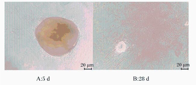
将肠道组织剪碎至3~5 mm3大小后平铺于T25培养瓶中,培养5 d后可见组织块边缘有肠上皮细胞群迁出,在200倍镜下呈现不规则的线条状,且大小不一,细胞之间紧密相连(图1-A);培养28 d后可见细胞铺满瓶底,细胞随着生长逐渐伸展为梭形,折光率高,中央的胞核清晰可见(图1-B)。
图1 组织块法细胞迁出图

Fig.1 Migration images of cells using tissue explant method (200×)

2.1.2 酶消化法

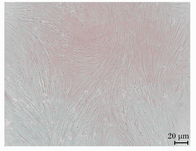
0.25 mg/mL胰蛋白酶(含0.91 mmol/L EDTA)可在15 min内快速消化肠道组织,得到的细胞悬液中有较多的单细胞与细胞团,细胞群体构成均一且无细胞碎屑,细胞折光率高,细胞膜完整,细胞核清晰可见,细胞活率达80%以上。培养12 d后可见细胞铺满瓶底,贴壁细胞单层生长,细胞间紧密连接,边界清晰,呈不规则的长梭形(图2)。
图2 酶消化法培养12 d后细胞贴壁生长图

Fig.2 Adhesion growth image of cells after 12 days cultured by enzymatic digestion method (200×)

2.2 细胞消化条件筛选

表2中数据显示,不同消化酶对细胞的解离效果各异,0.25 mg/mL胰蛋白酶(含0.91 mmol/L EDTA)消化15 min能够从4.79 g肠道组织中获得2.02×10⁷个细胞,其中1.78×10⁷个活细胞,细胞活率显著高于使用胶原酶Ⅰ或胶原酶Ⅰ+胶原酶Ⅳ消化时(P>0.05)。
表2 不同消化酶分离效果比对

Table 2 Comparison of separation effects of different digestive enzymes

项目
Items
胰蛋白酶 胶原酶Ⅰ
Collagenase Ⅰ
胶原酶Ⅰ+胶原酶Ⅳ Collagenase
Ⅰ+collagenase Ⅳ
肠道重量Intestinal weight/g 4.79±0.01 4.74±0.01 4.77±0.01
细胞总数Total cell count/×107 2.02±0.05a 1.54±0.03b 1.49±0.02b
活细胞数Viable cell count/×107 1.78±0.02a 1.02±0.02b 0.96±0.05b
细胞活率Cell viability/% 88.23±0.03a 66.20±0.02b 64.75±0.02b

同行数据肩标无字母或相同小写表示差异不显著(P>0.05),不同小写字母表示差异显著(P<0.05)。

No letter or the same letter on the shoulder of peer data indicated no significant difference (P>0.05), while different lowercase letters indicated significant difference (P<0.05).

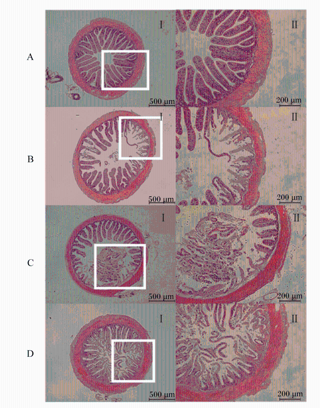
鉴于0.25 mg/mL胰蛋白酶(含0.91 mmol/L EDTA)较突出的消化效果,随后对胰蛋白酶作用不同时段的肠道组织进行HE染色。未经酶解处理的肠道组织可见绒毛结构致密完整并富含杯状细胞(图3-A),而经0.25 mg/mL胰蛋白酶(含0.91 mmol/L EDTA)消化5 min后,肠绒毛出现明显断裂,少量单细胞被消化下来(图3-B);消化15 min后,肠绒毛出现多处断裂,可见较多单细胞与细胞团(图3-C);消化25 min后,肠绒毛出现大面积断裂,大量细胞被分离但已受到损伤,组织已被过度消化(图3-D)。综上可知,0.25 mg/mL胰蛋白酶(含0.91 mmol/L EDTA)消化肠道组织15 min为最佳消化时间,既能够确保足够的细胞分离量,又能够保持细胞的完整性和活性。
图3 0.25 mg/mL胰蛋白酶(含0.91 mmol/L EDTA)对肠道组织消化效果HE染色切片图

Fig.3 HE staining section images of digestive effect of 0.25 mg/mL trypsin (containing 0.91 mmol/L EDTA) on intestinal tissue

A:0 min;B:5 min;C:15 min;D:25 min。

2.3 细胞培养条件优化

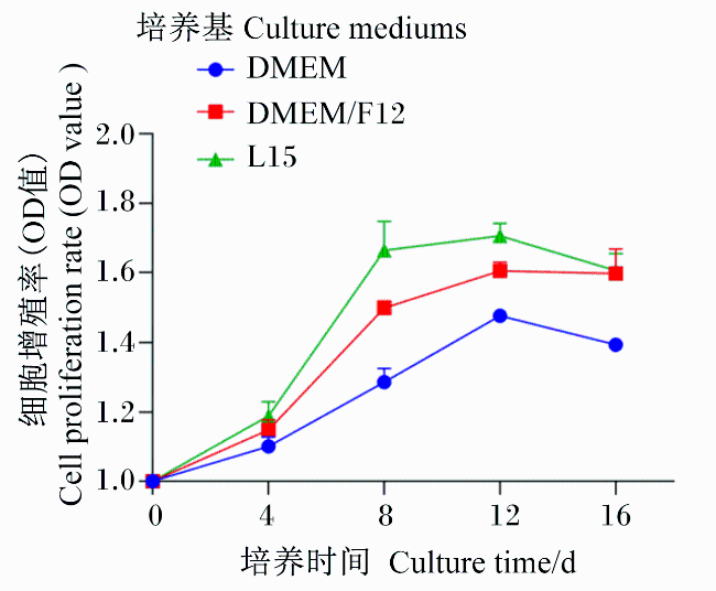
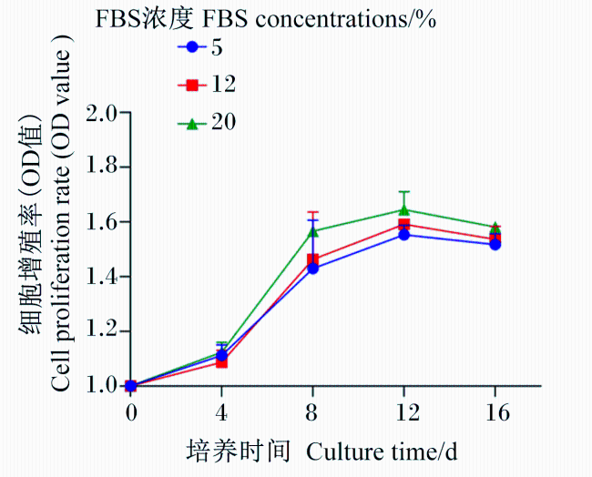
为筛选斜带石斑鱼肠上皮细胞体外培养的最佳条件,本试验探究了细胞对DMEM/F-12、DMEM和L-15这3种培养基的适应性,结果显示:接种4 d内,细胞在3种培养基中的增殖率无明显差异;接种5~12 d,细胞在L15培养基中的增殖率大于同期的DMEM/F-12与DMEM培养基(图4);在整个培养过程中,细胞在含20% FBS的L15培养基中的增殖率最大,15% FBS次之,5% FBS最小(图5)。
图4 不同培养基下细胞的增殖率

Fig.4 Cell proliferation rate under different culture mediums

图5 不同FBS浓度下细胞的增殖率

Fig.5 Cell proliferation rate under different FBS concentrations

2.4 生长曲线

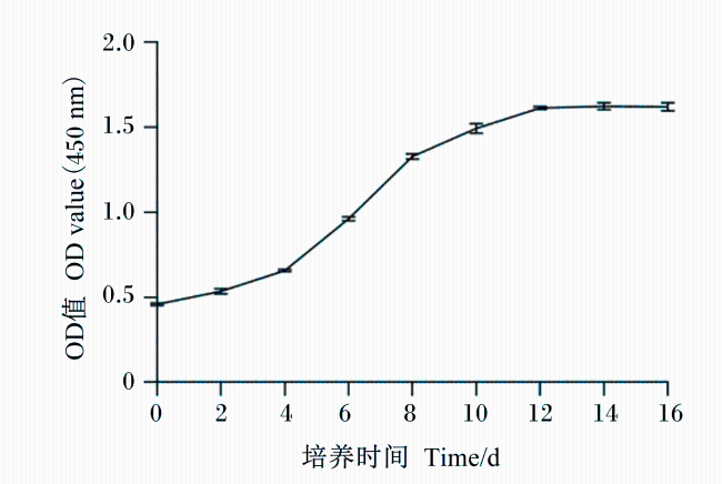
图6所示,细胞生长曲线呈典型的“S”形,接种后1~4 d生长速度较缓,为滞后期,5~8 d为细胞指数生长期,12 d达到最大值,随后进入平台期,细胞不再继续分裂增殖。
图6 细胞生长曲线(接种密度1×10⁶个/mL)

Fig.6 Cell growth curve (inoculum density 1×10⁶ cells/mL)

2.5 肠上皮细胞鉴定

2.5.1 碱性磷酸酶染色

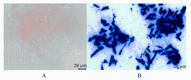
碱性磷酸酶染色结果(图7)显示:阴性对照不显色,而培养的细胞呈蓝紫色,提示所培养的细胞是分化完全、功能健全的成熟肠上皮细胞。
图7 肠上皮细胞碱性磷酸酶染色鉴定结果图

A:肠上皮细胞碱性磷酸酶染色阴性;B:肠上皮细胞碱性磷酸酶染色阳性。

Fig.7 Identification results of alkaline phosphatase staining in intestinal epithelial cells (200×)

A: negative staining for alkaline phosphatase in intestinal epithelial cells; B: positive staining for alkaline phosphatase in intestinal epithelial cells.

2.5.2 透射电镜观察

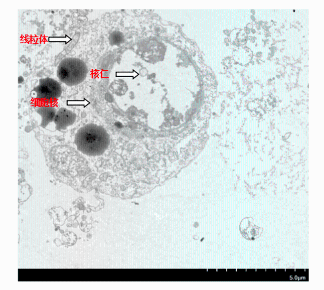
透射电镜图显示,细胞的边界清晰,细胞膜完整,细胞内含有丰富的线粒体,细胞核及核仁清晰可见且核较大(图8)。
图8 斜带石斑鱼肠上皮细胞电镜图

Fig.8 Electron micrograph of intestinal epithelial cells in Epinephelus coioides

2.5.3 序列分析

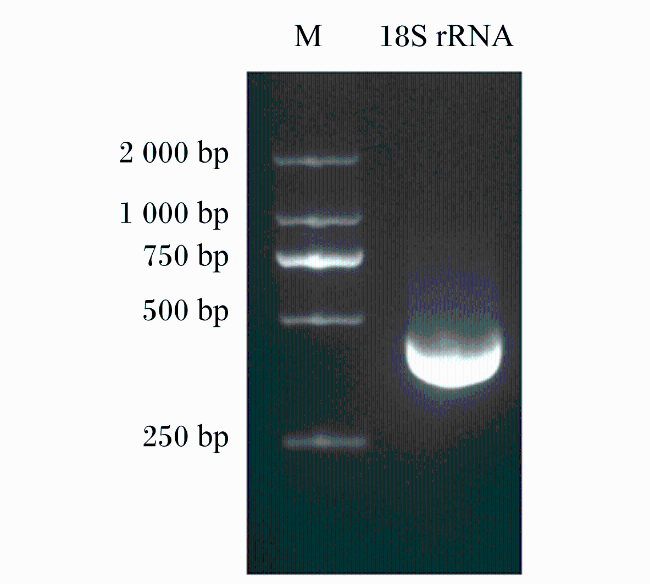
通过保守的18S rRNA基因分析证实了该细胞的起源。从提取的细胞cDNA中扩增18S rRNA,得到预期PCR产物,大小为455 bp(图9)。测序结果已提交到GenBank(登录号:UB14835833 Seq1 PQ555476),通过NCBI序列比对分析,所得序列与鞍带石斑鱼(Epinephelus lanceolatus,序列号:XR_004502792.1)的匹配度最高,说明所得细胞来源于石斑鱼(斜带石斑鱼的全基因组没有公布,而鞍带石斑鱼与斜带石斑鱼的物种相似度较高,在序列比对时具有较高的参考价值)。
图9 斜带石斑鱼肠上皮细胞18S rRNA扩增条带

Fig.9 18S rRNA amplification band in intestinal epithelial cells of Epinephelus coioides

M:DL2000 DNA Marker。

2.5.4 肠道细胞标志性基因表达

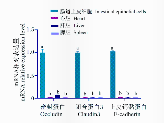
qRT-PCR结果表明,occludin、claudin3和E-cadherin等3种标志性基因在本试验所分离的斜带石斑鱼肠上皮细胞中高表达(P<0.05),而在斜带石斑鱼的其他组织如心脏、肝脏、脾脏中几乎不表达(图10),证明本试验中培养的细胞为肠上皮细胞。
图10 斜带石斑鱼肠道上皮细胞和心脏、肝脏、

脾脏中occludin、claudin3和E-cadherin基因的mRNA相对表达量

同一指标不同组织之间,数据柱标注不同字母表示差异显著(P<0.05)。

Fig.10 mRNA relative expression levels of occludin, claudin3 and E-cadherin genes in intestinal epithelial cells and heart, liver, spleen of Epinephelus coioides

For the same index among different tissues, the data columns marked with different letters indicate significant difference (P<0.05).

3 讨论

自1962年Wolf等[20]成功建立虹鳟生殖腺细胞系RTG-2以来,鱼类细胞培养技术开始受到广泛关注。相较于鱼体内其他组织的细胞,肠上皮细胞的研究相对滞后,这主要归因于肠道组织旺盛的代谢和复杂的微生态环境。作为肠黏膜营养代谢的关键功能单元,肠上皮细胞通过合成特定转运载体及消化酶主导三大营养素的吸收利用,其表面形成的微绒毛结构进一步扩大了营养摄取的表面积[21]。同时,肠上皮细胞通过紧密连接蛋白构建的物理屏障及分泌的免疫效应分子(如β-防御素、白细胞介素-8)调控肠道微环境稳态,进而介导营养物质利用与肠菌群代谢物产出的动态平衡[22]。因此,本研究重点探究了斜带石斑鱼肠上皮细胞原代培养及鉴定方法,以期为后续石斑鱼肠道营养相关研究提供离体细胞模型。据报道,原代细胞培养方法中组织块法简便高效,适用于组织量较少或生长较致密的组织,如鳍条、皮肤、肌肉和性腺等组织[23]。本试验用组织块法从斜带石斑鱼肠道组织中培养出肠上皮细胞,但培养5 d后才见细胞开始迁出,28 d时细胞密度可达培养瓶底80%。此外,本研究使用0.25 mg/mL胰蛋白酶(含0.91 mmo/L EDTA)消化肠道组织,接种的细胞2 d后开始贴壁,4 d进入指数生长期,12 d可达平台期。相较于组织块法,酶消化法在细胞增殖速度、产量和纯度上均有明显优势,可能是因为含EDTA的胰蛋白酶消化液能有效打破细胞间的连接,促进细胞分散、吸收养分和排出代谢废物,利于贴壁生长。值得注意的是,胶原酶Ⅰ、胶原酶Ⅳ混合物在本研究中的消化效果不佳,推测不同物种和组织对消化酶敏感性不同。有研究者在大口黑鲈性腺细胞系建立时采用0.25%胰蛋白酶消化效果良好[24],而使用胶原酶Ⅳ消化金头鲷(Sparus aurata)分离肝脏细胞时,所获得的活细胞数量显著少于胰蛋白酶处理[25],与本研究所得结果一致。
影响海水鱼类细胞体外培养的因素众多,其中培养基种类和血清浓度尤为关键[21]。本研究对比了L15、DMEM及DMEM/F12这3种培养基对斜带石斑鱼肠上皮细胞原代培养的效果,发现L15培养基效果最佳。这与众多海水鱼组织细胞培养的报道结果一致,包括青石斑鱼(Epinephelus awoara)的肾脏、肝脏细胞[26],赤点石斑鱼(Epinephelus akaara)的脾脏细胞[27],以及花鲈(Lateolabrax japonicus)的肠上皮细胞[2]。然而,也有研究指出,DMEM/F-12培养基是褐点石斑鱼(Epinephelus fuscoguttatus)鳍、心脏和鳔组织细胞系[28]及黄姑鱼(Nibea albiflora)肠上皮细胞[29]培养的最适选择;同时DMEM培养基也被用于大菱鲆[17]、银鲳(Pampus argenteus)[11]的肠上皮细胞培养中。氨基酸既可作为细胞蛋白质合成的前体和能量物质,还与细胞有丝分裂原的敏感性有关[30]。本研究发现,相较于DMEM/F12和DMEM培养基,L-15培养基因含更多的精氨酸、亮氨酸及赖氨酸含量,更适合促进斜带石斑鱼肠上皮细胞的贴壁生长。培养基仅为细胞生长构建基础环境,而为了有效促进细胞增殖,还需补充适量的血清。不同细胞类型对血清的需求量各异,其中原代肠道细胞因营养需求较高,对血清的要求尤为严格。在石斑鱼脑细胞原代培养中,15%与20%血清组细胞生长率显著高于5%与10%血清组[31]。在大黄鱼(Larimichthys crocea)性腺细胞培养的研究中,20% FBS表现出最佳效果[32]。本研究结果显示,随着培养时间的延长(4~16 d),20% FBS组细胞的增殖率最高,且FBS浓度上升促使细胞增殖峰值提前,该结果与戴定威[33]的研究一致。综上可知,斜带石斑鱼肠上皮细胞原代培养的适宜培养基为含20% FBS的L15培养基。
本研究中,原代培养的第1代斜带石斑鱼肠上皮细胞呈现成纤维细胞样和上皮细胞表型,这一特征与先前报道的大菱鲆肠上皮细胞的形态[34]相似。不同的是,本研究组织块法在培养28 d时得到典型上皮样细胞,而酶消化法在培养12 d时细胞却呈现纤维状。究其原因,一方面,酶消化的过程中因成纤维细胞相较于肠上皮细胞不耐受胰蛋白酶消化而使得细胞群体被纯化,但是组织块法中的细胞并未被纯化;另一方面,通过蛋白酶降解细胞外基质中的胶原蛋白和黏附蛋白,导致的细胞间连接(如紧密连接、桥粒)快速解离会影响细胞极性,而组织块迁出的细胞无化学刺激,仅受机械剪切力影响,细胞胞体扩展充分。另外,透射电镜观察显示,本试验培养的斜带石斑鱼肠上皮细胞内富含线粒体和内质网,且细胞边界清晰,含1~2个核仁,胞质丰富,与鲫鱼肠上皮细胞形态特征[35]一致。碱性磷酸酶特异性表达于肠上皮细胞表面,利用碱性磷酸酶染色技术可精确鉴定肠上皮细胞,此技术先前已成功应用于花鲈肠上皮细胞的鉴定[2]。本试验中,碱性磷酸酶染色结果显示酶消化法获得的斜带石斑鱼肠上皮细胞呈蓝紫色,表明细胞富含碱性磷酸酶,为成熟的斜带石斑鱼原代肠上皮细胞。定位于细胞表面连接处的occludin、claudin3及E-cadherin蛋白,对于维持肠道上皮细胞间连接及结构完整性扮演着不可或缺的角色[36-37]。特别是E-cadherin,近年来被证明可促进肠道细胞表面的黏附,并通过改变下游黏附信号而调控肠上皮细胞的增殖与分化[38]。本研究获得肠上皮细胞中occludin、claudin3及E-cadherin基因的mRNA相对表达量显著高于心脏、肝脏及脾脏中。Wei等[28]在黄姑鱼肠上皮细胞中也成功检测到occludin、claudin、E-cadherin等蛋白的高度表达,与本研究结果一致。此外,18S rRNA片段扩增结果证实本研究培养的细胞来源于,与其他细胞系特征[39-41]一致。

4 结论

① 相较于组织块法,酶消化法在斜带石斑鱼肠上皮细胞培养过程中展现出快速高效的优势。
② 0.25 mg/mL胰蛋白酶(含0.91 mmol/L EDTA)相较于胶原酶更适宜作为斜带石斑鱼肠上皮细胞分离的消化酶。
③ 在含20% FBS的L15培养基中,28 ℃恒温培养可获得生长旺盛、形态均一且状态良好的斜带石斑鱼肠上皮细胞。
[1]
PETERSON L W, ARTIS D. Intestinal epithelial cells:regulators of barrier function and immune homeostasis[J]. Nature Reviews Immunology, 2014, 14(3):141-153.

[2]
李莎, 刘紫严, 杨红玲, 等. 花鲈肠上皮细胞原代培养及鉴定方法的探讨研究[J]. 水产科学, 2024, 43(3):381-389.

LI S, LIU Z Y, YANG H L, et al. Primary culture and identification of intestinal epithelial cells in sea bass Lateolabrax maculatus[J]. Fisheries Science, 2024, 43(3):381-389. (in Chinese)

[3]
MINGHETTI M, DRIESCHNER C, BRAMAZ N, et al. A fish intestinal epithelial barrier model established from the rainbow trout (Oncorhynchus mykiss) cell line,RTgutGC[J]. Cell Biology and Toxicology, 2017, 33(6):539-555.

[4]
LIU Y P, GE X F, LI C, et al. Derivation and characterization of new cell line from intestine of turbot (Scophthalmus maximus)[J]. In Vitro Cellular & Developmental Biology-Animal, 2023, 59(2):153-162.

[5]
ZHANG Y J, CHEN W, MAI K S, et al. In vitro assay for evaluating the effects of three anti-nutritional factors on the primary-cultured intestinal epithelial cells isolated from Japanese flounder,Paralichthys olivaceus[J]. Aquaculture Research, 2015, 46(1):242-251.

[6]
宋鹏. JAK2介导瘦素调控草鱼肠道PepT1基因表达的研究[D]. 硕士学位论文. 长沙: 湖南农业大学, 2019.

SONG P. JAK2 Mediates the regualtion of PepTl by leptin in grass carp (Ctenopharyngodon idellus) intestinal[D]. Master’s Thesis. Changsha: Hunan Agricultural University, 2019. (in Chinese)

[7]
PASQUARIELLO R, VERDILE N, PAVLOVIC R, et al. New stable cell lines derived from the proximal and distal intestine of rainbow trout (Oncorhynchus mykiss) retain several properties observed in vivo[J]. Cells, 2021, 10(6):1555.

[8]
WANG Y B, YAN X X, FU L L. Effect of selenium nanoparticles with different sizes in primary cultured intestinal epithelial cells of crucian carp,Carassius auratus gibelio[J]. International Journal of Nanomedicine, 2013,8:4007-4013.

[9]
ZHOU X X, WANG Y B, PAN Y A, et al. Investigation of the cytotoxicity of apidaecin on intestinal epithelial cells of tilapia (Oreochromis niloticus) in vitro[J]. Aquaculture International, 2008, 16(6):591-600.

[10]
YU G J, OU W H, AI Q H, et al. In vitro study of sodium butyrate on soyasaponin challenged intestinal epithelial cells of turbot (Scophthalmus maximus L.) refer to inflammation,apoptosis and antioxidant enzymes[J]. Fish and Shellfish Immunology Reports, 2021,2:100031.

[11]
GAO Q X, GAO Q, MIN M H, et al. Ability of Lactobacillus plantarum lipoteichoic acid to inhibit Vibrio anguillarum-induced inflammation and apoptosis in silvery pomfret (Pampus argenteus) intestinal epithelial cells[J]. Fish & Shellfish Immunology, 2016,54:573-579.

[12]
PAN X, WU T, ZHANG L, et al. In vitro evaluation on adherence and antimicrobial properties of a candidate probiotic Clostridium butyricum CB2 for farmed fish[J]. Journal of Applied Microbiology, 2008, 105(5):1623-1629.

[13]
马博, 孙桂波, 杨志宏, 等. 肠上皮细胞模型不同培养条件的优化及适应性研究[J]. 中国实验方剂学杂志, 2011, 17(11):205-210.

MA B, SUN G B, YANG Z H, et al. Comparative study on different culture conditions for Caco-2 cell model[J]. Chinese Journal of Experimental Traditional Medical Formulae, 2011, 17(11):205-210. (in Chinese)

[14]
孙秀梅, 程帆, 刘维, 等. 4种小鼠肠上皮细胞分离培养方法的比较[J]. 西北农林科技大学学报(自然科学版), 2013, 41(5):25-31.

SUN X M, CHENG F, LIU W, et al. Comparison of four primary culture methods for mouse intestinal epithelial cells[J]. Journal of Northwest A&F University (Natural Science Edition), 2013, 41(5):25-31. (in Chinese)

[15]
GUZMAN-MURILLO M, MERINO-CONTRERAS M L, ASCENCIO F. Interaction between Aeromonas veronii and epithelial cells of spotted sand bass (Paralabrax maculatofasciatus) in culture[J]. Journal of Applied Microbiology, 2000, 88(5):897-906.

[16]
LANGAN L M, OWEN S F, JHA A N. Establishment and long-term maintenance of primary intestinal epithelial cells cultured from the rainbow trout,Oncorhynchus mykiss[J]. Biology Open, 2018, 7(3):bio032870.

[17]
姜俊. 谷氨酰胺对鲤鱼肠上皮细胞生长和代谢的影响[D]. 硕士学位论文. 雅安: 四川农业大学, 2005.

JIANG J. Effects of glutamine on the growth and metabolism of carp intestinal epithelial cells in primary culture[D]. Master’s Thesis. Ya’an: Sichuan Agricultural University, 2005. (in Chinese)

[18]
农业农村部渔业渔政管路局, 全国水产技术推广总站, 中国水产学会. 2024中国渔业统计年鉴[M]. 北京: 中国农业出版社, 2024.

China Fisheries Law Enforcement, National Fisheries Technology Extension Center, China Society of Fisheries. 2024 China fishery statistical yearbook[M]. Beijing: China Agriculture Press,2024. (in Chinese)

[19]
FAN J L, ZHENG J Y, LAI W J, et al. Establishment of a new intestinal cell line from the grouper (Epinephelus coioides) for aquatic virus pathogenesis[J]. Aquaculture, 2023,562:738771.

[20]
WOLF K, QUIMBY M C. Established eurythermic line of fish cells in vitro[J]. Science, 1962, 135(3508):1065-1066.

[21]
KIELA P R, GHISHAN F K. Physiology of intestinal absorption and secretion[J]. Best Practice & Research Clinical Gastroenterology, 2016, 30(2):145-159.

[22]
MORRISON D J, PRESTON T. Formation of short chain fatty acids by the gut microbiota and their impact on human metabolism[J]. Gut Microbes, 2016, 7(3):189-200.

[23]
毛美琴, 蓝桢宇, 李进, 等. 鱼类细胞培养历史、应用及培养条件研究概述[J]. 广东农业科学, 2022, 49(3):124-134.

MAO M Q, LAN Z Y, LI J, et al. Overview of the history,applications,and culture conditions of fish cell culture[J]. Guangdong Agricultural Sciences, 2022, 49(3):124-134. (in Chinese)

[24]
GETCHELL R G, GROOCOCK G H, CORNWELL E R, et al. Development and characterization of a largemouth bass cell line[J]. Journal of Aquatic Animal Health, 2014, 26(3):194-201.

[25]
FIGUEIREDO N, MATOS B, DINIZ M, et al. Marine fish primary hepatocyte isolation and culture: new insights to enzymatic dissociation pancreatin digestion[J]. International Journal of Environmental Research and Public Health, 2021, 18(4):1380.

[26]
LAI Y S, MURALI S, JU H Y, et al. Two iridovirus-susceptible cell lines established from kidney and liver of grouper,Epinephelus awoara (Temminck & Schlegel),and partial characterization of grouper iridovirus[J].Journal of Fish Diseases, 2000, 23(6):379-388.

[27]
HUANG X H, HUANG Y H, SUN J J, et al. Characterization of two grouper Epinephelus akaara cell lines:application to studies of Singapore grouper iridovirus (SGIV) propagation and virus-host interaction[J]. Aquaculture, 2009, 292(3/4):172-179.

[28]
WEI Y B, FAN T J, JIANG G J, et al. Establishment of a novel fin cell line from Brown-marbled grouper,Epinephelus fuscoguttatus (Forsskål),and evaluation of its viral susceptibility[J]. Aquaculture Research, 2009, 40(13):1523-1531.

[29]
王立改, 曾延清, 汪波, 等. 黄姑鱼肠道上皮细胞体外分离培养及鉴定[J]. 动物营养学报, 2022, 34(2):1296-1304.

WANG L G, ZENG Y Q, WANG B, et al. Isolation,culture and identification of intestinal epithelial cells of Nibea albiflora in vitro[J]. Chinese Journal of Animal Nutrition, 2022, 34(2):1296-1304. (in Chinese)

[30]
HASEGAWA K, MIYATA Y, CARR B I. Glutamic acid potentiates hepatocyte response to mitogens in primary culture[J]. Journal of Cellular Physiology, 1994, 158(2):365-373.

[31]
LIU X F, WU Y H, WEI S N, et al. Establishment and characterization of a brain-cell line from kelp grouper Epinephelus moara[J]. Journal of Fish Biology, 2018, 92(2):298-307.

[32]
郭一兰, 王志勇. 大黄鱼性腺原代细胞培养技术的建立[J]. 集美大学学报(自然科学版), 2020, 25(5):321-327.

GUO Y L, WANG Z Y. Establishment of primary cell culture technique for gonadal tissue of large yellow croaker[J]. Journal of Jimei University (Natural Science Edition), 2020, 25(5):321-327. (in Chinese)

[33]
戴定威, 吴圣楣, 廖贤平, 等. 人类胎儿和新生大鼠小肠上皮细胞的分离培养方法[J]. 中华儿科杂志, 1995(3):176.

DAI D W, WU S M, LIAO X P, et al. Method for isolation and culture of human fetal and neonatal rat small intestinal epithelial cells[J]. Chinese Journal of Pediatrics, 1995(3):176. (in Chinese)

[34]
SU L, GUO B S, FENG R X, et al. Establishment and characterization of an intestinal epithelial cell line from Japanese flounder (Paralichthys olivaceus)[J]. Aquaculture, 2024,580:740325.

[35]
宋增福, 吴天星, 潘晓东. 鲫肠道上皮细胞原代培养方法的研究[J]. 淡水渔业, 2008, 38(1):67-69,34.

SONG Z F, WU T X, PAN X D. Study on the primary culture method of intestinal epithelial cells from Carassius auratus[J]. Freshwater Fisheries, 2008, 38(1):67-69,34. (in Chinese)

[36]
LAI Y S, JOHN J A C, LIN C H, et al. Establishment of cell lines from a tropical grouper,Epinephelus awoara (Temminck & Schlegel),and their susceptibility to grouper irido- and nodaviruses[J].Journal of Fish Diseases, 2003, 26(1):31-42.

[37]
WEN C M, HUANG J Y, CIOU J H, et al. Immunochemical and molecular characterization of GBC4 as a tanycyte-like cell line derived from grouper brain[J]. Comparative Biochemistry and Physiology Part A:Molecular & Integrative Physiology, 2009, 153(2):191-201.

[38]
HUANG S M, KUO S T, KUO H C, et al. Assessment of fish iridoviruses using a novel cell line GS-1,derived from the spleen of orange-spotted grouper Epinephelus coioides (Hamilton) and susceptible to ranavirus and megalocytivirus[J]. Journal of Veterinary Medical Science, 2018, 80(11):1766-1774.

[39]
KYOKO O O, KONO H, ISHIMARU K, et al. Expressions of tight junction proteins Occludin and Claudin-1 are under the circadian control in the mouse large intestine:implications in intestinal permeability and susceptibility to colitis[J]. PLoS One, 2014, 9(5):e98016.

[40]
SCHNOOR M. E-cadherin is important for the maintenance of intestinal epithelial homeostasis under basal and inflammatory conditions[J]. Digestive Diseases and Sciences, 2015, 60(4):816-818.

[41]
CHEN S Y, ZHENG Y J, RAN X J, et al. Integrin αEβ7+ T cells direct intestinal stem cell fate decisions via adhesion signaling[J]. Cell Research, 2021, 31(12):1291-1307.

Outlines

/