REVIEW

Mechanism of Multi-Organ Damage Induced by Microplastic Exposure and Its Green Regulatory Strategies

  • CHEN Peng , 1 ,
  • YI Lalet 1, 2 ,
  • LI Wenting 2 ,
  • SUN Haizhou , 1, 2, *
Expand
  • 1 College of Animal Science, Inner Mongolia Agricultural University, Hohhot 010018, China
  • 2 Key Laboratory of Healthy Herbivorous Animal Breeding and Animal Product Quality Control of Ministry of Agriculture and Rural Affairs (Jointly Built by Ministry and Province), Inner Mongolia Key Laboratory of Herbivorous Animal Nutrition Science, Inner Mongolia Academy of Agriculture and Animal Husbandry Sciences, Hohhot 010031, China
*professor, E-mail:

Received date: 2025-03-20

  Online published: 2025-10-15

Abstract

As a kind of new environmental pollutant, microplastics have been widely distributed in water, soil and atmosphere worldwide because of their small particle size, strong adsorption and difficult degradation. Microplastics can be ingested by animals through the digestive tract, respiratory tract and other ways, and are concentrated in the intestine, liver, brain and other important organs. The damage mechanism mainly includes the destruction of intestinal barrier function, inducing oxidative stress, triggering inflammatory response and metabolic disorders, etc., and then has a wide impact on the digestive, respiratory, nervous, reproductive and immune systems of animals. At present, regulatory measures such as probiotics, microbial hydrolases, plant extracts and fecal microbiota transplantation have been proven to effectively reduce the toxicity of microplastics, thereby improving the health of animals. This paper systematically reviewed the mechanism of damage caused by microplastic exposure to animal intestinal tract, liver, brain, reproduction and other organs, and discussed the research progress of relevant regulatory measures, aiming to provide theoretical basis for in-depth understanding of the harm of microplastics to animal health, and provide reference for developing effective green regulatory strategies and promoting the green and sustainable development of livestock and poultry industry.

Cite this article

CHEN Peng , YI Lalet , LI Wenting , SUN Haizhou . Mechanism of Multi-Organ Damage Induced by Microplastic Exposure and Its Green Regulatory Strategies[J]. Chinese Journal of Animal Nutrition, 2025 , 37(10) : 6461 -6478 . DOI: 10.12418/CJAN2025.524

塑料因其化学性质稳定、重量轻和耐腐蚀等特点,被广泛应用于人类生产生活的各个领域[1]。然而,随着塑料制品的大规模使用,塑料废弃物问题日益严重。数据显示,当前全球塑料垃圾年产量为350万t,预计到2040年,塑料垃圾的产量将飙升至2.5亿t左右[2]。塑料垃圾在化学、物理和生物等外力作用下,逐渐分解破碎成粒径≤5 mm的颗粒、纤维或碎片,即微塑料[3]。美国国家海洋和大气管理局将这种微塑料定义为一种新兴的环境污染物,具有极强的持久性和缓慢的降解性,已引起全球范围内的广泛关注[4]。近年来,微塑料颗粒在全球范围内的水、土壤和大气中广泛分布,甚至在海鲜、饮料、水果和蔬菜中也被检测到[5]。这些微塑料颗粒通过各种环境介质(如水、土壤和空气)极易被动物摄入[6-7],并通过循环系统富集在肠道、血液、肝脏、脾脏、心脏、肺脏和卵巢等重要器官中,进而对动物的消化、呼吸、神经、泌尿、生殖和免疫系统产生损伤,最终通过食物链对人类健康构成潜在威胁[8-11]
为充分了解微塑料暴露毒性及其对动物健康的影响相关发展趋势和研究热点,本文基于Web of Science(WOS)核心数据库,以“micro-nano plastic”“microplastic”“micro-plastic”“micro-sized plastic”“nanoplastics”“nano-plastic”“nano-sized plastic”“micro (nano) plastics”与“toxicity”“toxic effect”“adverse effect”以及“animal” “pig”“cattle”“cow”“beef”“bird”“fish”“hen” “broiler”“chicken”“lamb”“goat”“sheep” “goose”“duck”“rat”“horse” “rabbit”“mice”“camel” 等关键词进行检索,共检索到1 298篇相关文献。如图1所示,近10年来,该领域的发文量和引用数量呈明显增长趋势,表明微塑料毒性及其对动物健康的影响已成为学术界的研究热点。通过CiteSpace 6.2.R4软件对文献数据的可视化分析发现,“聚丙烯微塑料”“饲料安全”“聚苯乙烯纳米塑料”“肾脏纤维化”“生化效应”“草甘膦除草剂共暴露毒性”和“人类健康”等关键词频繁出现,进一步证实了微塑料与动物健康之间的密切关联。
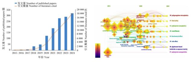
图1 2015—2024年微塑料暴露与动物健康相关论文发表及研究热点聚类

Fig.1 Papers related to microplastic exposure and animal health published from 2015 to 2024 and clustering of research hotspots

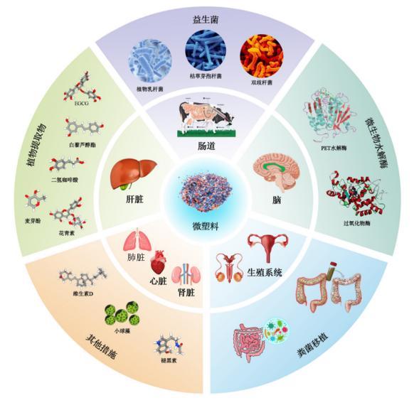
现有相关报道指出,微塑料暴露能够引起动物消化道发生严重损伤[4],但微塑料暴露对其他器官的损伤机制及绿色调控策略的研究相对较少。因此,本文通过系统梳理国内外相关研究进展,重点探讨微塑料暴露对动物肠道、肝脏、脑和生殖等器官的损伤机制,并总结益生菌、微生物水解酶、植物提取物和粪菌移植等调控措施在缓解微塑料毒性方面的作用(图2),旨在为降低微塑料在动物养殖生产过程中的危害提供理论基础,进一步推动畜禽养殖业的绿色可持续发展。
图2 微塑料暴露对动物多器官损伤的影响及调控策略

EGCG:表没食子儿茶素-3-没食子酸酯 epigallocatechin-3-gallate;PET:聚对苯二甲酸乙二醇酯 polyethylene terephthalate。

Fig.2 Effects of microplastic exposure on multiple organ damage of animals and regulatory strategies

1 微塑料暴露对动物器官的损伤机制

1.1 微塑料暴露对动物肠道的损伤机制

肠道既是动物消化吸收营养物质的主要场所,也是抵御肠道内的各种有害微生物及各类毒素和保障机体内环境稳态的重要屏障[12]。微塑料暴露能够破坏肠道微生物,导致生态失调[13-14]。研究发现,微塑料暴露导致小鼠肠道微生态失调,肠道菌群组成在门、属和种水平上发生显著变化,其中有益菌丰度显著降低,而破坏肠道屏障功能的有害菌(如梭菌属、拟杆菌属和埃希氏菌属)丰度显著提高[15-16]。李志青等[4]探讨了微塑料通过诱导氧化应激和炎症反应来严重影响肠道功能紊乱和菌群失调的毒性机制。此外,微塑料被动物摄取之后,其表面的“塑料际”可以在肠道微生物中富集潜在的致病菌,这些致病菌通过代谢活动产生毒素、炎症因子等有害代谢产物,从而改变周围环境中微生物的群落结构并对动物肠道造成损伤[17-18]。Cheng等[19]研究发现,有12个细菌分类群从“塑料际”向文昌鱼肠道微生物群转移,致病菌葡萄球菌属就是12个细菌分类群之一。同时,微塑料对肠道屏障的直接物理损伤和功能性破坏已被多项研究证实。例如,暴露于不同粒径(如70 nm、50 μm)和类型(如聚苯乙烯、聚乙烯)的微塑料会导致肠黏膜结构中断、上皮细胞脱落、黏液分泌减少以及杯状细胞数量下降[20-22]。微塑料在肠道中的积累与肠上皮细胞摩擦,诱发革兰氏阴性菌细胞壁中的脂多糖穿透体循环,刺激炎症因子[如肿瘤坏死因子-α(tumor necrosis factor-α,TNF-α)、白细胞介素(interleukin,IL)-1β和干扰素-γ(interferon-γ,IFN-γ)]的产生,进一步加重肠道屏障功能的损伤[23]。微塑料暴露还会导致肠道形态和紧密连接发生损伤。Lei等[24]研究发现,在10 d内接触70 μm大小的微塑料会导致斑马鱼和秀丽隐杆线虫的肠绒毛开裂和肠上皮细胞分裂。Su等[25]研究进一步表明,聚苯乙烯微塑料暴露破坏小鼠小肠的微观结构,降低十二指肠和空肠紧密连接基因的表达,并导致肠道菌群失调,且紧密连接基因表达与菌群中普雷沃氏菌科呈正相关。
值得注意的是,细胞对于微塑料(≤5 mm)和纳米塑料(≤1 μm)的吸附能力也不同。有研究表明,结肠癌Caco-2细胞对于不同粒径的聚苯乙烯微塑料(300 nm、500 nm、1 μm、3 μm、6 μm)的24 h吸收率分别为73%、71%、49%、43%和30%[26]。这表明微塑料的粒径越小,进入细胞的可能性越大,对细胞的损伤也越高。这在体内研究中也得到证实,粒径<10μm的微塑料可以从肠道转移到淋巴和循环系统,并在肝脏、肾脏和大脑等组织中积累;而粒径<0.1 μm的纳米塑料则能够穿过细胞膜,突破血脑屏障和胎盘屏障进入大脑和胎盘[27-29]。同时,不同类型的微塑料对动物的危害程度也不同。聚苯乙烯的苯乙烯单体可通过肠道在大脑、卵巢和睾丸等多个器官累积,会引发神经毒性、导致生殖器官细胞凋亡和激素异常、损伤肝脏组织形态和功能、诱导肾脏损伤以及加剧心肌炎症和肺脏纤维化[30-36]。聚乙烯的乙烯单体和聚酰胺的己二酸和己二胺可导致肠道通透性和炎症增加,能穿过肠道屏障和血脑屏障改变大脑神经递质水平,进而影响神经传递和行为模式,其中聚乙烯的乙烯单体还可诱导肝细胞氧化应激、炎症、凋亡和坏死[37-39]。聚丙烯的丙烯单体、聚氯乙烯的氯乙烯单体以及聚对苯二甲酸乙二醇酯(polyethylene terephthalate,PET)的对苯二甲酸和乙二醇均可导致肠道绒毛开裂和菌群失衡,并引发肠道炎症和组织损伤[24,40]。不同类型微塑料对动物器官的影响见表1
表1 不同类型微塑料对动物器官的影响

Table 1 Effects of different types of microplastics on animal organs

微塑料类型
Types of
microplastics
主要成分
Main
components
作用靶器官
Target organ
of action
危害程度和机制
Degree and mechanism
of harm
参考文献
References


聚乙烯
Polyethylene


乙烯单体
肠道 导致肠道隐窝深度增加,肠道通透性和炎症增加 [15]
肝脏 诱导肝细胞氧化应激、炎症、细胞凋亡和坏死 [37]
大脑 可以穿过血脑屏障并提高大脑中5-羟色胺、
多巴胺和乙酰胆碱的水平
[38]


聚苯乙烯
Polystyrene


苯乙烯单体
肠道 破坏小肠微观结构,降低紧密连接基因的
表达,并导致肠道菌群失调
[25]
肝脏 破坏肝脏组织形态,导致肝脏脂质紊乱和细胞凋亡 [30]
大脑 能够穿透血脑屏障,引发大脑神经毒性 [31]
睾丸 导致睾丸组织学改变、精子发生异常和激素分泌异常 [32]
卵巢 引起卵巢颗粒细胞凋亡和激素紊乱 [33]
肺脏 调节上皮-间质转化,加剧肺脏纤维化 [34]
肾脏 激活肾脏补体5a(C5a)/补体5a受体(C5aR)
通路来诱导肾脏损伤
[35]
心脏 触发细胞焦亡并加剧心肌炎症 [36]

聚酰胺
Polyamide

己二酸和己二胺
肠道 导致肠道有益菌丰度降低,有害菌丰度提高 [39]
大脑 改变神经递质合成,影响大脑神经传递和行为模式 [39]
聚丙烯
Polypropylene
丙烯单体 肠道 导致肠道绒毛开裂和菌群失衡 [24]
聚氯乙烯
Polyvinyl chloride
氯乙烯单体 肠道 导致结肠组织损伤,增加拟杆菌门与
厚壁菌门的比例
[40]
聚对苯二甲酸乙二醇酯
Polyethylene terephthalate
对苯二甲酸和
乙二醇
肠道 导致肠道炎症和组织损伤,诱导肠道菌群紊乱 [40]

1.2 微塑料暴露对动物肝脏的损伤机制

微塑料暴露通过破坏肠道屏障功能,增强内毒素等外源性有害物质的运输,进而通过肠-肝轴引发肝脏损伤[41]。例如,5 μm聚苯乙烯微塑料暴露6周后,肉鸡肠道紧密连接蛋白表达降低,Wnt/β-连环蛋白(β-catenin)信号传导被抑制,肠道血管屏障遭到破坏,导致有害细菌和代谢物通过肠-肝轴转移至肝脏,触发肝脏免疫反应,并促进脂质代谢紊乱和细胞凋亡[42]。此外,微塑料通过Toll样受体2(Toll like receptors 2,TLR2)/核因子-κB(nuclear factor-κB,NF-κB)/NOD样受体家族热蛋白结构域包含3(NOD-like receptor family pyrin domain containing 3,NLRP3)通路诱导肠道菌群紊乱,进一步加剧肝脏炎症反应[37,43]。微塑料暴露诱导的氧化应激是肝脏损伤的重要机制之一。例如,80 nm聚苯乙烯微塑料处理24 h后,斑马鱼肝脏中活性氧(reactive oxygen species,ROS)生成量显著增加,抗氧化剂谷胱甘肽过氧化酶1α(glutathione peroxidase 1α,GPX1α)基因表达下调,而细胞色素P450家族1亚家族A成员1(cytochrome P450 family 1 subfamily A member 1,CYP1A1)基因表达上调,最终导致肝脏萎缩[44]。类似地,高剂量聚苯乙烯微塑料在雄性后代小鼠肝脏中诱导了氧化应激和炎性细胞浸润,增加了促炎细胞因子的产生[45]。微塑料暴露驱动了多种形式的细胞死亡,包括细胞凋亡、细胞焦亡和铁死亡。研究表明,微塑料通过激活小鼠肝脏内源性细胞凋亡信号p53/B细胞淋巴瘤-2(B-cell lymphoma-2,Bcl-2)/B细胞淋巴瘤-2相关X蛋白(B-cell lymphoma-2 associated X protein,Bax)通路[30],以及蛋白激酶RNA样内质网激酶(protein kinase RNA-like endoplasmic reticulum kinase,PERK)和丝裂原活化蛋白激酶(mitogen activated protein kinase,MAPK)通路诱导斑马鱼肝细胞凋亡[46]。同时,微塑料通过NLRP3/凋亡相关斑点样蛋白(apoptosis-associated speck-like protein,ASC)和胱天蛋白酶-1(Caspase-1)依赖性途径诱导小鼠肝细胞焦亡[47-48],并通过诱导脂质过氧化调节铁死亡相关蛋白[如转铁蛋白受体(transferrin receptor,TFRC)、铁蛋白重链1(ferritin heavy chain 1,FTH1)和谷胱甘肽过氧化酶4(glutathione peroxidase 4,GPX4)]的表达,进一步加剧肝脏组织的损伤,导致肝脏功能失调,最终引发一系列代谢紊乱和炎症反应[47]
肝脏是控制与不同器官和组织相连的各种代谢途径的主要代谢器官之一,也是动物的主要解毒器官[49]。微塑料暴露已被证实可通过多种机制诱发肝脏的糖代谢、脂质代谢、氨基酸代谢和胆汁酸代谢紊乱,进而对动物机体健康产生消极影响。例如,2 μm聚氯乙烯微塑料暴露60 d后,小鼠肝细胞微观结构出现损伤,表现为肝细胞球囊扩大、细胞空泡化和细胞间隙扩张[50]。微塑料通过肠-肝轴代谢紊乱引起胰岛素分泌失调,并诱发肝脏胰岛素抵抗[51]。肝脏转录组学和代谢组学分析表明,微塑料可以扰乱单糖和脂质代谢,包括磷酸戊糖途径和糖异生[52]。此外,微塑料诱导金头鲷肝脏炎症、氧化损伤和脂质代谢紊乱,改变肠-肝轴稳态,引发肝脏损伤[53]。在氨基酸代谢方面,微塑料暴露导致鸡肝脏中谷氨酰胺和谷氨酸合成过量,过量的谷氨酰胺和谷氨酸通过破损的血脑屏障进入小脑组织,触发自噬依赖性铁死亡和细胞凋亡[54]。在胆汁酸代谢方面,聚苯乙烯微塑料暴露会破坏肠道屏障,导致微塑料在肝脏组织中积累,并引起胆汁酸合成和外排相关基因表达失调,最终导致胆汁淤积和代谢紊乱[55]。综上可知,微塑料暴露通过多种相互关联的机制对肝脏产生毒性作用,包括肠道屏障功能障碍、氧化应激、炎症反应、细胞死亡和代谢紊乱,这些机制共同导致肝脏损伤,进而对动物健康产生显著负面影响。

1.3 微塑料暴露对动物脑和神经的损伤机制

微塑料能够穿透动物的肠道屏障和血脑屏障,进入中枢神经系统,直接引发神经毒性反应,导致神经元损伤和炎症反应[56-57]。其中,粒径小于0.293 μm的微塑料颗粒可以被小鼠胃肠道吸收并穿透血脑屏障,进入大脑后引发一系列神经毒性反应[31,58]。这种暴露不仅能够引发大脑中的神经退行性病变,还可能导致特定神经元的损失,尤其是广泛分布于中枢神经系统并在认知功能和运动控制中发挥关键作用的胆碱能和多巴胺能神经元[59-60]。此外,短期暴露于聚苯乙烯微塑料即可对小鼠的学习、记忆和探索能力产生负面影响,且随着暴露时间的延长,这种认知缺陷会进一步加剧[61]。在神经元结构层面,聚苯乙烯微塑料暴露会显著影响神经元的轴突生长,表现为轴突缺失和突触密度降低[62]。例如,在斑马鱼原代运动神经元中,聚苯乙烯微塑料暴露不仅减少了轴突长度,还显著下调了与神经元生长相关基因的表达[63]。神经递质作为神经元之间或神经元与效应细胞之间交流的关键化学物质,其水平也受到微塑料的显著影响[64]。Yu等[38]研究表明,聚乙烯微塑料暴露会提高斑马鱼大脑中5-羟色胺、多巴胺和乙酰胆碱的水平,而聚苯乙烯微塑料则通过破坏这些神经递质的平衡来引发脑损伤[65]
除了直接作用外,微塑料暴露还能通过破坏肠道微生物群组成和扰乱肠-脑轴间接损害大脑[66-67]。具体而言,微塑料会破坏肠道菌群平衡,损害肠道屏障功能,引发炎症细胞因子[如IL-1、IL-6和脂多糖]的释放,进而改变血脑屏障的通透性,穿过血脑屏障并引发脑损伤[68-69]。Yang等[70]研究发现,小鼠摄入的微塑料会改变肠道微生物群并引发肠道免疫反应,导致巨噬细胞溶酶体损伤,释放IL-1等促炎细胞因子,这些因子可穿过血脑屏障并影响脑功能。此外,微塑料暴露还会诱导肠道微生物群的破坏,进一步影响其代谢物以及宿主产生的分子,从而加剧脑损伤[71]。例如,Huang等[39]研究表明,聚苯乙烯微塑料暴露会减少七彩神仙鱼肠道中乳酸杆菌和双歧杆菌数量,降低其代谢产物(γ-氨基丁酸)水平,进而干扰神经递质合成,影响神经传递和行为模式。同样,Zhang等[72]研究发现,斑马鱼暴露于聚苯乙烯微塑料30 d后,肠道中短链脂肪酸和5-羟色胺水平显著下降,同时5-羟色胺合成关键酶[色氨酸羟化酶(tryptophan hydroxylase,TPH)1和TPH2]及其大脑受体mRNA表达发生改变,最终导致斑马鱼运动和学习能力异常。微塑料对神经系统的损伤机制涉及直接穿透血脑屏障、神经元损伤、神经递质紊乱以及通过肠-脑轴的间接影响等多个层面。上述研究不仅揭示了微塑料的复杂毒性机制,也为未来进一步探索其神经毒性提供了重要方向。

1.4 微塑料暴露对动物生殖的损伤机制

微塑料对生殖系统的损伤作用主要通过干扰下丘脑-垂体-性腺轴实现,这一激素信号系统由下丘脑释放促性腺激素释放激素启动,进而刺激垂体产生黄体生成素和卵泡刺激素[73]。其中,微塑料能够穿透血脑屏障,直接作用于下丘脑弓状核的促性腺激素释放激素神经元,通过激活炎症信号通路(如NF-κB)或氧化应激(产生ROS),抑制促性腺激素释放激素的合成与释放[74]。多项研究表明,微塑料进入海马体并通过IL-1β、IL-6和TNF-α触发局部炎症,损伤中枢神经系统和神经信号传导[75];还通过引发氧化应激显著减少小鼠体内睾酮、黄体生成素和卵泡刺激素等关键生殖激素的产生[32,76-77]。在雌性生殖系统中,微塑料暴露会显著损害卵巢功能。Wu等[78]研究表明,暴露于100 mg/L聚苯乙烯微塑料和200 mg/kg邻苯二甲酸二(2-乙基己基)酯35 d会导致小鼠卵巢颗粒细胞层破坏,引发卵泡碎裂和闭锁,其机制是通过激活大麻素受体1(cannabinoid receptor 1,CNR1)/cereblon(CRBN)/yin yang 1(YY1)/细胞色素P450家族2亚家族E成员1(cytochrome P450 family 2 subfamily E member 1,CYP2E1)信号通路促进ROS过度产生和氧化应激,进而导致卵巢颗粒细胞氧化DNA损伤。此外,不同浓度[0、0.015、0.150和1.500 mg/(kg·d)]的0.5 μm聚苯乙烯微塑料会诱导大鼠卵巢颗粒细胞凋亡,导致卵巢储备能力下降和纤维化发生[33]。而对于雄性生殖系统,不同尺寸的聚苯乙烯微塑料均能损害精子质量和数量,破坏血睾屏障[79],并降低精子活力、睾丸激素水平,增加异常精子形态的发生率[76,80]。进一步研究发现,微塑料在睾丸中积累会诱导氧化应激,改变细胞凋亡、铁死亡和炎症相关基因表达,损害能量代谢,最终导致睾丸微结构和功能受损[81-82]
微塑料的生殖毒性不仅限于个体,其在生殖组织中的生物积累能够促使其穿过胎盘屏障,进一步影响母体和胎儿的健康[45]。Hu等[83]研究发现,口服微塑料导致怀孕小鼠的胚胎吸收率增加、子宫小动脉的数量和直径减少以及怀孕小鼠的免疫功能障碍,所有这些都最终损害了胚胎发育。微塑料能够在胎盘细胞内化和易位,显著影响胎盘代谢功能[84-85]。Aghaei等[86]研究发现,微塑料暴露会通过干扰生物素代谢、赖氨酸降解以及糖酵解/糖异生途径,诱导胎盘代谢谱的显著变化。值得注意的是,微塑料还能通过母体传播给后代[87]。Fournier等[88]研究表明,妊娠晚期母体肺部暴露于微塑料会导致其转移到胎盘和胎儿组织,使胎儿胎盘易受不利影响。类似地,在蛋鸡中,饲喂含微塑料的饲粮会导致鸡蛋的卵白和卵黄中均检出微塑料,其中邻苯二甲酸二正丁酯和邻苯二甲酸二(2-乙基己基)酯更易积累在卵黄中,而卵白中邻苯二甲酸单酯的浓度显著高于卵黄[89]。上述研究表明,微塑料暴露不仅能够危害精子和卵母细胞的产生和质量,还能够突破胎盘屏障来危害胚胎;更重要的是,微塑料可以通过母体传播给后代,进一步扩大其对生殖健康的潜在影响。
除此之外,微塑料对畜禽早期生长发育的负面影响逐渐受到关注。微塑料通过直接摄入或经母体传递进入胚胎或幼体后,干扰其关键发育阶段的生理过程,从而导致生长迟缓、器官发育异常及代谢功能障碍。研究表明,聚苯乙烯微塑料(100 μg/L)通过改变下丘脑-垂体-甲状腺轴相关基因的表达来降低甲状腺素的水平,从而导致斑马鱼幼体早期生长抑制[90],并且与全氟辛烷磺酸盐联合暴露后,对基因调控的影响更持久[91]。类似地,肉鸡试验显示,1日龄肉仔鸡持续28 d摄入含聚乙烯微塑料的饲粮后,体重显著降低,抗氧化能力减弱,并造成肝脏、肾脏、脾脏和肠道组织损伤和肠道菌群紊乱,表明微塑料可能通过抑制营养吸收或诱导炎症反应阻碍生长[5]。从生命发育的视角来看,生命早期阶段的健康状态对个体终生生理功能具有决定性影响。根据Barker提出的“疾病和健康的发育起源假说(DoHaD)”,胚胎期至幼龄早期的营养与环境暴露可通过表观遗传重编程等机制,永久性改变器官发育轨迹,进而提高成年期代谢综合征、免疫缺陷等慢性疾病风险[92]。该理论为解析微塑料的远期危害提供了重要框架。幼龄畜禽在关键发育窗口期接触微塑料,可能通过以下机制产生持续性影响:1)破坏甲状腺/生长激素轴信号传导,导致生长板软骨细胞增殖受抑[93];2)诱导肠道干细胞DNA甲基化异常,削弱屏障修复能力[93];3)改变脂肪组织代谢印记基因[如过氧化物酶体增殖物激活受体γ(peroxisome proliferator-activated receptor γ,PPARγ)和CCAAT增强子结合蛋白α(CCAAT enhancer binding protein α,C/EBPα)]表达模式,诱发程序性肥胖[94]。因此,微塑料对早期生长发育的损害不仅表现为即时的生长性能下降,更可能通过“发育编程”效应在成年期转化为不可逆的生理缺陷。

1.5 微塑料暴露对动物其他器官的损伤机制

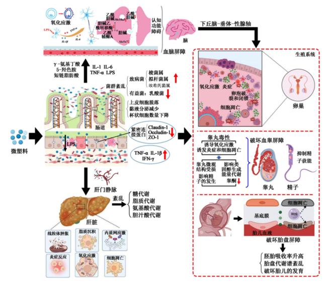
微塑料暴露对肺脏、肾脏和心脏等机体器官产生的不利影响已逐渐被揭示,其作用机制复杂且相互关联。Xuan等[34]研究发现,微塑料暴露不仅可诱发小鼠肺脏损伤,还会引起肠道黏膜和菌群受损。肠道微生物群(如乳酸菌)衍生的代谢物乳酸进入血液循环后,积聚在肺脏、肠道和血清中,通过触发低氧诱导因子1α(hypoxia inducible factor 1α,HIF1α)/多聚嘧啶区结合蛋白1(poly-pyrimidine tract binding protein 1,PTBP1)信号通路,调节上皮-间质转化,从而加剧微塑料诱导的肺脏纤维化。微塑料对肾脏的损伤作用同样不容忽视。Liang等[35]发现,聚苯乙烯微塑料通过损害肠道屏障功能,提高补体5a(complement 5a,C5a)水平,并激活肾脏中的C5a/补体5a受体(complement 5a receptor,C5aR)通路,最终诱导小鼠肾脏损伤。Wang等[95]研究发现,微塑料通过诱导氧化应激引起线粒体稳态失衡,导致线粒体DNA泄漏到细胞质中。随后,细胞质中的线粒体DNA激活环状单磷酸鸟苷-单磷酸腺苷合成酶(cyclic GMP-AMP synthase,cGAS)/干扰素基因刺激因子(stimulator of interferon genes,STING)信号通路,促进炎症反应,最终诱导心肌细胞衰老。这一机制与Zhang等[36]的研究相呼应,其发现5 μm聚苯乙烯微塑料通过NF-κB/NLRP3/gasdermin D(GSDMD)轴触发细胞焦亡,并加剧心肌炎症。这些研究共同揭示了微塑料通过不同信号通路对心脏功能的多重影响。此外,Wang等[96]研究发现,聚苯乙烯微塑料暴露不仅降低了肌肉纤维的平均横截面积和直径,还增加了脂质沉积。同时,微塑料加剧了细胞内ROS的产生和氧化应激,通过降低p38 MAPK的磷酸化抑制成肌分化,并通过上调NF-κB的表达促进成脂分化。图3汇总了微塑料暴露对动物器官的损伤机制[42,68,81,97-98]
图3 微塑料暴露对动物器官的损伤机制

LPS:脂多糖 lipopolysaccharide;TNF-α:肿瘤坏死因子-α tumor necrosis factor-α;IL:白细胞介素 interleukin;Slc18a3:溶质载体家族18成员A3 solute carrier family 18 member A3;Slc5a7:溶质载体家族5成员A7 solute carrier family 5 member A7;TJ/AJ:紧密连接/黏附连接 tight junction/adherens junction;Claudin-1:密封蛋白-1;Occludin-1:闭合蛋白-1;ZO-1:闭锁小带蛋白-1 zonula occludens-1;ROS:活性氧 reactive oxygen species。

Fig.3 Mechanism of microplastic exposure causing organ damage in animals[42,68,81,97-98]

2 微塑料暴露下动物健康改善的绿色调控措施

通过深入探究调控措施对微塑料暴露动物健康的影响,不仅能更好地理解两者间的复杂关系,还能为保护动物健康、维护生态平衡提供重要的理论依据与实践指导。目前,绿色调控措施的研究主要集中在益生菌、微生物水解酶、植物提取物和粪菌移植等方面,这些措施通过不同的机制发挥作用,为减轻微塑料毒性提供了多角度的解决方案(表2)。其中,益生菌、植物提取物和粪菌移植等措施在缓解微塑料暴露动物机体损伤方面有一定的研究进展,而微生物水解酶尚没有在动物机体上的研究,但具有极大的开发潜力。
表2 微塑料暴露下动物健康改善的调控措施

Table 2 Regulatory measures to improve animal health under microplastic exposure

调控措施
Regulatory measures
动物
Animals
微塑料类型和尺寸
Types and sizes of microplastics
作用效果
Action effects
参考文献
References


益生菌
Probiotics
鼠李糖乳杆菌GG
Lactobacillus rhamnosus GG
C57BL/6小鼠 聚苯乙烯
(100 nm)
缓解黏膜异常分泌、改善肠道菌群、调节胆汁酸
代谢和降低肝脏炎症来保护暴露微塑料小鼠肝脏
[105]
乳杆菌属、长双歧杆菌和肠球菌属
Lactobacillus, Bifidobacterium
longum, and Enterococcus
ICR小鼠 聚苯乙烯
(5 μm)
抑制肠道微生物驱动的白细胞介素-17A
(IL-17A)信号传导的激活,减轻微塑料引起的
炎症反应、精子质量下降
[106]
植物乳杆菌P101
Lactobacillus plantarum P101
C57BL/6小鼠 邻苯二甲酸二(2-乙基
己基)酯和聚苯乙烯
(500 nm)
抗炎、抗氧化功效和重塑肠道菌群结构来减轻微
塑料和邻苯二甲酸二(2-乙基己基)酯的联合毒性损伤
[103]


微生物水解酶
Microbial hydrolases
聚对苯二甲酸乙二醇酯
水解酶HG1-5和HG2-5
Polyethylene terephthalate
hydrolases HG1-5 and HG2-5
人类消化道 聚对苯二甲酸
乙二醇酯(400 μm)
HG1-5能够在微塑料表面形成凹槽,而HG2-5则
进一步将聚对苯二甲酸乙二醇酯水解为单(2-羟乙基)
对苯二甲酸和对苯二甲酸
[109]
过氧化物酶Peroxidase 黄粉虫 聚苯乙烯(1 μm) 将聚苯乙烯微塑料转化为2-苯基氢醇和2-羟基苯甲酸甲酯 [112]


植物提取物
Plant extracts
表没食子儿茶素-3-没食子酸酯
Epigallocatechin-3-gallate
C57BL/6小鼠 聚苯乙烯
(5 μm)
优化肠道微生物组成、改善肠道屏障功能、
减少外周炎症、重塑血清代谢(尤其是调节嘌呤代谢)
[116]
丁酸白藜芦醇酯
Resveratrol butyrate esters
大鼠 聚苯乙烯
(5 μm)
调节一氧化氮水平、肾素-血管紧张素系统、肠道
微生物群及其代谢物来保护大鼠免受高血压和肾脏损伤
[118]
二氢咖啡酸
Dihydrocaffeic acid
大鼠 聚苯乙烯
(80 nm)
通过环磷酸腺苷(cAMP)/蛋白激酶A(PKA)/
磷酸化环磷酸腺苷反应元件结合蛋白(p-CREB)/
钙离子转运ATP酶B2(Atp2b2)信号通路缓解
聚苯乙烯微塑料诱导的神经炎症和细胞凋亡
[119]
矢车菊素-3-O-葡萄糖苷
Cyanidin-3-O-glucoside
C57BL/6小鼠 聚苯乙烯
(5 μm)
增加粪便聚苯乙烯微塑料排出,吲哚-3-
乳酸、N-乙酰血清素和L-色氨酸与其外源性代谢高度相关
[123]


粪菌移植
Fecal microbiota
transplantation
健康小鼠粪便微生物群
Fecal microbiota from healthy mice
C57BL/6小鼠 聚苯乙烯
(5 μm)
通过重组肠道微生物来缓解微塑料暴露
引起的神经损伤,并恢复了小鼠的学习和记忆能力
[128]
健康人类粪便微生物群
Fecal microbiota from healthy human
秀丽隐杆线虫 聚苯乙烯
(5 μm)
通过激活PMK-1/SKN-1通路,增加细胞内
抗氧化谷胱甘肽的产生,从而减轻微塑料毒性
[135]


其他
Others
褪黑激素
Melatonin
白化大鼠 聚乙烯
(4~6 μm)
提高黏蛋白分泌和紧密连接蛋白表达量、改善
聚苯乙烯微塑料诱导的空肠组织病理学和超微结构变化
[139]
小球藻、柠檬酸、番茄红素
Chlorella, citric acid, lycopene
尖齿胡鲶 聚乙烯
(<100 nm)
改善暴露于微塑料非洲鲶鱼肝脏、
肾脏和肠道的组织学损伤
[137]
维生素D
Vitamin D
斑马鱼 聚苯乙烯
(80 nm)
定向改变肠道病毒组来减轻聚苯乙烯微塑料暴露
引起的神经毒性和免疫毒性
[57]

2.1 益生菌

益生菌被定义为随食物或水进入胃肠道的活性微生物,通过改善宿主内部微生物的平衡来产生有益影响[99]。近年来,益生菌(乳酸菌)已被证明能够直接吸附微塑料,减少其在肠道中的积累[100]。Xia等[101]研究发现,肠系膜明串珠菌DH通过增加粪便中聚乙烯微塑料的排泄,减少其在肠道中的积累,从而减轻对肠道屏障的损害,并降低血液中微塑料的浓度。此外,益生菌在缓解微塑料暴露对肝脏的损伤方面也表现出显著效果。Dong等[102]研究发现,由枯草芽孢杆菌、屎肠球菌、罗伊氏乳杆菌和乳酸片球菌组成的益生菌混合物,通过其抗氧化特性和调节MAPK信号通路,有效抑制了尼罗罗非鱼中微塑料诱导的肝脏氧化应激损伤。这一发现与游涛[103]的研究相呼应,进一步证实了益生菌在保护肝脏健康中的重要作用。在免疫调节方面,Li等[104]的研究揭示了短双歧杆菌M-16V的多重作用,该菌株不仅通过抑制辅助型T细胞(T helper cell,Th)2和Th17淋巴细胞亚群、激活髓样分化因子88(myeloid differentiation factor 88,MyD88)表达并促进IL-12的产生来调节免疫反应,还通过降低普雷沃氏菌属(Prevotella)、艾森伯格菌属(Eisenbergiella)和肠单胞菌属(Intestinimonas)等菌属的丰度,部分修复了微塑料暴露导致的肠道菌群失调。此外,鼠李糖乳杆菌GG和益生菌补充剂(乳杆菌属、长双歧杆菌和肠球菌属)通过缓解黏膜异常分泌、改善肠道菌群、调节胆汁酸代谢、抑制IL-17A信号传导以及降低肝脏炎症,有效保护了暴露于微塑料的小鼠肝脏,并减轻了炎症反应,改善了精子质量下降[105-106]。以上研究表明,益生菌通过吸附微塑料、减少肠道吸收、缓解氧化应激、调节免疫反应以及改善肠道菌群等多种机制,在减轻微塑料暴露引起的生物毒性中发挥了重要作用。

2.2 微生物水解酶

水解酶通过裂解聚合物表面的化学基团,将其转化为低聚物和单体[107],随后这些产物通过主动或被动运输进入细胞质中被同化[108]。以PET为例,Zhou等[109]研究发现,健康的人类肠道微生物群可能含有2种PET水解酶——HG1-5和HG2-5。其中,HG1-5能够在微塑料表面形成凹槽,而HG2-5则进一步将PET聚合物分解成单(2-羟乙基)对苯二甲酸和对苯二甲酸。同样,Zhang等[110]通过肠道宏基因组和宏转录组测序,从人类肠道中鉴定出一种新型PET水解酶HGMP01,该酶展现出高效水解PET纳米颗粒的能力。值得注意的是,Lopez-Lorenzo等[111]研究首次证实,PET水解酶WT和S238A能够将人血清中的PET微塑料解聚为无毒的单体对苯二甲酸和单(2-羟乙基)对苯二甲酸酯,从而缓解微塑料在机体内的毒性作用;体外试验同样表明,这2种水解酶对细胞活力无显著影响,验证了其安全性。含有苯环的聚苯乙烯是一种较难降解的微塑料类型。Kang等[112]研究表明,从黄粉虫肠道中分离的霍氏肠杆菌LG3能够利用多种过氧化物酶将聚苯乙烯微塑料转化为2-苯基氢醇和2-羟基苯甲酸甲酯。此外,来自昆虫幼虫肠道的克雷伯菌EMBL-1(Klebsiella sp. EMBL-1)则通过氢化酶破坏聚氯乙烯的表面结构[113],聚氯乙烯是一种具有高疏水性和结晶性的难降解微塑料,而克雷伯菌属的潜在的降解能力为处理这类微塑料提供了新的可能性。综上所述,不同微生物及其分泌的酶类在处理不同类型微塑料方面展现出显著的潜力,并且在机体肠道微生物中分离得到。但微生物水解酶在缓解动物机体毒性这方面尚没有研究,未来可以通过携带微塑料降解酶的工程菌或微藻来缓解微塑料对动物机体的损伤,其作用机制还需要进一步探究。

2.3 植物提取物

植物提取物来源广泛,是一种安全、绿色的天然产物,其生物活性物质包括多糖类、黄酮类、生物碱类、皂苷类和多酚类等,具有抗炎、抑菌和抗氧化等生物学功能[114]。绿茶中的主要活性成分表没食子儿茶素-3-没食子酸酯(epigallocatechin-3-gallate,EGCG)通过调节肠道菌群、抑制结肠、肝脏和全身炎症,以及减轻肝脏代谢紊乱,有效缓解微塑料暴露引起的损伤[115]。此外,EGCG还通过抑制海马体Toll样受体4(Toll-like receptor 4,TLR4)/MyD88/NF-κB信号通路发挥抗焦虑作用[116]。丁酸白藜芦醇和二氢咖啡酸作为天然多酚类化合物,在缓解微塑料诱导的损伤方面展现出显著的保护作用。丁酸白藜芦醇通过调节一氧化氮水平、肾素-血管紧张素系统以及肠道微生物群及其代谢物,在慢性肾病和微塑料联合暴露的幼年大鼠模型中有效预防高血压和肾脏损伤[117-118]。二氢咖啡酸则通过激活环磷酸腺苷(cyclic adenosine monophosphate,cAMP)/蛋白激酶A(protein kinase A,PKA)/磷酸化环磷酸腺苷反应元件结合蛋白(phosphorylated cAMP response element binding protein,p-CREB)/钙离子转运ATP酶B2(ATPase plasma membrane Ca2+ transporting 2,Atp2b2)信号通路,缓解聚苯乙烯微塑料诱导的神经炎症和细胞凋亡[119]。麦芽酚作为红参中的天然芳香类化合物,通过改善肠道屏障功能、维持肠道微生物稳态,并激活单磷酸腺苷活化蛋白激酶(AMP-activated protein kinase,AMPK)/哺乳动物雷帕霉素靶蛋白(mammalian target of rapamycin,mTOR)通路促进自噬,减少聚苯乙烯微塑料诱导的肠上皮细胞凋亡[120-121]。矢车菊素-3-O-葡萄糖苷(calycin-3-O-glucoside,C3G)是一种从食物中提取的花青素,具有抗氧化、抗炎、调节糖和脂代谢、保护肠屏障和调节肠道菌群的多重功能[122]。研究表明,C3G能够有效缓解聚苯乙烯微塑料暴露引发的氧化应激和炎症反应,同时调节肠道微生物组扰动,抑制与炎症相关的功能细菌(如脱硫弧菌、螺杆菌等),并减少与炎症和人类疾病相关的细菌功能基因[123-124]。此外,C3G通过上调色氨酸代谢(莽草酸盐、L-色氨酸、吲哚-3-乳酸和N-乙酰血清素)和胆汁酸代谢(3β-羟基-5-胆酸、鹅去氧胆酸盐、牛磺酸和石胆酸)相关代谢物,进一步增强其保护作用[125]。以上植物提取物通过多靶点机制,为缓解微塑料暴露引起的多器官损伤提供了潜在的调控策略。

2.4 粪菌移植

粪菌移植是一种将健康供体的完整微生物群落转移到受体肠道中的方法,旨在通过重塑肠道微生物组成和功能来治疗疾病。研究表明,肠道微生物在降解微塑料方面具有一定潜力。例如,Rajendran等[126]通过16S rRNA基因测序分析,从鱼肠道中鉴定出能够促进微塑料降解的假单胞菌属,这些细菌同时表现出益生菌特性。同样,Jang等[127]从人类肠道中分离出多种对低密度聚乙烯和聚丙烯具有降解活性的细菌[如产酸克雷伯菌(Klebsiella oxytoca)、肺炎克雷伯菌(Klebsiella pneumoniae)、路氏肠杆菌(Enterobacter ludwigii)和四川肠杆菌(Enterobacter sichuanensis)]。此外,Sun等[128]研究发现,多西环素和聚苯乙烯微塑料的共同暴露会破坏小鼠肠道微生物群稳态,导致脑损伤和炎症,并通过肠-脑轴损害学习和记忆功能;值得注意的是,粪菌移植成功逆转了这些神经损伤,并恢复了认知功能。在探索缓解微塑料毒性的方法中,黄粉虫、大蜡螟等昆虫幼虫展现出微塑料生物降解能力[129-134],这表明其肠道中可能含有降解微塑料相关的菌群或酶。此外,Chu等[135]研究发现,将健康人类供体的粪便微生物群移植到秀丽隐杆线虫中,能够通过激活PMK-1/SKN-1通路,增加细胞内抗氧化谷胱甘肽的产生,从而减轻微塑料毒性。粪菌移植不仅能够修复微塑料暴露引起的肠道微生物群紊乱和神经损伤,还可能通过移植具有降解能力的微生物群,为缓解其他物种中微塑料毒性提供新的解决方案。

2.5 其他措施

除此之外,还有小球藻、褪黑素和维生素D等外源性添加剂在缓解微塑料暴露引起的机体损伤方面展现出显著潜力,相关研究也逐步揭示了其具体作用机制。例如,Zheng等[136]研究发现,小球藻通过激活MAPK和过氧化物酶体增殖物激活受体(peroxisome proliferator-activated receptor,PPAR)信号通路,有效减轻了不同粒径微塑料对尼罗罗非鱼造成的肠道损伤。这一发现与Sayed等[137]的研究相呼应,后者通过膳食补充小球藻、番茄红素和柠檬酸,显著改善了暴露于微塑料的非洲鲶鱼在肝脏、肾脏和肠道中的组织学损伤。这些研究共同表明,小球藻及其相关成分在缓解微塑料毒性方面具有潜在的应用价值。同时,维生素D通过定向改变肠道病毒组,有效减轻聚苯乙烯微塑料暴露引起的神经毒性和免疫毒性[57]。此外,褪黑激素作为一种由松果体产生的激素,不仅控制昼夜节律,还具有免疫调节、抗炎和抗氧化特性[138]。El Gazzar等[139]研究发现,褪黑激素通过提高黏蛋白分泌和紧密连接蛋白的表达,显著改善了聚苯乙烯微塑料诱导的空肠组织病理学和超微结构变化。

3 小结与展望

微塑料污染对畜禽健康的危害呈现多系统、跨器官的复合毒性效应,其通过破坏肠道屏障、诱导氧化应激与炎症反应、干扰代谢稳态及神经内分泌调控等机制,对消化、免疫、生殖及神经系统造成深远危害,严重威胁畜牧业的可持续发展和畜产品安全体系。值得注意的是,微塑料对畜禽早期生长发育的损伤是生命全程毒性链的关键起点,其通过表观遗传重编程、菌群-器官轴紊乱和内分泌干扰,在胚胎期、哺乳期和幼年期的敏感窗口期诱发不可逆损伤,并通过跨代传递扩大健康威胁。这提示亟需将微塑料污染防控纳入畜禽“从配子到出栏”的全周期健康管理体系,以阻断“早期暴露-成年发病”的毒性传递路径。当前,益生菌、微生物水解酶、植物提取物和粪菌移植等绿色调控技术虽在缓解微塑料毒性方面展现出应用潜力,但仍存在问题。一方面,这些措施的应用效果受畜禽品种、年龄、饲养环境及微塑料污染程度等因素影响,需进一步开展精细化与精准化研究;另一方面,调控措施的作用机制尚未完全明确,尤其是多种物质协同对抗微塑料危害的机制有待深入挖掘。
未来研究应重点关注以下方向:1)微塑料暴露的毒性机制纵深解析。结合单细胞测序、空间代谢组学等前沿技术,系统揭示微塑料在器官-细胞-分子水平的动态损伤路径,尤其是其对线粒体功能、表观遗传修饰及免疫微环境的作用。2)畜禽早期生长发育的毒性机制解析。深入探究胚胎期、哺乳期和幼年期敏感窗口期内,微塑料通过表观遗传重编程、菌群-器官轴紊乱和内分泌干扰诱发不可逆损伤的具体路径,明确不同发育阶段的关键靶器官与分子靶点。3)绿色调控技术优化。开发基于合成生物学的工程菌(如携带塑料降解酶的益生菌),探索植物提取物纳米递送系统,并筛选高效降解酶与天然活性成分的组合方案。4)畜禽全周期健康管理体系构建。建立“从配子到出栏”的微塑料暴露监测网络,整合早期预警、实时干预和跨代风险评估技术,阻断“早期暴露-成年发病”的毒性传递链,制定涵盖养殖全流程的污染防控标准。
[1]
KALIA V C, PATEL S K S, KARTHIKEYAN K K, et al. Manipulating microbial cell morphology for the sustainable production of biopolymers[J]. Polymers, 2024, 16(3):410.

[2]
VUPPALADADIYAM S S V, VUPPALADADIYAM A K, SAHOO A, et al. Waste to energy:trending key challenges and current technologies in waste plastic management[J]. Science of the Total Environment, 2024,913:169436.

[3]
CRISTALDI A, FIORE M, ZUCCARELLO P, et al. Efficiency of wastewater treatment plants (WWTPs) for microplastic removal:a systematic review[J]. International Journal of Environmental Research and Public Health, 2020, 17(21):8014.

[4]
李志青, 刘磊, 沈维军, 等. 微塑料来源及其在动物体内的暴露途径与毒性作用研究进展[J]. 动物营养学报, 2023, 35(9):5453-5464.

DOI

LI Z Q, LIU L, SHEN W J, et al. Advances in research on sources of microplastics and their exposure routes and toxic effects in animals[J]. Chinese Journal of Animal Nutrition, 2023, 35(9):5453-5464. (in Chinese)

DOI

[5]
LI A Y, WANG Y L, KULYAR M F, et al. Environmental microplastics exposure decreases antioxidant ability,perturbs gut microbial homeostasis and metabolism in chicken[J]. Science of The Total Environment, 2023, 856(Pt 1):159089.

[6]
SRIDHARAN S, KUMAR M, SINGH L, et al. Microplastics as an emerging source of particulate air pollution:a critical review[J]. Journal of Hazardous Materials, 2021,418:126245.

[7]
SHI X, XU T, CUI W, et al. Combined negative effects of microplastics and plasticizer DEHP:the increased release of nets delays wound healing in mice[J]. Science of The Total Environment, 2023,862:160861.

[8]
LESLIE H A, VAN VELZEN M J M, BRANDSMA S H, et al. Discovery and quantification of plastic particle pollution in human blood[J]. Environment International, 2022,163:107199.

[9]
PEÑALVER R, PÉREZ-ÁLVAREZ M D, CAMPILLO N, et al. Static headspace-gas chromatography with mass spectrometry for the assessment of the bioaccumulation of volatile organic compounds associated with microplastics in animal tissues[J]. Analytica Chimica Acta, 2025,1333:343412.

[10]
HE Y C, YIN R C. The reproductive and transgenerational toxicity of microplastics and nanoplastics:a threat to mammalian fertility in both sexes[J]. Journal of Applied Toxicology, 2024, 44(1):66-85.

[11]
PRATA J C, DA COSTA J P, LOPES I, et al. Environmental exposure to microplastics:an overview on possible human health effects[J]. Science of The Total Environment, 2020,702:134455.

[12]
张德明. 日粮添加枯草芽孢杆菌对生长育肥猪肠道屏障功能的影响[D].硕士学位论文. 杨凌: 西北农林科技大学, 2022.

ZHANG D M. Effects of dietary Bacillus subtilis supplementation on intestinal barrier function of growing-finishing pigs[D].Master’s Thesis. Yangling: Northwest A&F University, 2022. (in Chinese)

[13]
程学英, 肖清婷, 柯志彬, 等. 微塑料暴露对小鼠肠道菌群分布及代谢途径的影响[J]. 中国微生态学杂志, 2024, 36(9):1000-1008.

CHENG X Y, XIAO Q T, KE Z B, et al. The effect of microplastic exposure on the distribution and metabolic pathways of gut microbiota in mice[J]. Chinese Journal of Microecology, 2024, 36(9):1000-1008. (in Chinese)

[14]
ALI S, PEÑA A N, LAFAZANOS Y S, et al. What gastroenterologists should know about microplastics and nanoplastics[J]. Journal of Clinical Gastroenterology, 2025, 59(2):105-109.

DOI PMID

[15]
DJOUINA M, VIGNAL C, DEHAUT A, et al. Oral exposure to polyethylene microplastics alters gut morphology,immune response,and microbiota composition in mice[J]. Environmental Research, 2022, 212 (Pt B):113230.

[16]
HUANG W T, YIN H, YANG A A, et al. Influence of the co-exposure of microplastics and tetrabromobisphenol A on human gut:simulation in vitro with human cell Caco-2 and gut microbiota[J]. Science of The Total Environment, 2021,778:146264.

[17]
THERY J, LI L L, DAS S, et al. Multigenerational exposure of microplastics on the microbiota of E.affinis (copepod):a comparative study between biodegradable and nonbiodegradable microplastics[J]. Frontiers in Ecology and Evolution, 2023,11:1231346.

[18]
SHI X D, CHEN Z J, WEI W, et al. Toxicity of micro/nanoplastics in the environment:roles of plastisphere and eco-corona[J]. Soil & Environmental Health, 2023, 1(1):100002.

[19]
CHENG J G, MEISTERTZHEIM A L, LEISTENSCHNEIDER D, et al. Impacts of microplastics and the associated plastisphere on physiological,biochemical,genetic expression and gut microbiota of the filter-feeder amphioxus[J]. Environment International, 2023,172:107750.

[20]
LU L, WAN Z Q, LUO T, et al. Polystyrene microplastics induce gut microbiota dysbiosis and hepatic lipid metabolism disorder in mice[J]. Science of The Total Environment, 2018,631/632:449-458.

[21]
YANG H, XIONG H R, MI K H, et al. Toxicity comparison of nano-sized and micron-sized microplastics to goldfish Carassius auratus larvae[J]. Journal of Hazardous Materials, 2020,388:122058.

[22]
LIMONTA G, MANCIA A, BENKHALQUI A, et al. Microplastics induce transcriptional changes,immune response and behavioral alterations in adult zebrafish[J]. Scientific Reports, 2019, 9(1):15775.

[23]
LIU S, LI H, WANG J, et al. Polystyrene microplastics aggravate inflammatory damage in mice with intestinal immune imbalance[J]. Science of the Total Environment, 2022,833:155198.

[24]
LEI L L, WU S Y, LU S B, et al. Microplastic particles cause intestinal damage and other adverse effects in zebrafish Danio rerio and nematode Caenorhabditis elegans[J]. Science of The Total Environment, 2018,619/620:1-8.

[25]
SU Q L, WU J, TAN S W, et al. The impact of microplastics polystyrene on the microscopic structure of mouse intestine,tight junction genes and gut microbiota[J]. PLoS One, 2024, 19(6):e0304686.

[26]
WANG Q Q, BAI J L, NING B A, et al. Effects of bisphenol A and nanoscale and microscale polystyrene plastic exposure on particle uptake and toxicity in human Caco-2 cells[J]. Chemosphere, 2020,254:126788.

[27]
YONG C Q Y, VALIYAVEETTIL S, TANG B L. Toxicity of microplastics and nanoplastics in mammalian systems[J]. International Journal of Environmental Research and Public Health, 2020, 17(5):1509.

[28]
GRUBER M M, HIRSCHMUGL B, BERGER N, et al. Plasma proteins facilitates placental transfer of polystyrene particles[J]. Journal of Nanobiotechnology, 2020, 18(1):128.

DOI PMID

[29]
PRÜST M, MEIJER J, WESTERINK R H S. The plastic brain:neurotoxicity of micro- and nanoplastics[J]. Particle and Fibre Toxicology, 2020, 17(1):24.

[30]
SHENG S, HAN N X, WEI Y F, et al. Liver injury induced by exposure to polystyrene microplastics alone or in combination with cadmium in mice is mediated by oxidative stress and apoptosis[J]. Biological Trace Element Research, 2024, 202(5):2170-2183.

[31]
LIU Z Y, SOKRATIAN A, DUDA A M, et al. Anionic nanoplastic contaminants promote Parkinson’s disease-associated α-synuclein aggregation[J]. Science Advances, 2023, 9(46):eadi8716.

[32]
JIN H B, YAN M H, PAN C, et al. Chronic exposure to polystyrene microplastics induced male reproductive toxicity and decreased testosterone levels via the LH-mediated LHR/cAMP/PKA/StAR pathway[J]. Particle and Fibre Toxicology, 2022, 19(1):13.

DOI PMID

[33]
AN R, WANG X F, YANG L, et al. Polystyrene microplastics cause granulosa cells apoptosis and fibrosis in ovary through oxidative stress in rats[J]. Toxicology, 2021,449:152665.

[34]
XUAN L H, XU Z, LUO J H, et al. Lactate exacerbates lung damage induced by nanomicroplastic through the gut microbiota-HIF1a/PTBP1 pathway[J]. Experimental & Molecular Medicine, 2023, 55(12):2596-2607.

[35]
LIANG Y R, LIU D H, ZHAN J, et al. Polystyrene microplastics induce kidney injury via gut barrier dysfunction and C5a/C5aR pathway activation[J]. Environmental Pollution, 2024,342:122909.

[36]
ZHANG Y, YIN K, WANG D X, et al. Polystyrene microplastics-induced cardiotoxicity in chickens via the ROS-driven NF-κB-NLRP3-GSDMD and AMPK-PGC-1α axes[J]. Science of The Total Environment, 2022,840:156727.

[37]
XU R, CAO J W, LV H L, et al. Polyethylene microplastics induced gut microbiota dysbiosis leading to liver injury via the TLR2/NF-κB/NLRP3 pathway in mice[J]. Science of the Total Environment, 2024,917:170518.

[38]
YU H R, CHEN Q N, QIU W H, et al. Concurrent water- and foodborne exposure to microplastics leads to differential microplastic ingestion and neurotoxic effects in zebrafish[J]. Water Research, 2022,219:118582.

[39]
HUANG J N, WEN B, XU L, et al. Micro/nano-plastics cause neurobehavioral toxicity in discus fish (Symphysodon aequifasciatus):insight from brain-gut-microbiota axis[J]. Journal of Hazardous Materials, 2022,421:126830.

[40]
XIE L L, CHEN T L, LIU J Y, et al. Intestinal flora variation reflects the short-term damage of microplastic to the intestinal tract in mice[J]. Ecotoxicology and Environmental Safety, 2022,246:114194.

[41]
LUO T, WANG D, ZHAO Y, et al. Polystyrene microplastics exacerbate experimental colitis in mice tightly associated with the occurrence of hepatic inflammation[J]. Science of The Total Environment, 2022,844:156884.

[42]
YIN K, WANG D X, ZHANG Y, et al. Dose-effect of polystyrene microplastics on digestive toxicity in chickens (Gallus gallus):multi-omics reveals critical role of gut-liver axis[J]. Journal of Advanced Research, 2022,52:3-18.

[43]
CHEN X W, XUAN Y, CHEN Y W, et al. Polystyrene nanoplastics induce intestinal and hepatic inflammation through activation of NF-κB/NLRP3 pathways and related gut-liver axis in mice[J]. Science of The Total Environment, 2024,935:173458.

[44]
WANG Q P, CHEN G L, TIAN L Y, et al. Neuro- and hepato-toxicity of polystyrene nanoplastics and polybrominated diphenyl ethers on early life stages of zebrafish[J]. Science of The Total Environment, 2023, 857(Pt 2):159567.

[45]
HUANG T, ZHANG W J, LIN T T, et al. Maternal exposure to polystyrene nanoplastics during gestation and lactation induces hepatic and testicular toxicity in male mouse offspring[J]. Food and Chemical Toxicology, 2022,160:112803.

[46]
HU Q L, WANG H, HE C, et al. Polystyrene nanoparticles trigger the activation of p38 MAPK and apoptosis via inducing oxidative stress in zebrafish and macrophage cells[J]. Environmental Pollution, 2021,269:116075.

[47]
MU Y W, SUN J Y, LI Z Y, et al. Activation of pyroptosis and ferroptosis is involved in the hepatotoxicity induced by polystyrene microplastics in mice[J]. Chemosphere, 2022, 291(Pt 2):132944.

[48]
ZHONG G L, RAO G, TANG L X, et al. Combined effect of arsenic and polystyrene-nanoplastics at environmentally relevant concentrations in mice liver:activation of apoptosis,pyroptosis and excessive autophagy[J]. Chemosphere, 2022,300:134566.

[49]
TIAN R, GUAN M, CHEN L, et al. Mechanism insights into the histopathological changes of polypropylene microplastics induced gut and liver in zebrafish[J]. Ecotoxicology and Environmental Safety, 2024,280:116537.

[50]
CHEN X B, ZHUANG J S, CHEN Q L, et al. Chronic exposure to polyvinyl chloride microplastics induces liver injury and gut microbiota dysbiosis based on the integration of liver transcriptome profiles and full-length 16S rRNA sequencing data[J]. Science of The Total Environment, 2022,839:155984.

[51]
SHI C Z, HAN X H, GUO W, et al. Disturbed gut-liver axis indicating oral exposure to polystyrene microplastic potentially increases the risk of insulin resistance[J]. Environment International, 2022,164:107273.

[52]
WANG C L, HOU M M, SHANG K Y, et al. Microplastics (polystyrene) exposure induces metabolic changes in the liver of rare minnow (Gobiocypris rarus)[J]. Molecules, 2022, 27(3):584.

[53]
DEL PIANO F, ALMROTH B C, LAMA A, et al. Subchronic oral exposure to polystyrene microplastics affects hepatic lipid metabolism,inflammation,and oxidative balance in gilthead seabream (Sparus aurata)[J]. Ecotoxicology and Environmental Safety, 2024,279:116455.

[54]
YIN K, WANG D X, ZHAO H J, et al. Polystyrene microplastics up-regulates liver glutamine and glutamate synthesis and promotes autophagy-dependent ferroptosis and apoptosis in the cerebellum through the liver-brain axis[J]. Environmental Pollution, 2022,307:119449.

[55]
WEN J, SUN H, YANG B W, et al. Environmentally relevant concentrations of microplastic exposure cause cholestasis and bile acid metabolism dysregulation through a gut-liver loop in mice[J]. Environmental Science & Technology, 2024, 58(4):1832-1841.

[56]
SHAN S, ZHANG Y F, ZHAO H W, et al. Polystyrene nanoplastics penetrate across the blood-brain barrier and induce activation of microglia in the brain of mice[J]. Chemosphere, 2022,298:134261.

[57]
TENG M M, LI Y X, ZHAO X L, et al. Vitamin D modulation of brain-gut-virome disorder caused by polystyrene nanoplastics exposure in zebrafish (Danio rerio)[J]. Microbiome, 2023, 11(1):266.

DOI PMID

[58]
KOPATZ V, WEN K, KOVÁCS T, et al. Micro- and nanoplastics breach the blood-brain barrier (BBB):biomolecular corona’s role revealed[J]. Nanomaterials, 2023, 13(8):1404.

[59]
BEKDASH R A. The cholinergic system,the adrenergic system and the neuropathology of Alzheimer’s disease[J]. International Journal of Molecular Sciences, 2021, 22(3):1273.

[60]
HAN S W, CHOI J, RYU K Y. Recent progress and future directions of the research on nanoplastic-induced neurotoxicity[J]. Neural Regeneration Research, 2024, 19(2):331-335.

[61]
WANG S W, HAN Q, WEI Z L, et al. Polystyrene microplastics affect learning and memory in mice by inducing oxidative stress and decreasing the level of acetylcholine[J]. Food and Chemical Toxicology, 2022,162:112904.

[62]
ZHOU R R, ZHOU D, YANG S X, et al. Neurotoxicity of polystyrene nanoplastics with different particle sizes at environment-related concentrations on early zebrafish embryos[J]. Science of The Total Environment, 2023,872:162096.

[63]
ZHANG Y, ZHANG X, YAN Q, et al. Melatonin attenuates polystyrene microplastics induced motor neurodevelopmental defect in zebrafish (Danio rerio) by activating nrf2-isl2a axis[J].Ecotoxicology and Environmental Safety, 2022,241:113754.

[64]
TELEANU R I, NICULESCU A G, ROZA E, et al. Neurotransmitters-key factors in neurological and neurodegenerative disorders of the central nervous system[J]. International Journal of Molecular Sciences, 2022, 23(11):5954.

[65]
JEONG S, JANG S, KIM S S, et al. Size-dependent seizurogenic effect of polystyrene microplastics in zebrafish embryos[J]. Journal of Hazardous Materials, 2022,439:129616.

[66]
KUAI Y, CHEN Z N, XIE K, et al. Long-term exposure to polystyrene microplastics reduces macrophages and affects the microbiota-gut-brain axis in mice[J]. Toxicology, 2024,509:153951.

[67]
李佳熙, 姬向荣, 刘芙瑶, 等. 微塑料暴露引起“肠道菌群-肠-脑轴”毒性及作用机制研究进展[J]. 环境化学, 2025, 44(2):419-431.

LI J X, JI X R, LIU F Y, et al. Research progress on toxicity and mechanism of gut microbiota-gut-brain axis induced by microplastics and nanoplastics exposure[J]. Environmental Chemistry, 2025, 44(2):419-431. (in Chinese)

[68]
WANG J, YANG Y, SHI Y P, et al. Oxidized/unmodified-polyethylene microplastics neurotoxicity in mice:perspective from microbiota-gut-brain axis[J]. Environment International, 2024,185:108523.

[69]
YAN M Y, MAN S L, SUN B Y, et al. Gut liver brain axis in diseases:the implications for therapeutic interventions[J]. Signal Transduction and Targeted Therapy, 2023, 8(1):443.

[70]
YANG Q Y, DAI H X, CHENG Y, et al. Oral feeding of nanoplastics affects brain function of mice by inducing macrophage IL-1 signal in the intestine[J]. Cell Reports, 2023, 42(4):112346.

[71]
TENG M M, ZHAO X L, WANG C J, et al. Polystyrene nanoplastics toxicity to zebrafish:dysregulation of the brain-intestine-microbiota axis[J]. ACS Nano, 2022, 16(5):8190-8204.

[72]
ZHANG C, LI Y Y, YU H B, et al. Co-exposure of nanoplastics and arsenic causes neurotoxicity in zebrafish (Danio rerio) through disrupting homeostasis of microbiota-intestine-brain axis[J]. Science of The Total Environment, 2024,912:169430.

[73]
ZHANG Y Y, LUO C Y, HUANG P X, et al. Effects of chronic exposure to a high fat diet,nutritive or non-nutritive sweeteners on hypothalamic-pituitary-adrenal (HPA) and -gonadal (HPG) axes of male Sprague-Dawley rats[J]. European Journal of Nutrition, 2024, 63(6):2209-2220.

[74]
WANG M, WU Y, LI G G, et al. The hidden threat:unraveling the impact of microplastics on reproductive health[J]. Science of The Total Environment, 2024,935:173177.

[75]
张亚萍, 田蕾, 谢晓倩, 等. 妊娠期纳米聚苯乙烯纳米塑料暴露对胎鼠发育及神经毒性研究[J]. 中国应用生理学杂志, 2022, 38(6):760-765.

ZHANG Y P, TIAN L, XIE X Q, et al. Effects of nanopolystyrene nanoplastic exposure on the development and neurotoxicity of fetal rats during gestation[J]. Chinese Journal of Applied Physiology, 2022, 38(6):760-765. (in Chinese)

[76]
JIN H B, MA T, SHA X X, et al. Polystyrene microplastics induced male reproductive toxicity in mice[J]. Journal of Hazardous Materials, 2021,401:123430.

[77]
ZHAO T X, SHEN L J, YE X, et al. Prenatal and postnatal exposure to polystyrene microplastics induces testis developmental disorder and affects male fertility in mice[J]. Journal of Hazardous Materials, 2023,445:130544.

[78]
WU H, LIU Q H, YANG N X, et al. Polystyrene-microplastics and DEHP co-exposure induced DNA damage,cell cycle arrest and necroptosis of ovarian granulosa cells in mice by promoting ROS production[J]. Science of The Total Environment, 2023,871:161962.

[79]
WEI Y X, ZHOU Y, LONG C L, et al. Polystyrene microplastics disrupt the blood-testis barrier integrity through ROS-Mediated imbalance of mTORC1 and mTORC2[J]. Environmental Pollution, 2021,289:117904.

[80]
AMEREH F, BABAEI M, ESLAMI A, et al. The emerging risk of exposure to nano (micro) plastics on endocrine disturbance and reproductive toxicity:from a hypothetical scenario to a global public health challenge[J]. Environmental Pollution, 2020,261:114158.

[81]
XU W Q, YUAN Y Y, TIAN Y, et al. Oral exposure to polystyrene nanoplastics reduced male fertility and even caused male infertility by inducing testicular and sperm toxicities in mice[J]. Journal of Hazardous Materials, 2023,454:131470.

[82]
王佳博, 李榕, 娜孜拉·赛提尼亚孜, 等. 聚苯乙烯微塑料致雄性小鼠生殖损伤及铁死亡的作用机制[J]. 环境与职业医学, 2025, 42(2):224-231.

WANG J B, LI R, NAZIRA S, et al. Mechanisms of reproductive toxicity and ferroptosis induced by polystyrene microplastics in male mice[J]. Journal of Environmental & Occupational Medicine, 2025, 42(2):224-231. (in Chinese)

[83]
HU J N, QIN X L, ZHANG J W, et al. Polystyrene microplastics disturb maternal-fetal immune balance and cause reproductive toxicity in pregnant mice[J]. Reproductive Toxicology, 2021,106:42-50.

[84]
DUSZA H M, KATRUKHA E A, NIJMEIJER S M, et al.Uptake,transport,and toxicity of pristine and weathered micro- and nanoplastics in human placenta cells[J]. Environmental Health Perspectives, 2022, 130(9):97006.

[85]
ENYOH C E, DURU C E, OVUORAYE P E, et al. Evaluation of nanoplastics toxicity to the human placenta in systems[J]. Journal of Hazardous Materials, 2023,446:130600.

[86]
AGHAEI Z, MERCER G V, SCHNEIDER C M, et al. Maternal exposure to polystyrene microplastics alters placental metabolism in mice[J]. Metabolomics, 2022, 19(1):1.

DOI PMID

[87]
BARCELÓ D, PICÓ Y, ALFARHAN A H. Microplastics:detection in human samples,cell line studies,and health impacts[J]. Environmental Toxicology and Pharmacology, 2023,101:104204.

[88]
FOURNIER S B, D’ERRICO J N, ADLER D S, et al. Nanopolystyrene translocation and fetal deposition after acute lung exposure during late-stage pregnancy[J]. Particle and Fibre Toxicology, 2020, 17(1):55.

DOI PMID

[89]
徐嘉萍. 微塑料对有机化合物的吸附/解吸及对鸡毒性的研究[D].博士学位论文. 天津: 南开大学, 2022.

XU J P. Sorption/desorption of organic chemicals on microplastics and the toxicity on Gallus gallus domesticus[D]. Ph.D.Thesis. Tianjin: Nankai University, 2022. (in Chinese)

[90]
ZUO J L, HUO T B, DU X, et al. The joint effect of parental exposure to microcystin-LR and polystyrene nanoplastics on the growth of zebrafish offspring[J]. Journal of Hazardous Materials, 2021,410:124677.

[91]
SHU Q S, XIE S L, JUNAID M, et al. MPs and PFOS single and combined exposure significantly alter genetic expressions of growth hormone and insulin growth factor-related biomarkers during zebrafish embryonic development[J]. Science of The Total Environment, 2024,949:174925.

[92]
蔡威. 生命早期营养精典[M]. 上海: 上海交通大学出版社, 2019:1-21.

CAI W. Early life nutrition[M]. Shanghai: Shanghai Jiao Tong University Press, 2019:1-21. (in Chinese)

[93]
DE OLIVEIRA C G, BOAVENTURA T P, SANTOS F A C D, et al. Effects of polypropylene microparticles ingestion on the growth and physiology of Piaractus brachypomus,and its accumulation and tissue damage[J]. Aquatic Toxicology, 2025,283:107338.

[94]
CHEN J H, CHEN G H, PENG H Q, et al. Microplastic exposure induces muscle growth but reduces meat quality and muscle physiological function in chickens[J]. Science of The Total Environment, 2023,882:163305.

[95]
WANG K H, DU Y P, LI P X, et al. Nanoplastics causes heart aging/myocardial cell senescence through the Ca2+/mtDNA/cGAS-STING signaling cascade[J]. Journal of Nanobiotechnology, 2024, 22(1):96.

[96]
WANG S C, LIU J, YAO Y J, et al. Polystyrene microplastics-induced ROS overproduction disrupts the skeletal muscle regeneration by converting myoblasts into adipocytes[J]. Journal of Hazardous Materials, 2021,417:125962.

[97]
BALALI H, MORABBI A, KARIMIAN M. Concerning influences of micro/nano plastics on female reproductive health: focusing on cellular and molecular pathways from animal models to human studies[J]. Reproductive Biology and Endocrinology, 2024, 22(1):141.

DOI PMID

[98]
YANG D Q, ZHU J D, ZHOU X S, et al. Polystyrene micro- and nano-particle co-exposure injures fetal thalamus by inducing ROS-mediated cell apoptosis[J]. Environment International, 2022,166:107362.

[99]
MOHAPATRA S K, RANVIR S, NIKAM P, et al. Alleviation of fertility disorders based on probiotics induced immune system improvement:a review[J]. Critical Reviews in Biomedical Engineering, 2019, 47(5):427-436.

[100]
ZHAO L L, DOU Q N, CHEN S Y, et al. Adsorption abilities and mechanisms of Lactobacillus on various nanoplastics[J]. Chemosphere, 2023,320:138038.

[101]
XIA X H, MA X Y, LIANG N, et al. Damage of polyethylene microplastics on the intestine multilayer barrier,blood cell immune function and the repair effect of Leuconostoc mesenteroides DH in the large-scale loach (Paramisgurnus dabryanus)[J]. Fish & Shellfish Immunology, 2024,147:109460.

[102]
DONG R, ZHOU C L, WANG S Y, et al. Probiotics ameliorate polyethylene microplastics-induced liver injury by inhibition of oxidative stress in Nile tilapia (Oreochromis niloticus)[J]. Fish & Shellfish Immunology, 2022,130:261-272.

[103]
游涛. 植物乳杆菌P101缓解微塑料和邻苯二甲酸二(2-乙基己基)酯的联合毒性及机制研究[D].硕士学位论文. 南昌: 南昌大学, 2024.

YOU T. Study on Lactiplantibacillus plantarum P101 ameliorates thecombined toxicity of microplastics and di (2-ethylhexyl) phthalate and its mechanism[D].Master’s Thesis. Nanchang: Nanchang University, 2024. (in Chinese)

[104]
LI N, WANG J, LIU P, et al. Multi-omics reveals that Bifidobacterium breve M-16V may alleviate the immune dysregulation caused by nanopolystyrene[J]. Environment International, 2022,163:107191.

[105]
YU C H, XU Y W, WEI Y P, et al. Gut microbiota and liver metabolomics reveal the potential mechanism of Lactobacillus rhamnosus GG modulating the liver toxicity caused by polystyrene microplastics in mice[J]. Environmental Science and Pollution Research, 2024, 31(4):6527-6542.

[106]
ZHANG Y C, HOU B L, LIU T, et al. Probiotics improve polystyrene microplastics-induced male reproductive toxicity in mice by alleviating inflammatory response[J]. Ecotoxicology and Environmental Safety, 2023,263:115248.

[107]
ALI S S, ELSAMAHY T, AL-TOHAMY R, et al. Plastic wastes biodegradation:mechanisms,challenges and future prospects[J]. Science of The Total Environment, 2021,780:146590.

[108]
KOTHAWALE S S, KUMAR L, SINGH S P. Role of organisms and their enzymes in the biodegradation of microplastics and nanoplastics:a review[J]. Environmental Research, 2023,232:116281.

[109]
ZHOU H Q, SHI S B, YOU Q H, et al. Polyethylene terephthalate hydrolases in human gut microbiota and their implications for human health[J]. Microorganisms, 2024, 12(1):138.

[110]
ZHANG G Q, DU J K, ZHANG C S, et al. Identification of a PET hydrolytic enzyme from the human gut microbiome unveils potential plastic biodegradation in human digestive tract[J]. International Journal of Biological Macromolecules, 2024, 283(Pt 3):137732.

[111]
LOPEZ-LORENZO X, HUETING D, BOSSHARD E, et al. Degradation of PET microplastic particles to monomers in human serum by PETase[J]. Faraday Discussions, 2024,252:387-402.

[112]
KANG M G, KWAK M J, KIM Y. Polystyrene microplastics biodegradation by gut bacterial Enterobacter hormaechei from mealworms under anaerobic conditions:anaerobic oxidation and depolymerization[J]. Journal of Hazardous Materials, 2023,459:132045.

[113]
ZHANG Z, PENG H R, YANG D C, et al. Addendum: polyvinyl chloride degradation by a bacterium isolated from the gut of insect larvae[J]. Nature Communications, 2025, 16(1):3665.

[114]
黎容, 张桢, 许娟, 等. 植物提取物对家禽免疫功能的调控及其作用机制[J]. 湖南畜牧兽医, 2024(5):53-56.

LI R, ZHANG Z, XU J, et al. Regulation and mechanism of plant extracts on immune function of poultry[J]. Hunan Journal of Animal Science & Veterinary Medicine, 2024(5):53-56. (in Chinese)

[115]
ZHANG K K, YANG J Z, CHEN L J, et al. Gut microbiota participates in polystyrene microplastics-induced hepatic injuries by modulating the gut-liver axis[J]. ACS Nano, 2023, 17(15):15125-15145.

[116]
YANG J Z, ZHANG K K, LIU Y, et al. Epigallocatechin-3-gallate ameliorates polystyrene microplastics-induced anxiety-like behavior in mice by modulating gut microbe homeostasis[J]. Science of The Total Environment, 2023,892:164619.

[117]
HSU C N, HOU C Y, TAIN Y L. Preventive aspects of early resveratrol supplementation in cardiovascular and kidney disease of developmental origins[J]. International Journal of Molecular Sciences, 2021, 22(8):4210.

[118]
HUANG Y N, HSU C N, HOU C Y, et al. Resveratrol butyrate esters reduce hypertension in a juvenile rat model of chronic kidney disease exacerbated by microplastics[J]. Nutrients, 2024, 16(23):4076.

[119]
JIANG W B, HU C, CHEN Y Y, et al. Dysregulation of the microbiota-brain axis during long-term exposure to polystyrene nanoplastics in rats and the protective role of dihydrocaffeic acid[J]. Science of The Total Environment, 2023,874:162101.

[120]
靳明慧. 麦芽酚对聚苯乙烯纳米塑料致肠道损伤的保护作用及机制研究[D].硕士学位论文. 长春: 吉林农业大学, 2023.

JIN M H. Protective effect and mechanisms of maltol on polystyrene nanoplastic-induced intestinal injury[D].Master’s Thesis. Changchun: Jilin Agricultural University, 2023. (in Chinese)

[121]
JIN M H, HU J N, ZHANG M, et al. Maltol attenuates polystyrene nanoplastic-induced enterotoxicity by promoting AMPK/mTOR/TFEB-mediated autophagy and modulating gut microbiota[J]. Environmental Pollution, 2023,322:121202.

[122]
BAEK H, SANJAY, PARK M, et al. Cyanidin-3-O-glucoside protects the brain and improves cognitive function in APPswe/PS1ΔE9 transgenic mice model[J]. Journal of Neuroinflammation, 2023, 20(1):268.

DOI PMID

[123]
CHEN W, TU P C, YE X, et al. Cyanidin-3-O-glucoside impacts fecal discharge of polystyrene microplastics in mice:potential role of microbiota-derived metabolites[J]. Toxicology and Applied Pharmacology, 2022,453:116212.

[124]
CHEN W, ZHU R Y, YE X, et al. Food-derived cyanidin-3-O-glucoside reverses microplastic toxicity via promoting discharge and modulating the gut microbiota in mice[J]. Food & Function, 2022, 13(3):1447-1458.

[125]
CHEN W, ZHENG X D, YAN F J, et al. Modulation of gut microbial metabolism by cyanidin-3-O-glucoside in mitigating polystyrene-induced colonic inflammation:insights from 16S rRNA sequencing and metabolomics[J]. Journal of Agricultural and Food Chemistry, 2024, 72(13):7140-7154.

[126]
RAJENDRAN K, RAJENDIRAN R, RAVICHANDRAN R, et al. Investigation of microplastic accumulation in Rastrelliger kanagurta fish gut and microplastic degradation behaviour of existing gut bacteria Pseudomonas sp.[J]. Archives of Microbiology, 2022, 204(10):626.

[127]
JANG Y J, NYAMJAV I, KIM H R, et al. Identification of plastic-degrading bacteria in the human gut[J]. Science of The Total Environment, 2024,929:172775.

[128]
SUN H, YANG B W, ZHU X K, et al. Oral exposure of polystyrene microplastics and doxycycline affects mice neurological function via gut microbiota disruption:the orchestrating role of fecal microbiota transplantation[J]. Journal of Hazardous Materials, 2024,467:133714.

[129]
WANG Q J, CHEN H J, GU W Q, et al. Biodegradation of aged polyethylene (PE) and polystyrene (PS) microplastics by yellow mealworms (Tenebrio molitor larvae)[J]. Science of The Total Environment, 2024,927:172243.

[130]
DING M Q, DING J, ZHANG Z R, et al. Biodegradation of various grades of polyethylene microplastics by Tenebrio molitor and Tenebrio obscurus larvae:effects on their physiology[J]. Journal of Environmental Management, 2024,358:120832.

[131]
MATYAKUBOV B, LEE T J. Optimizing polystyrene degradation,microbial community and metabolite analysis of intestinal flora of yellow mealworms,Tenebrio molitor[J]. Bioresource Technology, 2024,403:130895.

[132]
DING M Q, DING J, YANG S S, et al. Effects of plastic aging on biodegradation of polystyrene by Tenebrio molitor larvae:insights into gut microbiome and bacterial metabolism[J]. Science of The Total Environment, 2024,953:176130.

[133]
JANAKIEV T, MILOŠEVIC D, PETROVIC M, et al. Chironomus riparius larval gut bacteriobiota and its potential in microplastic degradation[J]. Microbial Ecology, 2023, 86(3):1909-1992.

[134]
WANG S, SHI W, HUANG Z C, et al. Complete digestion/biodegradation of polystyrene microplastics by greater wax moth (Galleria mellonella) larvae:direct in vivo evidence,gut microbiota independence,and potential metabolic pathways[J]. Journal of Hazardous Materials, 2022, 423(Pt B):127213.

[135]
CHU Q, ZHANG S, YU X, et al. Fecal microbiota transplantation attenuates nano-plastics induced toxicity in Caenorhabditis elegans[J]. Science of The Total Environment, 2021,779:146454.

[136]
ZHENG Y, LU X X, HU J W, et al. Chlorella alleviates the intestinal damage of tilapia caused by microplastics[J]. Chemosphere, 2024,353:141644.

[137]
SAYED A E H, HANA M N, HAMED M, et al. Protective efficacy of dietary natural antioxidants on microplastic particles-induced histopathological lesions in African catfish (Clarias gariepinus)[J]. Environmental Science and Pollution Research, 2023, 30(9):24424-24440.

[138]
XU S Q, LI L L, WU J, et al. Melatonin attenuates sepsis-induced small-intestine injury by upregulating SIRT3-mediated oxidative-stress inhibition,mitochondrial protection,and autophagy induction[J]. Frontiers in Immunology, 2021,12:625627.

[139]
EL GAZZAR W B, SLIEM R E, BAYOUMI H B, et al. Melatonin alleviates intestinal barrier damaging effects induced by polyethylene microplastics in albino rats[J]. International Journal of Molecular Sciences, 2023, 24(17):13619.

Outlines

/