REVIEW

Research Progress on Central Mechanisms of Heat Stress Affecting Animal Appetite

  • YE Ziyuan ,
  • WANG Li’na , *
Expand
  • Guangdong Provincial Key Laboratory of Animal Nutrition Regulation, College of Animal Science, South China Agricultural University, Guangzhou 510642, China
*associate professor, E-mail:

Received date: 2025-03-18

  Online published: 2025-10-15

Abstract

Heat stress refers to a series of non-specific physiological responses triggered by animals to cope with high-temperature environments, leading to reduced feed intake, subsequent disease susceptibility, and compromised production performance. Feeding behavior in animals is regulated by the central nervous system (CNS), which integrates appetite-regulating signals and stress stimuli through key hypothalamic nuclei including the arcuate nucleus (ARC), preoptic area (POA), and paraventricular nucleus (PVN). This integration modulates the expression of appetite-related neuropeptides and the activation of neural projections, ultimately governing feeding behavior. The mechanisms underlying heat stress-induced appetite alterations involve complex CNS regulatory networks, primarily mediated by specific neural circuit activation and neurotransmitter dynamics that influence feeding patterns and dietary preferences. This review summarized recent research progress in molecular mechanisms and neural circuitry of CNS-mediated appetite regulation under heat stress, with a focus on hypothalamic nuclei projections and their functional connectivity. It aims to provide references for elucidating the biological basis of heat stress-induced appetite alterations in animals and developing anti-stress interventions.

Cite this article

YE Ziyuan , WANG Li’na . Research Progress on Central Mechanisms of Heat Stress Affecting Animal Appetite[J]. Chinese Journal of Animal Nutrition, 2025 , 37(10) : 6594 -6600 . DOI: 10.12418/CJAN2025.536

环境温度一直被视为是影响动物生长和健康的重要的因素之一。温度过高引起的热应激主要是指动物机体处于高温环境下产生的非特异性应答反应的总和,常伴随着各种生理和行为的不利变化[1-2]。摄食是动物生存的本能。在热应激情况下,动物摄食量和体重会明显下降,饲料转化率增加,食欲也会受到影响,导致死亡率和发病率升高,生产性能下降[3]。热应激对食欲的影响机制涉及复杂调控网络,包括能量稳态调节、代谢平衡维持、摄食行为改变及感觉信息整合等[4-5]。这些调控信号最终汇集到中枢神经系统(central nervous system,CNS),经CNS进行整合处理,进而影响摄食相关的神经环路,实现对食欲的调控。近年研究表明,热应激主要通过影响边缘系统的关键核团[如弓状核(ARC)、视前区(POA)、室旁核(PVN)和臂旁核(PBN)等],改变下丘脑食欲相关神经肽的表达与神经投射激活程度,最终抑制动物摄食行为。因此,本文主要就近年来报道的热应激影响动物食欲的中枢机制进行综述,以期为畜禽生产中应对高温环境带来的不利影响提供理论参考。

1 热应激对动物摄食及相关行为的影响

众所周知,热应激会使动物的摄食行为受到抑制。据已有的研究表明,当暴露于应激刺激时,动物首先倾向于改变它们的行为以适应新的环境条件[6-7]。热应激条件下,肉鸡优先表现出“栖息”、“抬翅”和“喘息”等典型热调节行为,其次是“饮水”和“摄食”等基础代谢活动调节[8];猪会更倾向选择躺卧在湿度较高的区域,靠水分蒸发帮助其散热,尽管猪的日摄食次数增加,但由于单次进食时长和摄食量同步减少,最终导致每天的总进食时间缩短,日摄食量也随之降低[9-10]。热应激按对动物的影响可分为两大类,即“急性”和“慢性”。急性热应激是指环境温度短暂和快速的上升,持续时间相对较短(数小时)。慢性热应激是指持续的高环境温度,或每天只在相同时间段内存在高温,持续时间相对较长(数天至数周),允许动物适应环境。急性与慢性热应激都会对动物的摄食及相关行为产生影响,进而影响动物的摄食量和日增重[11-12]。有研究发现,与常温[(20±1) ℃]条件下相比,慢性(7 d)热应激[(35±1) ℃]处理导致猪摄食量降低了约36%,而急性(24 h)热应激[(35±1) ℃]条件下摄食量降低了64%[13]
另外,可口的食物能够刺激食欲,还能缓解压力等给人带来的不适反应[14]。然而,在热应激环境下,这种积极作用却会被抑制。研究发现,急性热应激限制了鸡的认知表现,使其对获得可口食物的动机减弱[15]。研究表明,急性(1 h)热应激(35 ℃)条件下肉鸡对可口食物黄粉虫的摄食偏好显著降低,表明热应激可以抑制动物对可口食物的摄食[16]。与此相似的是,在急性热应激条件下小鼠对高脂食物的摄入也受到明显抑制[17]
研究表明,热应激不仅会改变动物的摄食行为,影响动物的摄食量,还可能通过抑制动物对食物的偏好性来进一步影响其食欲,并对动物机体产生不利影响。其具体中枢调节机制有待进一步研究来揭示。

2 热应激对动物摄食相关核团的影响及调控环路

下丘脑区域包括多个功能性核团,是调节机体食物摄入的最关键部分[18]。除此之外,在下丘脑以外的区域,如脑桥和边缘系统,也在调节摄食行为和能量平衡中发挥重要作用。以往研究通常集中在常温条件下,而热应激下这些核团之间如何连接以及发挥怎样的功能来影响动物的摄食,仍有待探究。

2.1 下丘脑信号传导

2.1.1 ARC

下丘脑ARC是中枢神经系统中调控食欲的核心区域,其中存在2个具有拮抗功能的神经元群:共表达刺鼠相关蛋白(agouti-related protein,AgRP)和神经肽Y(neuropeptide Y,NPY)的神经元具有促进摄食的作用,而共表达阿片-促黑素细胞皮质素原(pro-opiomelanocortin,POMC)和可卡因-苯丙胺转录调节肽(cocaine and amphetamine regulate transcript,CART)的神经元则具有抑制摄食的作用,二者共同调控动物的摄食行为,是中枢神经系统内调控食欲的一级神经元[19]
研究发现,ARCAgRP神经元靶向各种下游脑区,如下丘脑室旁核(paraventricular nucleus of hypothalamic,PVN)、终纹床核(bed nucleus of the stria terminalis,BNST)、下丘脑外侧区(lateral hypothalamic area,LHA)和丘脑室旁核(paraventricular thalamic nucleus,PVT)等具有协调进食功能的核团,来驱动机体的进食行为[20]。在慢性(10 h/d,21 d)热应激[(36±1) ℃]情况下,动物下丘脑AgRP/NPY mRNA表达水平显著降低[21],食欲抑制信号黑皮质素受体4(melanocortin 4 receptor,MC4R)和POMC mRNA的表达水平都显著增加[21-23]。然而也有报道称,急性(2或5 h)热应激[(40±1) ℃]会引起雏鸡下丘脑中NPY基因的表达水平显著升高[24]。NPY具有“趋利避害”作用,在抵抗应激、缓解焦虑和恐惧以及调节摄食行为和能量稳态过程中发挥重要作用[25-27]。此外,NPY的升高可能有助于热应激期间的体温调节,因为有研究表明,NPY可以在正常环境温度下降低雏鸡的体温[28]。此外,也有研究发现,急性(48 min或6 h)热应激(35 ℃)并没有导致小鼠或肉鸡下丘脑POMC/CART mRNA表达水平变化[29-30]。研究发现,急性(1 h)热应激(39 ℃)条件下,ARCPOMC神经元中的辣椒素受体1(transient receptor potential vanilloid 1,TRPV1)样受体被激活,并且TRPV1样受体的表达导致食物摄入显著减少[31]

2.1.2 POA

POA位于下丘脑的前端,约有30%的神经元是热敏神经元,这些神经元能够感知大脑温度的变化,并通过调节体温来维持体温稳态[32]。在热应激条件下,γ-氨基丁酸(γ-aminobutyric acid,GABA)能神经元从POA投射至背内侧核(dorsomedial nucleus,DMN),抑制大鼠的颤抖与非颤抖性产热[33]。同时,POA内的多巴胺(dopamine,DA)神经元可诱导小鼠皮肤血管舒张[34]。POA中这些神经元群的激活也导致了摄食行为的变化。例如,激活内侧视前区(medial preoptic area,mPOA)神经元可引起小鼠进入冬眠,并表现出体温过低和厌食行为[35]。对mPOA神经元的顺行标记研究显示,mPOA神经元向多个食欲中枢投射,如ARC、PVN、LHA和DMN等[36]。mPOA核团中的温度敏感性神经元通过与ARC和PVN的突触连接,调节摄食和体温。在应对高环境温度时,这些神经元通过调节食物摄入和能量消耗来维持动物机体体温稳态[37-38]
有研究发现,急性(5 min)高温(35或40 ℃)环境下,内侧视前区的前腹侧和脑室周围部分(the anteroventral and periventricular portions of medial preoptic area,apmPOA)GABA能神经元可接受来自外侧臂旁核背侧亚核(dorsal subnucleus of the lateral parabrachial nucleus,LPBd)的谷氨酸(glutamic acid,GLU)能神经元传递的升温信号而被激活,抑制机体产热,而apmPOA投射到下游PVN的神经元不仅对高温敏感,而且对食物摄入有抑制作用[35]。研究表明,由apmPOA神经元调控的协调进食行为作为行为性体温调节机制,主要作用于维持能量稳态的生理过程,该核团仅在可能破坏能量稳态的环境条件下被特异性激活,而对能量稳态没有影响的局部热刺激通常不会做出反应[39]

2.1.3 PVN

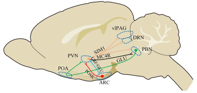
PVN不仅是启动并调节应激反应的关键核团,也是协调食欲的整合中心(图1)。PVN可接受来自AgRP和POMC神经元整合的多种激素和营养元素信号以调控能量平衡,如PVN中含有大量的MC4R,可接受来自POMC产生的α-促黑素细胞激素(α-melanocyte-stimulating hormone,α-MSH)从而起到抑制食欲的作用[40]。此外,PVN中的神经元还可向其他核团发出投射以调控摄食。有研究发现,PVN中的促甲状腺激素释放激素(thyrotropin-releasing hormone,TRH)神经元和垂体腺苷酸环化酶激活肽(pituitary adenylate cyclase-activating peptide,PACAP)神经元可通过GLU选择性地驱动AgRPARC神经元促进饥饿摄食,但不驱动促进饱腹感的POMCARC神经元[41]。PVN中表达专一转录因子1(single-minded 1,SIM1)的神经元投射到腹外侧中脑导水管周围灰质(ventrolateral periaqueductal gray matter,vIPAG)、中缝背核(dorsal raphe nucleus,DRN)或PBN也会引起食物摄入量下降[42]。然而热应激下PVN如何参与调控摄食的报道较少。
图1 下丘脑室旁核调控动物摄食的投射神经环路

PVN:下丘脑室旁核paraventricular nucleus of hypothalamic;ARC:弓状核arcuate nucleus;POA:视前区preoptic area;PBN:臂旁核parabrachial nucleus;vlPAG:腹外侧中脑导水管周围灰质ventrolateral periaqueductal gray;DRN:中缝背核dorsal raphe nucleus;SIM1:专一转录因子1 single-minded 1;MC4R:黑皮质素受体4 melanocortin 4 receptor;GLU:谷氨酸glutamic acid;CRF:促肾上腺皮质激素释放因子corticotropin-releasing factor;POMC:阿片-促黑素细胞皮质素原pro-opiomelanocortin。

Fig.1 Hypothalamic paraventricular nucleus regulates projection neural circuits in animal feeding

急性(3 h)热应激[(38±1) ℃]可引起雏鸡海马连合核(nucleus of the hippocampal commissure,NHpC)释放抑食因子促肾上腺皮质激素释放因子(corticotropin-releasing factor,CRF),启动应激反应,随后PVN释放CRF以维持应激反应,并可能对ARC中的NPY神经元产生负反馈,从而减少NPY的表达并导致食物摄入量的减少[43]。此外,PVN中包含表达CRF的神经元[44];有研究发现,中枢注射CRF时,大鼠的食物摄入被抑制并减轻体重,而应激反应过程中,PVN内神经元也会分泌CRF[45],推测PVNCRF可能参与了热应激引起的动物摄食量下降。
此外,热应激还能上调PVN中应激激素受体——糖皮质激素受体和盐皮质激素受体的表达,增强下丘脑-垂体-肾上腺轴(hypothalamic-pituitary-adrenal axis,HPA轴)负反馈,进一步抑制食欲[45]

2.2 下丘脑外其他核团信号传导

2.2.1 PBN

PBN主要介导皮肤热感信号的传导,也被认为介导食欲抑制[46-47]。有报道称,孤束核(nucleus tractus solitarius,NTS)中的GLU能神经元可以激活PBN神经元的兴奋性并抑制进食[48];此外,臂旁外侧核(lateral parabrachial nucleus,LPBN)可接受来自PVNMC4R的投射,通过GLU激活LPBN的神经元活性从而抑制食欲[49],但在热应激条件下PBN/LPBN如何影响摄食报道较少。研究表明,急性(1 h)热应激(40 ℃)条件下,PBN中的温度响应型GLU能神经元可支配伸长细胞,产生血管内皮生长因子A(vascular endothelial growth factor A,VEGFA),抑制ARC中AgRP和酪氨酸羟化酶(tyrosine hydroxylase,TH)神经元的兴奋性,导致动物摄食量减少,并可能抑制动物在热应激下对偏好食物的摄食[50]

2.2.2 杏仁核(amygdala)

研究表明,中央杏仁核(central nucleus of the amygdala,CeA)也与摄食控制有关[51]。CeA中有丰富的NPY表达,热应激可能会通过抑制NPY的促食欲作用而引起摄食量下降[52]。有研究发现,内侧杏仁核(medial amygdala,MeA)可以对应激做出反应,并且ARCPOMC→MeA回路的激活可减少短期食物摄入[53],因此ARCPOMC→MeA回路也可能导致应激诱导的食欲减退。此外,还有研究发现,慢性束缚应激也可增加ARCPOMC神经元的活性[54],因此推测热应激可能会通过激活ARCPOMC→MeA回路而引起动物摄食减少。

2.2.3 中脑腹侧被盖区(ventral tegmental area,VTA)

研究发现,中脑边缘奖赏系统VTA中的DA神经元可接受ARC中的NPY的激活,进而投射到伏隔阂(nucleus accumbens,NAc),刺激NAc释放DA,而DA会促使动物产生愉悦摄食行为,促进摄食量增加[55]
目前,热应激对动物摄食偏好的影响及机制鲜有报道。但已有证据表明,应激可以通过中枢神经系统来减弱机体对奖励敏感性的反应[56]。急性(12 h)热应激(37 ℃)通过抑制VTA内TH的表达,降低DA合成,从而削弱奖赏系统对摄食行为的正向调控[46]。最新研究发现,急性(12 h)热应激(37 ℃)主要通过影响NAc而不是VTA来调节动物的摄食偏好,可能是通过降低NAc区域的DA水平来影响奖赏系统,进而抑制动物对偏好食物的摄入。此外,NAc中的促肾上腺皮质激素释放因子受体2——环磷腺苷效应元件结合蛋白(cAMP-responsive element binding protein,CREB)通路可能也介导了热应激减少摄食的过程[17]

3 小结与展望

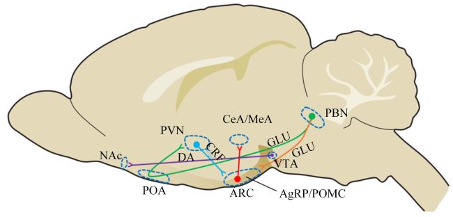
作为机体能量代谢在中枢的整合器,下丘脑通过协调食欲调控与体温调节两大生理功能,在动物热应激状态下的摄食行为调控中占据核心地位。随着神经环路示踪技术和光遗传学等研究手段的进步,在小鼠和大鼠等模式动物中,热应激下参与调控动物摄食行为的神经环路正被逐步揭示(图2)。
图2 热应激下参与调控动物摄食行为的神经环路

ARC:弓状核 arcuate nucleus;PVN:下丘脑室旁核 paraventricular nucleus of hypothalamic;POA:视前区 preoptic area;PBN:臂旁核 parabrachial nucleus;CeA:中央杏仁核 central nucleus of the amygdala;MeA:内侧杏仁核 medial amygdala;VTA:中脑腹侧被盖区 ventral tegmental area;NAc:伏隔阂 nucleus accumbens;GLU:谷氨酸 glutamic acid;DA:多巴胺 dopamine;CRF:促肾上腺皮质激素释放因子corticotropin-releasing factor;AgRP:刺鼠相关蛋白 agouti-related protein;POMC:阿片-促黑素细胞皮质素原 pro-opiomelanocortin。

Fig.2 Neural circuits involved in regulation of feeding behavior in animals under heat stress

值得注意的是,这些特定的中枢食欲调控环路既可通过协同作用维持摄食稳态,亦可独立响应代谢信号实现对摄食行为的调控。然而,在热应激条件下,影响畜禽摄食的研究主要集中在与摄食相关的基因或蛋白表达水平上,具体中枢神经环路机制研究尚不充分,仍处于起步阶段,具有广阔的研究前景。因此,未来的研究工作有必要进一步深入探究CNS对畜禽摄食行为调控的潜在中枢调控机制,这将为建立热应激条件下精准营养调控方案提供理论依据,也将为开发新型抗应激饲料添加剂奠定神经生物学基础。
[1]
SERVIENTO A M, LABUSSIÈRE E, CASTEX M, et al. Effect of heat stress and feeding management on growth performance and physiological responses of finishing pigs[J]. Journal of Animal Science, 2020, 98(12):skaa387.

[2]
OUCHI Y, TANIZAWA H, SHIRAISHI J I, et al. Repeated thermal conditioning during the neonatal period affects behavioral and physiological responses to acute heat stress in chicks[J]. Journal of Thermal Biology, 2020,94:102759.

[3]
KATIYAR R, GONMEI C, DEORI S, et al. Effect of heat stress on pig production and its mitigation strategies:a review[J]. Tropical Animal Health and Production, 2025, 57(3):139.

[4]
CROSS A J, BROWN-BRANDL T M, KEEL B N, et al. Feeding behavior of grow-finish swine and the impacts of heat stress[J]. Translational Animal Science, 2020, 4(2):txaa023.

[5]
GONZALEZ-RIVAS P A, CHAUHAN S S, HA M, et al. Effects of heat stress on animal physiology,metabolism,and meat quality:a review[J]. Meat Science, 2020,162:108025.

[6]
MACK L A, FELVER-GANT J N, DENNIS R L, et al. Genetic variations alter production and behavioral responses following heat stress in 2 strains of laying hens[J]. Poultry Science, 2013, 92(2):285-294.

DOI PMID

[7]
COLLIER R J, BAUMGARD L H, ZIMBELMAN R B, et al. Heat stress:physiology of acclimation and adaptation[J]. Animal Frontiers, 2019, 9(1):12-19.

[8]
CARTONI MANCINELLI A, BALDI G, SOGLIA F, et al. Impact of chronic heat stress on behavior,oxidative status and meat quality traits of fast-growing broiler chickens[J]. Frontiers in Physiology, 2023,14:1242094.

[9]
MARTÍNEZ-MACIPE M, MAINAU E, MANTECA X, et al. Environmental and management factors affecting the time budgets of free-ranging Iberian pigs reared in Spain[J]. Animals, 2020, 10(5):798.

[10]
POULLET N, RAUW W M, RENAUDEAU D, et al. Plasticity of feeding behaviour traits in response to production environment (temperate vs.tropical) in group-housed growing pigs[J]. Scientific Reports, 2022, 12(1):847.

[11]
HAO Y, FENG Y J, YANG P G, et al. Nutritional and physiological responses of finishing pigs exposed to a permanent heat exposure during three weeks[J]. Archives of Animal Nutrition, 2014, 68(4):296-308.

DOI PMID

[12]
XIONG Y, YI H, WU Q, et al. Effects of acute heat stress on intestinal microbiota in grow-finishing pigs,and associations with feed intake and serum profile[J]. Journal of Applied Microbiology, 2020, 128(3):840-852.

[13]
QU H, AJUWON K M. Adipose tissue-specific responses reveal an important role of lipogenesis during heat stress adaptation in pigs[J]. Journal of Animal Science, 2018, 96(3):975-989.

DOI PMID

[14]
LEOW S, DIMMOCK J A, GUELFI K J, et al. Understanding the determinants of stress-induced eating-a qualitative study[J]. Appetite, 2021,165:105318.

[15]
IYASERE O S, OYETUNJI D E, WHETO M, et al. Effect of acute heat stress on cognitive performance of chickens in a feed-related discriminant task[J]. Journal of Thermal Biology, 2021,98:102914.

[16]
FENG X J, YE Z Y, XIE K L, et al. Effects of heat stress on the feeding preference of yellow-feathered broilers and its possible mechanism[J]. Journal of Thermal Biology, 2024,124:103959.

[17]
CHEN L S, WU H Y, LI Y X, et al. Corticotropin-releasing factor receptor type 2 in the midbrain critically contributes to the hedonic feeding behavior of mice under heat stress[J]. Biochemical and Biophysical Research Communications, 2022,602:77-83.

[18]
SOUSA-FERREIRA L, DE ALMEIDA L P, CAVADAS C. Role of hypothalamic neurogenesis in feeding regulation[J]. Trends in Endocrinology and Metabolism, 2014, 25(2):80-88.

[19]
BRÜNING J C, FENSELAU H. Integrative neurocircuits that control metabolism and food intake[J]. Science, 2023, 381(6665):eabl7398.

[20]
BETLEY J N, CAO Z F H, RITOLA K D, et al. Parallel,redundant circuit organization for homeostatic control of feeding behavior[J]. Cell, 2013, 155(6):1337-1350.

[21]
LI Z Q, ZHAO Y L, ZHUANG Y, et al. Effects of N-acetyl-L-cysteine on serum indices and hypothalamic AMPK-related gene expression under chronic heat stress[J]. Frontiers in Veterinary Science, 2022,9:936250.

[22]
ZHAO N, MU L, CHANG X Y, et al. Effects of varying intensities of heat stress on neuropeptide Y and proopiomelanocortin mRNA expression in rats[J]. Biomedical Reports, 2020, 13(5):39.

DOI PMID

[23]
GREENE E S, ARDAKANI M A, DRIDI S. Effects of an herbal adaptogen feed-additive on feeding-related hypothalamic neuropeptides in chronic cyclic heat-stressed chickens[J]. Neuropeptides, 2024,106:102439.

[24]
ITO K, BAHRY M A, HUI Y, et al. Acute heat stress up-regulates neuropeptide Y precursor mRNA expression and alters brain and plasma concentrations of free amino acids in chicks[J]. Comparative Biochemistry and Physiology Part A:Molecular & Integrative Physiology, 2015,187:13-19.

[25]
NISHIMURA H, WANG Y, ELHUSSINY M Z, et al. Central administration of neuropeptide Y reduces the cellular heat stress response and may enhance spleen antioxidative functions in heat-exposed chicks[J]. Neuroscience Letters, 2022,784:136749.

[26]
SAH R, GERACIOTI T D. Neuropeptide Y and posttraumatic stress disorder[J]. Molecular Psychiatry, 2013, 18(6):646-655.

DOI PMID

[27]
ZHANG L, BIJKER M S, HERZOG H. The neuropeptide Y system:pathophysiologicaL and therapeutic implications in obesity and cancer[J]. Pharmacology & Therapeutics, 2011, 131(1):91-113.

[28]
TACHIBANA T, SATO M, OIKAWA D, et al. Intracerebroventricular injection of neuropeptide Y modifies carbohydrate and lipid metabolism in chicks[J]. Regulatory Peptides, 2006, 136(1/2/3):1-8.

[29]
HEPLER C, FOY C E, HIGGINS M R, et al. The hypophagic response to heat stress is not mediated by GPR109A or peripheral β-OH butyrate[J]. American Journal of Physiology.Regulatory,Integrative and Comparative Physiology, 2016, 310(10):R992-R998.

[30]
LEI L, HEPENG L, XIANLEI L, et al. Effects of acute heat stress on gene expression of brain-gut neuropeptides in broiler chickens[J]. Journal of Animal Science, 2013, 91(11):5194-5201.

DOI PMID

[31]
JEONG J H, LEE D K, LIU S M, et al. Activation of temperature-sensitive TRPV1-like receptors in ARC POMC neurons reduces food intake[J]. PLoS Biology, 2018, 16(4):e2004399.

[32]
ZHOU Q, FU X, XU J H, et al. Hypothalamic warm-sensitive neurons require TRPC4 channel for detecting internal warmth and regulating body temperature in mice[J]. Neuron, 2023, 111(3):387-404.e8.

[33]
CONCEIÇÃO E P S, MADDEN C J, MORRISON S F. Neurons in the rat ventral lateral preoptic area are essential for the warm-evoked inhibition of brown adipose tissue and shivering thermogenesis[J]. Acta Physiologica, 2019, 225(4):e13213.

[34]
ABBOTT S B G, SAPER C B. Median preoptic glutamatergic neurons promote thermoregulatory heat loss and water consumption in mice[J]. The Journal of Physiology, 2017, 595(20):6569-6583.

DOI PMID

[35]
QIAN S W, YAN S M, PANG R Q, et al. A temperature-regulated circuit for feeding behavior[J]. Nature Communications, 2022, 13(1):4229.

[36]
ZHANG G W, SHEN L, TAO C, et al. Medial preoptic area antagonistically mediates stress-induced anxiety and parental behavior[J]. Nature Neuroscience, 2021, 24(4):516-528.

[37]
ALCANTARA I C, TAPIA A P M, APONTE Y, et al. Acts of appetite:neural circuits governing the appetitive,consummatory,and terminating phases of feeding[J]. Nature Metabolism, 2022, 4(7):836-847.

[38]
YU S, FRANÇOIS M, HUESING C, et al. The hypothalamic preoptic area and body weight control[J]. Neuroendocrinology, 2018, 106(2):187-194.

DOI PMID

[39]
FLOURIS A D. Functional architecture of behavioural thermoregulation[J]. European Journal of Applied Physiology, 2011, 111(1):1-8.

DOI PMID

[40]
KRASHES M J, LOWELL B B, GARFIELD A S. Melanocortin-4 receptor-regulated energy homeostasis[J]. Nature Neuroscience, 2016, 19(2):206-219.

DOI PMID

[41]
KRASHES M J, SHAH B P, MADARA J C, et al. An excitatory paraventricular nucleus to AgRP neuron circuit that drives hunger[J]. Nature, 2014, 507(7491):238-242.

[42]
STERNSON S M, EISELT A K. Three pillars for the neural control of appetite[J]. Annual Review of Physiology, 2017,79:401-423.

[43]
BOHLER M, GILBERT E R, CLINE M A. Reduced food intake during exposure to high ambient temperatures is associated with molecular changes in the nucleus of the hippocampal commissure and the paraventricular and arcuate hypothalamic nuclei[J]. General and Comparative Endocrinology, 2020,298:113576.

[44]
RICHARDSON R D, OMACHI K, KERMANI R, et al. Intraventricular insulin potentiates the anorexic effect of corticotropin releasing hormone in rats[J]. American Journal of Physiology:Regulatory, Integrative and Comparative Physiology, 2002, 283(6):R1321-R1326.

[45]
VOM BERG-MAURER C M, TRIVEDI C A, BOLLMANN J H, et al. The severity of acute stress is represented by increased synchronous activity and recruitment of hypothalamic CRH neurons[J]. Journal of Neuroscience, 2016, 36(11):3350-3362.

[46]
吴汉宇. 热应激对小鼠愉悦采食行为的影响及机制[D].硕士学位论文. 广州: 华南农业大学, 2020.

WU H Y. Effects of heat stress on hedonic feeding behavior in mice and its mechanism[D].Master’s Thesis. Guangzhou: South China Agricultural University, 2020. (in Chinese)

[47]
CARTER M E, SODEN M E, ZWEIFEL L S, et al. Genetic identification of a neural circuit that suppresses appetite[J]. Nature, 2013, 503(7474):111-114.

[48]
WU Q, CLARK M S, PALMITER R D. Deciphering a neuronal circuit that mediates appetite[J]. Nature, 2012, 483(7391):594-597.

[49]
JAIS A, BRÜNING J C. Arcuate nucleus-dependent regulation of metabolism-pathways to obesity and diabetes mellitus[J]. Endocrine Reviews, 2022, 43(2):314-328.

[50]
BENEVENTO M, ALPÁR A, GUNDACKER A, et al. A brainstem-hypothalamus neuronal circuit reduces feeding upon heat exposure[J]. Nature, 2024, 628(8009):826-834.

[51]
CAI H J, HAUBENSAK W, ANTHONY T E, et al. Central amygdala PKC-δ+ neurons mediate the influence of multiple anorexigenic signals[J]. Nature Neuroscience, 2014, 17(9):1240-1248.

[52]
MITCHELL C S, BEGG D P. The regulation of food intake by insulin in the central nervous system[J]. Journal of Neuroendocrinology, 2021, 33(4):e12952.

[53]
KWON E, JO Y H. Activation of the ARCPOMC→MeA projection reduces food intake[J]. Frontiers in Neural Circuits, 2020,14:595783.

[54]
QU N, HE Y L, WANG C M, et al. A POMC-originated circuit regulates stress-induced hypophagia,depression,and anhedonia[J]. Molecular Psychiatry, 2020, 25(5):1006-1021.

[55]
REZITIS J, HERZOG H, IP C K. Neuropeptide Y interaction with dopaminergic and serotonergic pathways:interlinked neurocircuits modulating hedonic eating behaviours[J]. Progress in Neuro-Psychopharmacology & Biological Psychiatry, 2022,113:110449.

[56]
IRONSIDE M, KUMAR P, KANG M S, et al. Brain mechanisms mediating effects of stress on reward sensitivity[J]. Current Opinion in Behavioral Sciences, 2018,22:106-113.

Outlines

/