RESEARCH PAPER

Study on Effects of Chitosan Oligosaccharides in Alleviating Liver Injury in Heat-Stressed Broilers

  • WANG Haoxuan ,
  • GAO Jinren ,
  • ZHANG Jia ,
  • LAN Ruixia , *
Expand
  • College of Coastal Agriculture Sciences, Guangdong Ocean University, Zhanjiang 524088, China
*lecturer, E-mail:

Received date: 2025-04-18

  Online published: 2025-11-14

Abstract

This experiment aimed to investigate the alleviating effects of chitosan oligosaccharides (COS) on liver injury in heat-stressed broilers. A total of 144 male 28-day-old Arbor Acres (AA) broilers with similar body weight were selected and randomly divided into 3 groups, with 6 replicates in each group and 8 broilers in each replicate. The broilers in the control group (CON group) were raised in a chicken house at (24±1) ℃, and those in the heat stress group (HS group) and the heat stress+200 mg/kg COS group (HSC200 group) were raised in chicken houses with a temperature of (34±1) ℃ from 08:00 to 18:00 and (24±1) ℃ at the other time. Meanwhile, the broilers in CON group and HS group were fed basal diets, while those in HSC200 group were fed the basal diet supplemented with 200 mg/kg COS. The experiment lasted for 14 days. The results showed as follows: 1) compared with CON group, the serum alanine aminotransferase (ALT) activity in HS group was significantly increased (P<0.05); compared with HS group, the serum ALT activity in HSC200 group was significantly decreased (P<0.05). 2) The histopathological analysis of liver tissues showed that compared with CON group, the hepatocytes in HS group were damaged, with certain bleeding, the cytoplasm was loose and transparent, with severe vacuolation, and some cells had inflammatory infiltration; compared with HS group, the swelling, bleeding and inflammatory infiltration of hepatocytes in HSC200 group were improved. 3) Compared with CON group, the malondialdehyde (MDA) content in liver in HS group was significantly increased (P<0.05), and the activities of total superoxide dismutase (T-SOD) and glutathione peroxidase (GSH-Px) as well as the mRNA relative expression levels of nuclear factor E2-related factor 2 (Nrf2), heme oxygenase-1 (HO-1) and catalase (CAT) in liver were significantly decreased (P<0.05); compared with HS group, the MDA content in liver in HSC200 group was significantly decreased (P<0.05), and the activities of T-SOD and GSH-Px as well as the mRNA relative expression levels of Nrf2, HO-1 and CAT in liver were significantly increased (P<0.05). 4) Compared with CON group, the mRNA relative expression levels of nuclear factor-κB p65 subunit (NF-κB p65) and interleukin-1β (IL-1β) in liver in HS group were significantly increased (P<0.05); compared with HS group, the NF-κB p65 mRNA relative expression level in liver in HSC200 group was significantly decreased (P<0.05). 5) Compared with CON group, the apoptosis rate of hepatocytes in HS group was significantly increased (P<0.05), the mRNA relative expression levels of heat shock protein 90 (HSP90), Caspase9 and B-cell lymphoma-2 associated X protein (Bax) were significantly increased (P<0.05), and the ratio of B-cell lymphoma-2 (Bcl-2) mRNA relative expression level to Bax mRNA relative expression level (Bcl-2/Bax) was significantly decreased (P<0.05); compared with HS group, the apoptosis rate of hepatocytes in HSC200 group was significantly decreased (P<0.05), and the mRNA relative expression levels of HSP90 and Caspase9 in liver were significantly decreased (P<0.05). In conclusion, dietary 200 mg/kg COS can alleviate the liver oxidative damage and inflammatory response in broilers induced by heat stress, reduce the apoptosis rate of hepatocytes, and thereby alleviate the histopathological damage of the liver.

Cite this article

WANG Haoxuan , GAO Jinren , ZHANG Jia , LAN Ruixia . Study on Effects of Chitosan Oligosaccharides in Alleviating Liver Injury in Heat-Stressed Broilers[J]. Chinese Journal of Animal Nutrition, 2025 , 37(11) : 7534 -7546 . DOI: 10.12418/CJAN2025.613

肉鸡品种的持续选育、高密度饲养模式、夏季极端天气的频发和全球气候变暖等多重诱因,使肉鸡在快速生长的同时,在生产过程中也极易发生热应激问题,导致肉鸡生长性能和肉品质下降、代谢紊乱、肠道功能损伤及免疫抑制,降低养殖效益[1-2]。热应激导致肠道通透性增加,伴随着肠道消化吸收功能抑制、肠道菌群结构变化及肝脏功能受损和代谢变化[3-4]。正常生理情况下,完整的肠道屏障功能保护肝脏免受肠道毒素、病原菌及其代谢物的侵袭,且肝脏自身的生物解毒功能和巨噬细胞等产生的抗炎因子,可以有效抑制肠道有害菌及其代谢产物诱导的炎症反应和氧化损伤[4-5]。但热应激损伤肠道屏障功能,增加肠道通透性和有害菌的数量,使有害菌及其代谢产物通过血液循环进入肝脏,导致氧化损伤并诱发炎症反应,损伤肝脏功能[6]。此外,肝脏是热应激的靶器官,热应激肉鸡肝脏出现组织病理损伤、肝细胞凋亡率增加以及氧化应激和炎症反应[7-9]。肝脏是机体重要的能量代谢器官,调节糖、脂和蛋白质的合成和分解[4]。肝脏炎症导致能量代谢异常,表现为骨骼肌蛋白质分解代谢增加,合成代谢减少,抑制肉鸡体重增加[9-12]。因此,缓解热应激肉鸡肝脏损伤可能是提高热应激肉鸡生长性能的途径之一。热应激引起的肉鸡肝脏氧化损伤和炎症反应在肝细胞凋亡和肝脏损伤中发挥重要作用,因此,缓解热应激肉鸡肝脏损伤的关键是提高肝脏的抗氧化能力和炎症反应[13-16]。壳寡糖(chitosan oligosaccharides,COS)是以甲壳素为原料,经过一系列处理得到的聚合度为2~10,由2~20个氨基葡萄糖通过β-1,4糖苷键连接而成的碱性氨基寡糖,具有抗炎、抗氧化、免疫调节、抗凋亡以及组织再生等多种生物学功能[17-19],并且已被证实可以缓解肝脏功能损伤[16,20-21]。虽然本研究团队前期的研究已经证实COS能改善热应激肉鸡生长性能,改善肠道健康和肉品质[22-24],但关于COS能否缓解热应激肉鸡肝脏损伤还未做相关研究。因此,本试验旨在通过研究COS对热应激肉鸡肝脏组织病理学损伤、抗氧化能力、炎症反应及细胞凋亡的影响,解析COS对热应激肉鸡肝脏损伤的缓解作用,为COS作为缓解热应激的饲料添加剂在肉鸡生产上的应用提供理论依据。

1 材料与方法

1.1 试验设计和饲粮

本试验方案经广东海洋大学滨海农业学院动物福利委员会批准(批准编号:SYXK-2018-0147)。试验选用144只体重接近的雄性28日龄爱拔益加(AA)肉鸡,随机分为3组,每组6个重复,每个重复8只鸡。其中,对照组(CON组)肉鸡饲养在(24±1) ℃的鸡舍,热应激组(HS组)和热应激+200 mg/kg COS组(HSC200组)饲养在08:00—18:00温度为(34±1) ℃、其余时间温度为(24±1) ℃的鸡舍;同时,CON组和HS组饲喂基础饲粮,HSC200组在基础饲粮的基础上添加200 mg/kg COS。试验期14 d。COS纯度为93.6%,平均分子质量为1 768 u,脱乙酰度≥90%。COS添加量依据前期预试验生长性能结果,饲粮添加200和400 mg/kg COS分别提高7.46%和1.78%平均日增重(ADG),提高1.61%和3.45%平均日采食量(ADFI),并降低12.66%和4.80%料重比(F/G)。
基础饲粮参照《鸡营养标准》(NY/T 33—2004)配制,其组成及营养水平见表1。试验期间通过加热器、空调和加湿器调控温湿度,肉鸡自由采食和饮水,按肉鸡饲养管理和免疫程序进行管理和免疫接种。
表1 基础饲粮组成及营养水平(风干基础)

Table 1 Composition and nutrient levels of the basal diet (air-dry basis)%

项目Items 含量Content
原料Ingredients
玉米Corn 55.00
小麦麸Wheat bran 2.00
豆粕Soybean meal 34.82
植物油Vegetable oil 5.00
石粉Limestone 0.50
食盐NaCl 0.30
磷酸氢钙CaHPO4 1.60
氯化胆碱Choline chloride 0.10
L-赖氨酸L-Lys 0.10
DL-蛋氨酸DL-Met 0.18
维生素预混料Vitamin premix1) 0.20
矿物质预混料Mineral premix2) 0.20
合计Total 100.00
营养水平Nutrient levels3)
代谢能ME/(MJ/kg) 12.82
粗蛋白质CP 19.92
总磷TP 0.44
钙Ca 0.93
赖氨酸Lys 0.45
蛋氨酸Met 1.30

1)维生素预混料为每千克饲粮提供 The vitamin premix provided the following per kg of the diet:VA 9 000 IU,VD3 3 240 IU,VE 6 IU,VK3 0.75 mg,VB1 1.5 mg,VB2 4.5 mg,VB6 1.5 mg,VB12 10 mg,生物素 biotin 0.2 mg,烟酰胺 nicotinamide 10.5 mg,叶酸 folic acid 0.45 mg,泛酸 pantothenic acid 9 mg。

2)矿物质预混料为每千克饲粮提供 The mineral premix provided the following per kg of the diet:Cu (as copper sulfate) 5~10 mg,Fe (as ferrous sulfate) 45~120 mg,Mn (as manganese sulfate) 45~85 mg,Zn (as zinc sulfate) 50~80 mg,Se (as sodium selenite) 0.2 mg,I (as potassium iodide) 0.15 mg.

3)粗蛋白质、总磷和钙含量分别参照GB/T 6432—2018、GB/T 6437—2018和GB/T 6436—2018进行测定;代谢能以及赖氨酸和蛋氨酸含量为计算值,参照《中国饲料成分及营养价值表(2024年第35版)》进行计算。The contents of CP, TP and Ca were determined in accordance with GB/T 6432—2018, GB/T 6437—2018 and GB/T 6436—2018, respectively; ME and contents of Lys and Met were calculated values in accordance with Tables of Feed Composition and Nutritive Values in China (35th edition, 2024).

1.2 样品采集

试验结束当日,禁食8 h后,每个重复随机选取1只鸡称重后,翅下采血并分离血清,用于检测血清谷草转氨酶(AST)和谷丙转氨酶(ALT)活性。随后放血屠宰,分离肝脏称重后,剪取2份1 g左右样品,液氮速冻后-80 ℃保存,分别用于肝脏抗氧化指标和相关基因表达测定;另取1 cm3左右肝脏组织固定于4%多聚甲醛溶液,用于肝脏组织病理学分析和TUNEL法检测组织细胞凋亡。

1.3 检测指标及方法

1.3.1 肝脏指数

肝脏指数计算公式为:
肝脏指数(g/kg)=肝脏重(g)/活重(kg)。

1.3.2 肝脏抗氧化指标

采用试剂盒(南京建成生物工程研究所)测定肝脏丙二醛(MDA)含量以及过氧化氢酶(CAT)、总超氧化物歧化酶(T-SOD)和谷胱甘肽过氧化物酶(GSH-Px)活性,操作步骤按照试剂盒说明书进行。

1.3.3 肝脏组织病理学分析

肝脏组织病理学分析参照Lan等[25]的方法,样品固定24 h后,经石蜡包埋、切片、苏木精-伊红(HE)染色和封片后,在显微镜下观察肝脏组织病理损伤情况并拍照。

1.3.4 肝脏相关基因表达

肝脏样品RNA提取、cDNA合成以及荧光定量PCR检测参照本课题组之前的方法[25],目的基因mRNA相对表达量的计算参照Livak等[26]的方法,以β-肌动蛋白(β-actin)为内参基因。引物序列见表2
表2 引物序列

Table 2 Primer sequences

基因
Genes
登录号
Accession number
引物序列
Primer sequences (5'—3')
β-肌动蛋白
β-actin
NM_205518.1 F:ATCCGGACCCTCCATTGTC
R:AGCCATGCCAATCTCGTCTT
核因子E2相关因子2
Nrf2
NM_205117.1 F:ATCACGAGCCCTGAAACCAA
R:GGCTGCAAAATGCTGGAAAA
血红素加氧酶-1
HO-1
XM_205344.1 F:ACTTCTATGGCAGCAACT
R:AATAGCGGGTGTAGGC
谷胱甘肽过氧化物酶1
GPX1
NM_000581.4 F:GATGAGATCCTGAGAGTGGTGGAC
R:TCATCAGGTAAGGTGGGCACAA
铜/锌超氧化物歧化酶
Cu/ZnSOD
NM_205064.1 F:TTGTCTGATGGAGATCATGGCTTC
R:TGCTTGCCTTCAGGATTAAAGTGAG
过氧化氢酶
CAT
NM_001031215.2 F:TACGGTTCTCCACTGTTGCTG
R:TGGATGAAGGATGGAAACAAC
Toll样受体4
TLR4
NM_001397379.1 F:CCTGCAACGGTCATCTCAG
R:GTCTCAGGGCTTGTTCTTCAG
核因子-κB p65亚基
NF-κB p65
NM_001396038.1 F:TGAAGAAACGGGAACTGGAAG
R:GGCACGGTTGTCATAGATGG
肿瘤坏死因子-α
TNF-α
XM_046927265.1 F:TGTGTATGTGCAGCAACCCGTAGT
R:GGCATTGCAATTTGGACAGAAGT
白细胞介素-1β
IL-1β
XM_046931582.1 F:GCTCTACATGTCGTGTGTGATGAG
R:TGTCGATGTCCCGCATGA
白细胞介素-6
IL-6
NC_052533.1 F:GCGAGAACAGCATGGAGATG
R:GTAGGTCTGAAAGGCGAACAG
白细胞介素-10
IL-10
NM_001004414.2 F:AGCTGACGGTGGACCTATTATT
R:GGCTTTGCGCTGGATTC
半胱天冬酶3
Caspase3
NM_204725.2 F:CCACCGAGATACCGGACTGT
R:AACTGCTTCGCTTGCTGTGA
半胱天冬酶9
Caspase9
XM_040689238.1 F:GTGTACCAGCTGCGAGCAGACC
R:GCTTTGAGGTTCCGCAGGGTC
B细胞淋巴瘤-2相关X蛋白
Bax
XM_040693909.1 F:ACTCTGCTGCTGCTCTCCTCTC
R:ATCCACGCAGTGCCAGATGTAATC
B细胞淋巴瘤-2
Bcl-2
NM_205339.3 F:ATCCACGCAGTGCCAGATGTAATC
R:CAGGAGAAATCGAACAAAGGC
热休克蛋白70
HSP70
NM_001006685.1 F:CGTCAGTGCTGTGGACAAGAGTA
R:CCTATCTCTGTTGGCTTCATCCT
热休克蛋白90
HSP90
NM_001109785.1 F:GAGTTTGACTGACCCGAGCA
R:TCCCTATGCCGGTATCCACA

1.3.5 肝细胞凋亡

TUNEL法检测肝细胞凋亡参照试剂盒(南京建成生物工程研究所)说明书进行,在荧光显微镜下对切片进行观察并拍照。每张切片随机选择10个视野,蓝色荧光为正常细胞,绿色荧光为凋亡细胞,统计阳性细胞(绿色)和阴性细胞(蓝色)的数量,计算肝细胞凋亡率。

1.4 数据统计分析

试验数据采用SAS 9.1.3软件进行单因素方差分析,并采用Duncan氏法进行组间差异的多重比较,结果以“平均值±标准误”形式表示,P<0.05表示差异显著。

2 结果与分析

2.1 COS对热应激肉鸡肝脏指数和肝脏功能损伤的影响

表3可知,HS组肉鸡血清ALT活性显著高于CON组(P=0.010),HSC200组血清ALT活性显著低于HS组(P=0.008)。
表3 COS对热应激肉鸡肝脏指数和肝脏功能损伤的影响

Table 3 Effects of COS on liver index and function impairment in heat-stressed broilers

项目
Items
组别Groups P
P-value
CON HS HSC200
肝脏重Liver weight/g 54.55±1.02 58.91±2.18 54.12±2.45 0.278
肝脏指数Liver index/(g/kg) 21.24±0.63 24.12±0.82 22.31±0.92 0.059
血清谷丙转氨酶Serum ALT/(U/L) 6.95±0.52b 9.24±0.54a 6.86±0.89b 0.048
血清谷草转氨酶Serum AST/(U/L) 5.17±0.83 8.38±1.60 4.60±0.92 0.096

同行数据肩标不同字母表示差异显著(P<0.05),相同字母或无字母表示差异不显著(P>0.05)。

In the same row, values with different letter superscripts indicated significant difference (P<0.05), while with the same letter or no letter superscripts indicated no significant difference (P>0.05). The same as below.

2.2 COS对热应激肉鸡肝脏组织病理损伤的影响

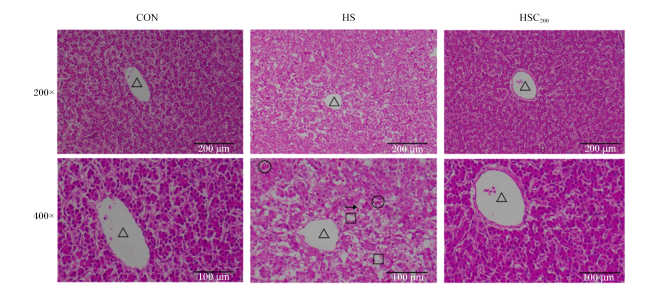
图1所示,肉鸡肝脏组织病理学分析结果显示,CON组肝脏无明显病理变化,肝细胞细胞质完好,无明显坏死现象;HS组肝细胞出现损伤,有一定出血,胞浆疏松化、透明,空泡化严重,部分细胞有炎性浸润;与HS组相比,HSC200组肝细胞肿胀、出血及炎症浸润状况得以改善。
图1 COS对热应激肉鸡肝脏组织病理损伤的影响

CON:对照组 control group;HS:热应激组 heat stress group;HSC200:热应激+200 mg/kg COS组 heat stress+200 mg/kg COS group。下图同 the same as below。

△:中央静脉 central vein;○:出血 bleeding;→:炎症浸润 inflammatory infiltration;□:细胞肿胀 cellular swelling。

Fig.1 Effects of COS on pathological injury of liver tissues in heat-stressed broilers

2.3 COS对热应激肉鸡肝脏抗氧化能力的影响

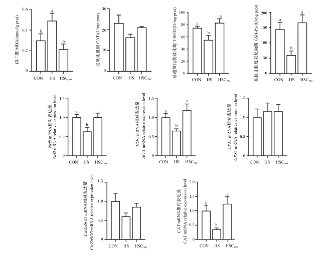
图2所示,与CON组相比,HS组肉鸡肝脏MDA含量显著提高(P=0.046),肝脏T-SOD(P=0.049)和GSH-Px(P=0.012)活性显著降低,肝脏核因子E2相关因子2(Nrf2)(P=0.042)、血红素加氧酶-1(HO-1)(P=0.045)和CAT(P=0.021)mRNA相对表达量显著降低。与HS组相比,HSC200组肉鸡肝脏MDA含量显著降低(P=0.008),肝脏T-SOD(P<0.001)和GSH-Px(P=0.003)活性显著提高,肝脏Nrf2(P=0.042)、HO-1(P=0.005)和CAT(P=0.002)mRNA相对表达量显著提高。
图2 COS对热应激肉鸡肝脏抗氧化能力的影响

数据柱标记不同字母表示差异显著(P<0.05)。下图同。

Fig.2 Effects of COS on liver antioxidant capacity in heat-stressed broilers

Value columns marked with different letters indicated significant difference (P<0.05). The same as below.

2.4 COS对热应激肉鸡肝脏炎症反应的影响

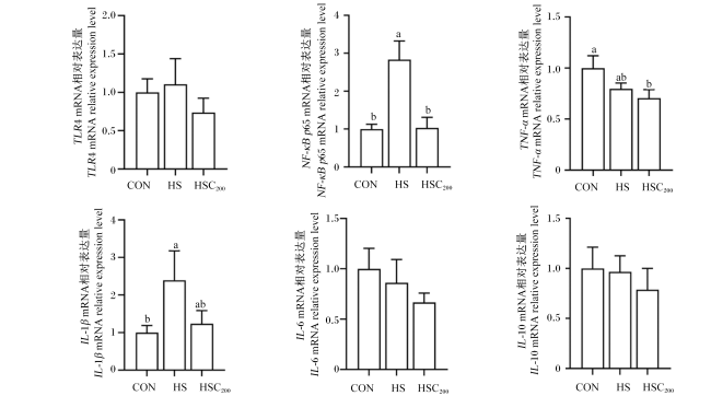
图3所示,与CON组相比,HS组肉鸡肝脏核因子-κB p65亚基(NF-κB p65)(P=0.002)和白细胞介素-1β(IL-1β)(P=0.031)mRNA相对表达量显著提高。与HS组相比,HSC200组肉鸡肝脏NF-κB p65 mRNA相对表达量显著降低(P=0.002)。
图3 COS对热应激肉鸡肝脏炎症反应的影响

Fig.3 Effects of COS on liver inflammatory response in heat-stressed broilers

2.5 COS对热应激肉鸡肝细胞凋亡的影响

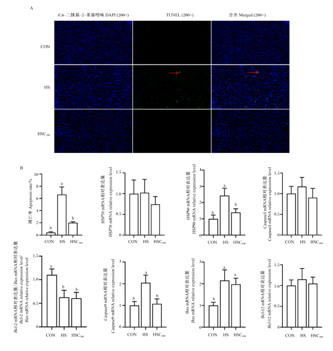
图4所示,与CON组相比,HS组肉鸡肝细胞凋亡率显著提高(P<0.001),肝脏热休克蛋白90(HSP90)(P=0.002)、半胱天冬酶9(Caspase9)(P=0.034)和B细胞淋巴瘤-2相关X蛋白(Bax)(P=0.018)mRNA相对表达量显著提高,肝脏B细胞淋巴瘤-2(Bcl-2)mRNA相对表达量/Bax mRNA相对表达量(Bcl-2/Bax)值显著降低(P=0.020)。与HS组相比,HSC200组肉鸡肝细胞凋亡率显著降低(P<0.001),肝脏HSP90(P=0.015)和Caspase9(P=0.046)mRNA相对表达量显著降低。
图4 COS对热应激肉鸡肝细胞凋亡及凋亡相关基因表达的影响

箭头处表示阳性细胞。

Fig.4 Effects of COS on hepatocyte apoptosis and apoptosis-related gene expression in heat-stressed broilers

The arrow indicates positive cells.

3 讨论

3.1 COS对热应激肉鸡肝脏指数和肝脏功能损伤的影响

肝脏是家禽营养代谢的核心器官,也是重要的解毒器官,极易受热应激影响,肝脏损伤破坏全身代谢平衡,影响家禽健康[9]。本研究结果显示,热应激提高肉鸡肝脏指数。Lu等[27]和Lan等[28]同样报道,热应激提高肉鸡肝脏指数,这与热应激导致肉鸡肝脏脂质代谢紊乱,肝脏脂质沉积增加相关;也可能是热应激造成肝脏损伤,引发肝脏病理性的代偿性肥大[9,27]。COS具有缓解应激和降脂的作用[20],Tao等[16]报道,COS降低非酒精脂肪肝小鼠肝脏重量;Deng等[29]报道,COS降低肥胖小鼠肝脏重量和肝脏指数。本研究结果显示,COS具有降低热应激肉鸡肝脏指数的趋势,这可能是因为COS具有抗氧化和抗炎功能,能够缓解肝脏损伤造成的病理性的代偿性肥大,同时降低肝脏脂质代谢导致的脂质异常沉积[29]
ALT和AST等血清转氨酶是评估肝脏损伤的指标[30]。Ma等[9]报道,热应激提高肉鸡血清ALT和AST活性,这是因为肝细胞受到损伤和刺激,细胞膜的通透性增大使ALT和AST释放进入血液。COS具有缓解肝脏损伤的功能,Chen等[31]报道,COS能有效缓解黄曲霉毒素诱导的小鼠肝脏氧化损伤,降低小鼠血清ALT和AST活性。Fathi等[30]和Lan等[32]报道,COS能分别降低冷应激和热应激肉鸡血清ALT和AST活性。本研究结果同样表明,COS能够降低热应激肉鸡血清ALT和AST活性,表明COS对热应激肉鸡肝脏损伤具有缓解作用。

3.2 COS对热应激肉鸡肝脏组织病理损伤的影响

肝脏是热应激损伤的靶器官,在热应激条件下,肝脏发生一系列的病理变化,包括细胞坏死、结构损伤、炎性浸润、凋亡增加及脂质异常沉积[7,9]。Tang等[8]和Ma等[9]报道,热应激肉鸡肝脏表面均出现明显病理性变化,表现为肝细胞有一定出血,细胞肿胀明显,胞浆呈现疏松化,部分细胞有炎性浸润的情况,这与本试验研究结果一致。COS具有缓解肝脏损伤的作用,Tao等[16]和Liu等[33]分别报道,COS通过缓解非酒精性脂肪肝小鼠和酒精性肝病小鼠肝脏的氧化损伤和炎性浸润以及减少脂质沉积进而缓解肝脏组织病理损伤。此外,Feng等[34]报道,COS改善非酒精性脂肪肝小鼠的肝细胞肿胀、细胞间界限模糊和脂质过度沉积。Tufan等[35]报道,COS降低日本鹌鹑肝脏脂滴沉积。本研究结果与上述研究结果相似,COS能够缓解肝细胞肿胀、出血及炎症浸润的状况,这可能是因为COS具有抗氧化和抗炎功能,通过提高热应激肉鸡肝脏的抗氧化能力,并抑制炎性损伤,进而缓解肝细胞凋亡和肝脏组织病理损伤。此外,COS具有细胞保护功能,可能通过在肝细胞的细胞膜外层形成保护膜,进而缓解肝细胞的损伤[36]

3.3 COS对热应激肉鸡肝脏抗氧化能力的影响

热应激导致氧化和抗氧化系统失衡,使机体产生过量的活性氧(ROS),诱导肝脏氧化损伤[37-38]。MDA是脂质过氧化的最终产物,是氧化应激的标志物[39]。抗氧化剂酶如T-SOD、CAT、GSH-Px等与自由基直接作用,是抵抗氧化损伤的第1道屏障[39]。本研究结果显示,热应激提高肉鸡肝脏MDA含量,降低肝脏T-SOD和GSH-Px活性,并下调肝脏Nrf2、HO-1和CAT mRNA相对表达量,表明热应激导致肝脏氧化损伤。Ding等[14]的报道同样表明,热应激提高肉鸡肝脏MDA含量,降低肝脏超氧化物歧化酶(SOD)和GSH-Px活性,下调肝脏Nrf2和HO-1 mRNA和蛋白表达量。张成等[40]也报道,热应激降低肉鸡肝脏CAT、GSH-Px和SOD活性,并提高肝脏MDA含量,下调肝脏Nrf2、HO-1和SOD mRNA相对表达量。COS具有抗氧化和清除自由基能力,其结构中的氨基、C2、C3和C6位的伯羟基和仲羟基,及还原端的羟基均能与自由基直接作用[41]。此外,COS还能通过激活Nrf2信号通路,上调下游抗氧化相关基因表达,提高抗氧化能力[41]。Wang等[42]报道,COS显著提高半乳糖腹腔注射大鼠肝脏Nrf2和CAT mRNA相对表达量。Liu等[33]报道,COS显著降低酒精性肝病小鼠肝脏MDA含量,提高肝脏SOD和CAT活性,上调肝脏Nrf2和HO-1蛋白表达量。Lan等[22,43]报道,COS显著提高胸肌铜/锌超氧化物歧化酶(Cu/ZnSOD)、CAT和谷胱甘肽过氧化物酶1(GPX1)以及十二指肠Nrf2、HO-1、Cu/ZnSODCATGPX1 mRNA相对表达量。本研究结果与之前的报道相似,COS降低热应激肉鸡肝脏MDA含量,提高肝脏GSH-Px和T-SOD活性,上调肝脏Nrf2、HO-1和CAT mRNA相对表达量,表明COS通过提高抗氧化酶活性,激活Nrf2信号通路上调抗氧化相关基因的表达,从而缓解热应激引起的肉鸡肝脏氧化损伤,进而有效缓解肝脏的组织病理损伤。

3.4 COS对热应激肉鸡肝脏炎症反应的影响

热应激导致肉鸡免疫失衡,并诱发炎症反应导致IL-1β、白细胞介素-6(IL-6)和肿瘤坏死因子-α(TNF-α)等促炎细胞因子过度产生,抑制白细胞介素-10(IL-10)等抗炎细胞因子产生,破坏炎症因子间的动态平衡,从而导致炎性损伤[44-45]。Toll样受体4(TLR4)/核因子-κB(NF-κB)信号通路是经典的炎症反应通路,正常情况下,NF-κB处于抑制状态,热应激上调TLR4的表达,启动TLR4/NF-κB信号通路的级联反应,促进NF-κB p65的核转位,从而激活相关炎症因子的表达[15,46]。Liu等[7]报道,热应激激活TLR4/NF-κB信号通路,上调肉鸡肝脏TLR4、NF-κB、TNF-α和IL-6的蛋白表达量。Lan等[22]报道,热应激上调肉鸡十二指肠IL-1βIL-6和TNF-α mRNA相对表达量,同时下调十二指肠IL-10 mRNA相对表达量。本试验结果表明,热应激上调肉鸡肝脏NF-κB p65和IL-1β mRNA相对表达量。COS具有抗炎功能,通过抑制TLR4信号通路激活,抑制NF-κB的核转位,进而抑制促炎细胞因子的表达,发挥抗炎功能[47]。Lan等[22]报道,COS下调热应激肉鸡十二指肠IL-1βIL-6的mRNA相对表达量,上调IL-10的mRNA相对表达量。本试验结果同样表明,COS下调热应激肉鸡肝脏NF-κB p65 mRNA相对表达量,并有降低肝脏IL-1β mRNA相对表达量的趋势,表明COS通过抑制TLR4/NF-κB信号通路的激活,下调相关炎症因子的表达,从而缓解肝脏炎性损伤,这与COS具有提升巨噬细胞和淋巴活性,增强免疫调节和抗炎功能相关[33]

3.5 COS对热应激肉鸡肝细胞凋亡的影响

肝细胞异常凋亡导致肝脏损伤,热应激导致的细胞凋亡与线粒体凋亡途径密切相关,大量研究证实,热应激显著提高细胞凋亡率[9,48-49]。热应激诱导ROS过量产生,并与脂类发生过氧化反应,损伤细胞膜、DNA及蛋白质,导致细胞凋亡[50]。此外,ROS可直接作为诱导凋亡的介质,破坏线粒体膜结构的完整性,导致膜上孔道开放,使线粒体细胞色素C释放,促进Caspase9的形成和半胱天冬酶3(Caspase3)活化,上调凋亡相关基因的表达,导致细胞凋亡[51]。Zhao等[13]报道,热应激提高肉鸡肝脏BaxCaspase9 mRNA相对表达量。周婉婷等[52]报道,急性热应激显著提高鸭肝细胞凋亡率,上调肝脏热休克蛋白70(HSP70)、HSP90和Caspase3 mRNA相对表达量,并下调Bcl-2蛋白表达量。刘峰成等[49]报道,热应激显著提高鸡脾脏淋巴细胞凋亡率和Bax蛋白表达量,并降低脾脏Bcl-2蛋白表达量。李建凤等[53]报道,热应激显著提高奶牛乳腺上皮细胞凋亡率,上调HSP70、HSP90、BaxCaspase3 mRNA相对表达量,下调Bcl-2 mRNA相对表达量以及Bcl-2/Bax值。本研究结果同样表明,热应激显著提高肉鸡肝细胞凋亡率,上调肝脏HSP90、BaxCaspase9 mRNA相对表达量,下调肝脏Bcl-2/Bax值,表明热应激通过激活半胱天冬酶信号通路诱导肝细胞凋亡,且该通路的激活与热应激导致的肝脏氧化损伤相关。HSP70和HSP90是重要的分子伴侣,在维持热应激细胞蛋白质结构稳定和保护细胞免受损伤中有积极作用[54],HSP90 mRNA相对表达量提高可能是热应激导致的代偿性增加,以缓解ROS对肝细胞的损伤[55]。Li等[17]报道,COS具有抗凋亡作用,能缓解过氧化氢诱导的氧化损伤细胞的凋亡,上调Bcl-2蛋白表达量,下调Bax蛋白表达量。Guo等[19]报道,COS显著降低溃疡性结肠炎小鼠结肠细胞凋亡率。本试验研究结果表明,COS降低热应激肉鸡肝细胞凋亡率,下调肝脏HSP90和Caspase9 mRNA相对表达量,表明COS通过下调凋亡相关基因的表达,缓解热应激导致的肝细胞凋亡,进而缓解肝脏的组织病理损伤。

4 结论

饲粮添加200 mg/kg COS可以缓解热应激诱导的肉鸡肝脏氧化损伤和炎症反应,降低肝细胞凋亡率,进而缓解肝脏的组织病理损伤。
[1]
APALOWO O O, EKUNSEITAN D A, FASINA Y O. Impact of heat stress on broiler chicken production[J]. Poultry, 2024, 3(2):107-128.

[2]
NAWAZ A H, AMOAH K, LENG Q Y, et al. Poultry response to heat stress:its physiological,metabolic,and genetic implications on meat production and quality including strategies to improve broiler production in a warming world[J]. Frontiers in Veterinary Science, 2021,8:699081.

[3]
郑雨萱, 黄嘉亮, 王翔, 等. 猴头菇多糖对热应激肉鸡生长性能、血液指标及物质代谢的影响[J]. 动物营养学报, 2023, 35(6):3708-3723.

ZHENG Y X, HUANG J L, WANG X, et al. Effects of Hericium erinaceus polysaccharide on growth performance,blood indices and substance metabolism of broilers under heat stress[J]. Chinese Journal of Animal Nutrition, 2023, 35(6):3708-3723. (in Chinese)

[4]
RINGSEIS R, EDER K. Heat stress in pigs and broilers:role of gut dysbiosis in the impairment of the gut-liver axis and restoration of these effects by probiotics,prebiotics and synbiotics[J]. Journal of Animal Science and Biotechnology, 2022, 13(1):126.

[5]
MEENA A S, SHUKLA P K, SHETH P, et al. EGF receptor plays a role in the mechanism of glutamine-mediated prevention of alcohol-induced gut barrier dysfunction and liver injury[J]. The Journal of Nutritional Biochemistry, 2019,64:128-143.

[6]
TRANAH T H, EDWARDS L A, SCHNABL B, et al. Targeting the gut-liver-immune axis to treat cirrhosis[J]. Gut, 2021, 70(5):982-994.

[7]
LIU Y L, DING K N, SHEN X L, et al. Chronic heat stress promotes liver inflammation in broilers via enhancing NF-κB and NLRP3 signaling pathway[J]. BMC Veterinary Research, 2022, 18(1):289.

[8]
TANG L P, LIU Y L, ZHANG J X, et al. Heat stress in broilers of liver injury effects of heat stress on oxidative stress and autophagy in liver of broilers[J]. Poultry Science, 2022, 101(10):102085.

[9]
MA B B, XING T, LI J L, et al. Chronic heat stress causes liver damage via endoplasmic reticulum stress-induced apoptosis in broilers[J]. Poultry Science, 2022, 101(10):102063.

[10]
MA B B, ZHANG L, LI J L, et al. Heat stress alters muscle protein and amino acid metabolism and accelerates liver gluconeogenesis for energy supply in broilers[J]. Poultry Science, 2021, 100(1):215-223.

[11]
ZOICO E, ROUBENOFF R. The role of cytokines in regulating protein metabolism and muscle function[J]. Nutrition Reviews, 2002, 60(2):39-51.

[12]
YUNIANTO V D, HAYASHI K, KANEDA S, et al. Effect of environmental temperature on muscle protein turnover and heat production in tube-fed broiler chickens[J]. British Journal of Nutrition, 1997, 77(6):897-909.

[13]
ZHAO Z X, YUAN Y M, ZHAO Z H, et al. Phlorotannin alleviates liver injury by regulating redox balance,apoptosis,and ferroptosis of broilers under heat stress[J]. Antioxidants, 2024, 13(9):1048.

[14]
DING K N, LU M H, GUO Y N, et al. Resveratrol relieves chronic heat stress-induced liver oxidative damage in broilers by activating the Nrf2-Keap1 signaling pathway[J]. Ecotoxicology and Environmental Safety, 2023,249:114411.

[15]
CHEN Y P, CHENG Y F, WEN C, et al. Protective effects of dietary mannan oligosaccharide on heat stress-induced hepatic damage in broilers[J]. Environmental Science and Pollution Research, 2020, 27(23):29000-29008.

[16]
TAO W J, SUN W J, LIU L J, et al. Chitosan oligosaccharide attenuates nonalcoholic fatty liver disease induced by high fat diet through reducing lipid accumulation,inflammation and oxidative stress in C57BL/6 mice[J]. Marine Drugs, 2019, 17(11):645.

[17]
LI Q Y, SHI W R, HUANG Y L. Comparison of the protective effects of chitosan oligosaccharides and chitin oligosaccharide on apoptosis,inflammation and oxidative stress[J]. Experimental and Therapeutic Medicine, 2024, 28(2):310.

[18]
NAVEED M, PHIL L, SOHAIL M, et al. Chitosan oligosaccharide (COS):an overview[J]. International Journal of Biological Macromolecules, 2019,129:827-843.

[19]
GUO J, LIAO M F, ZHU Y J, et al. The protective role of Chitooligosaccharides against chronic ulcerative colitis induced by dextran sulfate sodium in mice[J]. Journal of Functional Foods, 2021,87:104809.

[20]
LAN R X, LUO H W, WU F, et al. Chitosan oligosaccharides alleviate heat-stress-induced lipid metabolism disorders by suppressing the oxidative stress and inflammatory response in the liver of broilers[J]. Antioxidants, 2023, 12(8):1497.

[21]
EL-HABASHY A-Z, EL-ADL M, EL DAYEM G A, et al.Biochemical histopathological, and immunological effects of mannan and/or chitosan oligosaccharides in broilers intoxicated with aflatoxins[J]. Alexandria Journal of Veterinary Sciences, 2024, 80(1):67-75.

[22]
LAN R X, WU F, WANG Y C, et al. Chitosan oligosaccharide improves intestinal function by promoting intestinal development,alleviating intestinal inflammatory response,and enhancing antioxidant capacity in broilers aged d 1 to 14[J]. Poultry Science, 2024, 103(2):103381.

[23]
LAN R X, WANG Y C, WANG H X, et al. Dietary chitosan oligosaccharide supplementation improves meat quality by improving antioxidant capacity and fiber characteristics in the thigh muscle of broilers[J]. Antioxidants, 2024, 13(3):366.

[24]
LAN R X, LI Y X, CHANG Q Q, et al. Dietary chitosan oligosaccharides alleviate heat stress-induced intestinal oxidative stress and inflammatory response in yellow-feather broilers[J]. Poultry Science, 2020, 99(12):6745-6752.

[25]
LAN R X, LIU F, HE Z B, et al. Immunolocalization of GnRHRI,gonadotropin receptors,PGR,and PGRMCI during follicular development in the rabbit ovary[J]. Theriogenology, 2014, 81(8):1139-1147.

[26]
LIVAK K J, SCHMITTGEN T D. Analysis of relative gene expression data using real-time quantitative PCR and the 2-ΔΔCT method[J]. Methods, 2001, 25(4):402-408.

[27]
LU Z, HE X F, MA B B, et al. Increased fat synthesis and limited apolipoprotein B cause lipid accumulation in the liver of broiler chickens exposed to chronic heat stress[J]. Poultry Science, 2019, 98(9):3695-3704.

[28]
LAN R X, WANG Y C, WEI L L, et al. Heat stress exposure changed liver lipid metabolism and abdominal fat deposition in broilers[J]. Italian Journal of Animal Science, 2022, 21(1):1326-1333.

[29]
DENG X Y, YE Z J, CAO H, et al. Chitosan oligosaccharide ameliorated obesity by reducing endoplasmic reticulum stress in diet-induced obese rats[J]. Food & Function, 2020, 11(7):6285-6296.

[30]
FATHI M, SAEIDIAN S, BAGHAEIFAR Z, et al. Chitosan oligosaccharides in the diet of broiler chickens under cold stress had anti-oxidant and anti-inflammatory effects and improved hematological and biochemical indices,cardiac index,and growth performance[J]. Livestock Science, 2023,276:105338.

[31]
CHEN L, YAN J H, SHI H J, et al. Intervention mechanism of marine-based chito-oligosaccharide on acute liver injury induced by AFB1 in rats[J]. Bioresources and Bioprocessing, 2024, 11(1):13.

[32]
LAN R X, WEI L L, CHANG Q Q, et al. Effects of dietary chitosan oligosaccharides on oxidative stress and inflammation response in liver and spleen of yellow-feather broilers exposed to high ambient temperature[J]. Italian Journal of Animal Science, 2020, 19(1):1508-1517.

[33]
LIU Y L, SUN J W, YAN Q H, et al. Chitosan oligosaccharides prevent alcohol-induced liver disease by attenuating inflammation and oxidative stress[J]. Marine Drugs, 2025, 23(3):134.

[34]
FENG J Y, LIU Y J, CHEN J J, et al. Marine chitooligosaccharide alters intestinal flora structure and regulates hepatic inflammatory response to influence nonalcoholic fatty liver disease[J]. Marine Drugs, 2022, 20(6):383.

[35]
TUFAN T, ARSLAN C, SARI M, et al. Effects of chitosan oligosaccharides addition to Japanese quail's diets on growth,carcass traits,liver and intestinal histology,and intestinal microflora[J]. Kafkas University Veteriner Fakultesi Dergisi, 2015, 21(5):665-671.

[36]
CHEN J J, YANG Y C, XU Z Z, et al. Characterization of effects of chitooligosaccharide monomer addition on immunomodulatory activity in macrophages[J]. Food Research International, 2023,163:112268.

[37]
EMAMI N K, JUNG U, VOY B, et al. Radical response:effects of heat stress-induced oxidative stress on lipid metabolism in the avian liver[J]. Antioxidants, 2020, 10(1):35.

[38]
VANDANA G D, SEJIAN V, LEES A M, et al. Heat stress and poultry production:impact and amelioration[J]. International Journal of Biometeorology, 2021, 65(2):163-179.

[39]
DEL VESCO A P, KHATLAB A S, GOES E S R, et al. Age-related oxidative stress and antioxidant capacity in heat-stressed broilers[J]. Animal, 2017, 11(10):1783-1790.

[40]
张成, 王驰, 王孝成, 等. 白藜芦醇对热应激肉鸡肝脏抗氧化功能和Nrf2通路相关基因表达的影响[J]. 南京农业大学学报, 2021, 44(6):1154-1161.

ZHANG C, WANG C, WANG X C, et al. Effects of resveratrol on antioxidant function and the expression of Nrf2 signaling pathway-related genes in liver of heat-stressed broilers[J]. Journal of Nanjing Agricultural University, 2021, 44(6):1154-1161. (in Chinese)

[41]
ZHOU J W, WEN B J, XIE H Y, et al. Advances in the preparation and assessment of the biological activity of chitosan oligosaccharides with different structural characteristics[J]. Food & Function, 2021, 12(3):926-951.

[42]
WANG Y M, XIONG Y L, ZHANG A P, et al. Oligosaccharide attenuates aging-related liver dysfunction by activating Nrf2 antioxidant signaling[J]. Food Science & Nutrition, 2020, 8(7):3872-3881.

[43]
LAN R X, CHEN X C, ZHANG Y H, et al. Effects of dietary chitosan oligosaccharides supplementation on meat quality,chemical composition and anti-oxidant capacity in frizzled chickens[J]. Italian Journal of Animal Science, 2023, 22(1):639-650.

[44]
吴怡佳, 杨茹洁, 邓谭杰, 等. 豆蔻明对热应激儋州鸡肝脏抗氧化和抗炎性能的影响[J]. 中国饲料, 2025(3):99-105.

WU Y J, YANG R J, DENG T J, et al. Effect of cardamonin on antioxidant and anti-inflammatory properties of heat stress Danzhou chicken liver[J]. China Feed, 2025(3):99-105. (in Chinese)

[45]
KAGAN J C, MEDZHITOV R. Phosphoinositide-mediated adaptor recruitment controls Toll-like receptor signaling[J]. Cell, 2006, 125(5):943-955.

[46]
TANG L P, LI W H, LIU Y L, et al. Heat stress aggravates intestinal inflammation through TLR4-NF-κB signaling pathway in Ma chickens infected with Escherichia coli O157:H7[J]. Poultry Science, 2021, 100(5):101030.

[47]
MOHYUDDIN S G, QAMAR A, HU C Y, et al. Effect of chitosan on blood profile,inflammatory cytokines by activating TLR4/NF-κB signaling pathway in intestine of heat stressed mice[J]. Scientific Reports, 2021, 11(1):20608.

[48]
LUO M, LI L, XIAO C, et al. Heat stress impairs mice granulosa cell function by diminishing steroids production and inducing apoptosis[J]. Molecular and Cellular Biochemistry, 2016, 412(1/2):81-90.

[49]
刘峰成, 孟田田, 郅可欣, 等. 白藜芦醇对热应激诱导肉鸡脾脏淋巴细胞增殖及凋亡的作用研究[J]. 动物营养学报, 2023, 35(7):4604-4616.

LIU F C, MENG T T, ZHI K X, et al. Effects of resveratrol on proliferation and apoptosis of spleen lymphocytes in broilers under heat stress[J]. Chinese Journal of Animal Nutrition, 2023, 35(7):4604-4616. (in Chinese)

[50]
LI C M, WANG Y R, LI L, et al. Betaine protects against heat exposure-induced oxidative stress and apoptosis in bovine mammary epithelial cells via regulation of ROS production[J]. Cell Stress and Chaperones, 2019, 24(2):453-460.

[51]
GU Z T, WANG H, LI L, et al. Heat stress induces apoptosis through transcription-independent p53-mediated mitochondrial pathways in human umbilical vein endothelial cell[J]. Scientific Reports, 2014, 4(1):4469.

[52]
周婉婷, 杨晨, 彭翠甜, 等. 白藜芦醇对急性热应激条件下鸭肝抗氧化能力和细胞凋亡的影响[J]. 畜牧兽医学报, 2023, 54(1):239-251.

ZHOU W T, YANG C, PENG C T, et al. Effects of resveratrol on anti-oxidation and anti-apoptosis of hepatocytes of ducks on exposure to acute heat stress[J]. Acta Veterinaria et Zootechnica Sinica, 2023, 54(1):239-251. (in Chinese)

[53]
李建凤, 赵佳桢, 沈义媛, 等. 竹叶黄酮对热应激诱导奶牛乳腺上皮细胞氧化损伤的保护作用[J]. 动物营养学报, 2022, 34(1):626-639.

LI J F, ZHAO J Z, SHEN Y Y, et al. Protective effects of bamboo leaf flavonoids on oxidative damage of bovine mammary epithelial cells induced by heat stress[J]. Chinese Journal of Animal Nutrition, 2022, 34(1):626-639. (in Chinese)

[54]
CEDRAZ H, GROMBONI J G G, GARCIA A A P JUNIOR, et al. Heat stress induces expression of HSP genes in genetically divergent chickens[J]. PLoS One, 2017, 12(10):e0186083.

[55]
FUJIKAWA T, MUNAKATA T, KONDO S I, et al. Stress response in the ascidian Ciona intestinalis:transcriptional profiling of genes for the heat shock protein 70 chaperone system under heat stress and endoplasmic reticulum stress[J]. Cell Stress & Chaperones, 2010, 15(2):193-204.

Outlines

/