RESEARCH PAPER

Establishment of an in Vitro Heat Stress Model in Bovine Mammary Epithelial Cells

  • QI Honghui , 1, 2, 3 ,
  • KANG Jinhe 2 ,
  • GAO Shuxin , 1, * ,
  • WU Duanqin , 3, *
Expand
  • 1 College of Animal Science and Technology, Inner Mongolia Minzu University, Tongliao 028000, China
  • 2 Institute of Subtropical Agriculture, Chinese Academy of Sciences, Changsha 410125, China
  • 3 Institute of Bast Fiber Crops, Chinese Academy of Agricultural Sciences, Changsha 410205, China
* GAO Shuxin, professor, E-mail: ;
WU Duanqing, professor, E-mail:

Received date: 2025-05-09

  Online published: 2025-12-13

Abstract

This study aimed to establish an in vitro heat stress model of bovine mammary epithelial cells (BMECs) by simulating a high-temperature environment, and to investigate the changes in various indicators in BMECs under defined conditions of heat stress. The experiment included one control group (CON group) and three heat stress groups. BMECs in the CON group were maintained at 37 ℃, while those in the three heat stress groups were exposed to 41 (HS1 group), 42 (HS2 group), and 43 ℃ (HS3 group), respectively. At 1, 3, 6 and 12 h post-treatment, cell viability, reactive oxygen species (ROS) level, and cell morphology were examined to screen for the optimal treatment temperature and time. Based on the screened optimal temperature and time conditions, further measurements of intracellular indicators were performed. The results showed that compared with the CON group, the cell viability of the HS1 group was significantly increased at 1 h of treatment (P<0.05), but there was no significant difference between the two groups as time increased (P>0.05); there was no significant difference in cell viability between HS2 group and CON at all treatment time (P>0.05); with the increase of treatment time, the cell viability of HS3 group showed a downward trend, and it dropped below the critical value of 80% at 6 h of treatment (P<0.05). The intracellular ROS level in both the HS2 group and HS3 group was significantly higher than those in the CON group after more than 3 h of treatment (P<0.05), with the greatest difference observed between the HS3 group and CON group. Examination of cell morphology revealed that in the HS3 group, treated for 3 to 6 h, the cell density decreased obviously, the number of cells gradually reduced, and intercellular spaces increased. Based on the above results, 43 ℃ treatment for 6 h was selected as the BMECs in vitro heat stress modeling condition. Compared with CON, heat stress treatment (43 ℃ treatment for 6 h) significantly decreased the activities of antioxidant enzymes such as total superoxide dismutase (T-SOD), glutathione peroxidase (GSH-Px) and copper and zinc superoxide dismutase (CuZn-SOD) (P<0.05), and significantly increased the content of malondialdehyde (MDA) in cells (P<0.05); furthermore, it significantly increased the contents of heat shock protein (HSP) 70 and HSP90 (P<0.05), and also significantly upregulated the mRNA relative expression levels of heat stress-related genes HSPA6, HSPA1A, HSPAA1 and HSPA8 in cells (P<0.05). This study systematically evaluates the response of BMECs to temperature and time changes, and successfully establishes an in vitro heat stress model of BMECs by treating them at 43 ℃ for 6 h. This modeling condition effectively induces typical oxidative stress phenotypes of BMECs, including inhibition of antioxidant enzyme activities, accumulation of lipid peroxidation products, and dynamic imbalance of ROS.

Cite this article

QI Honghui , KANG Jinhe , GAO Shuxin , WU Duanqin . Establishment of an in Vitro Heat Stress Model in Bovine Mammary Epithelial Cells[J]. Chinese Journal of Animal Nutrition, 2025 , 37(12) : 8627 -8638 . DOI: 10.12418/CJAN2025.702

据世界气象组织(WMO)报道,2024年为有记录以来最热的一年,全球平均地表温度相较于1850—1900年的平均值高出了1.55 ℃,预计全球气温将在2030—2052年间上升1.5 ℃。地球温度的不断升高对奶牛养殖业造成了巨大冲击,热应激已经成为制约我国奶牛业发展的重要环境因素之一。当动物暴露于高于正常范围的环境温度下时,会导致生理功能紊乱和细胞损伤,特别是我国的南方地区,高温和潮湿的气候叠加,更易使动物体温升高,从而加剧对动物的伤害[1]。热应激不仅影响奶牛的生理健康,还会引发一系列动物福利问题,最终给养殖业带来巨大的经济损失[1-2]
热应激对荷斯坦奶牛的健康状况产生了多方面的负面影响,给奶牛养殖业带来了严峻挑战。首先,热应激严重损害了奶牛的免疫系统,降低其对疾病的抵抗力,进而导致乳房炎、蹄病等疾病的发病率显著上升[3-5]。不仅如此,热应激还会导致牛奶品质下降,具体表现为乳脂率和乳蛋白率降低,体细胞数量增加,极大地影响了牛奶的营养价值[6-7]。其次,热应激会引起奶牛的氧化应激,导致机体代谢紊乱,影响瘤胃微生物的多样性,甚至可能引发瘤胃酸中毒[8]。临床研究发现,暴露于高温环境的动物易患牛呼吸道疾病(business requirements document,BRD),通过影响免疫系统增加了奶牛对BRD相关病原体的易感性[9]。热应激还会影响奶牛血浆中皮质激素、黄体生成素和孕酮水平,进而影响下丘脑和垂体的功能,最终导致生殖激素分泌的变化[10-12]。综上所述,热应激对奶牛的健康状况、生产性能和繁殖性能均造成了严重的危害。因此,采取有效的降温措施和科学的管理策略,降低HS对奶牛的影响,是当前奶牛养殖行业亟待解决的问题。
奶牛乳腺上皮细胞(bovine mammary epithelial cells,BMECs)是用于体外研究奶牛乳腺生理机制常用的细胞系。众多试验在开展体外建模研究时,多选用BMECs作为研究对象,这得益于其稳定性强、不易被污染等显著优点[13-15]。研究表明,当细胞活力下降、活性氧(reactive oxygen species,ROS)水平升高且抗氧化能力降低时,意味着细胞进入了热应激状态[10,16];同时,超氧化物歧化酶(superoxide dismutase,SOD)、过氧化氢酶(catalase,CAT)和谷胱甘肽过氧化物酶(glutathione peroxidase,GSH-Px)作为细胞内抗氧化防御系统的主要抗氧化酶,与细胞的热应激损伤密切相关[17]
热应激会诱导细胞内蛋白质变性、聚集,进而干扰细胞的正常功能。为应对这一应激状况,细胞进化出一套复杂的应激反应机制,其中热休克蛋白(heat shock proteins,HSP)的表达是关键的保护性反应[18-19]。HSP70和HSP90是HSP家族中的2个重要成员,在热应激条件下,HSP70的表达能够迅速上调,结合到变性蛋白质上,防止蛋白质聚集,并协助其重新折叠;而HSP90主要参与蛋白质的成熟、稳定和功能维持,对一些关键的信号转导蛋白和客户蛋白尤为重要[20-21]。此外,HSP70和HSP90的表达还受到多种基因的调控,例如,HSPA6、HSPA1AHSPAA1和HSPA8等基因分别编码不同的HSP70和HSP90家族成员,在热应激下表达上调,反映了细胞对热应激的分子响应机制[22]。基于上述研究背景,本试验通过模拟高温环境诱导BMECs产生热应激反应,再现热应激状态下的细胞生理变化,为系统解析热应激状态下乳腺细胞分子响应机制提供可靠细胞平台,对预防奶牛热应激性生产性能下降具有重要应用价值。

1 材料与方法

1.1 试验材料

BMECs为课题组长期传代保存;细胞培养所需胎牛血清(FBS)、DMEM-F12培养基、0.25%胰蛋白酶及200 U/mL青链霉素均购自美国Gibco公司;细胞活力(CCK-8)检测试剂盒购自美国ApexBio生物科技有限公司;ROS检测试剂盒(AKCE002-1)购自北京盒子生工科技有限公司;丙二醛(MDA)检测试剂盒(S013S)、GSH-Px检测试剂盒(S0058)、铜锌超氧化物歧化酶(CuZn-SOD)检测试剂盒(S0103)和总超氧化物歧化酶(T-SOD)检测试剂盒(S0101S)均购自上海碧云天生物技术有限公司;牛HSP70酶联免疫吸附试验(ELISA)检测试剂盒和牛HSP90 ELISA检测试剂盒均购自于深圳市优品生物科技有限公司;反转录试剂盒和荧光定量PCR检测试剂盒均购自江苏百时美生物科技有限公司。

1.2 试验设计

试验设1个对照组(CON组)和3个高温处理组,CON组BMECs的处理温度为37 ℃,3个高温处理组BMECs的处理温度分别为41(HS1组)、42(HS2组)和43 ℃(HS3组),分别在体外培养1、3、6和12 h后测定细胞活力、细胞ROS水平并观察细胞形态,每个时间点每组设3个重复,筛选适宜的作用温度及时间,确定建立BMECs体外热应激的建模条件。基于筛选出适宜的作用温度及时间条件,进一步测定细胞内GSH-Px、MDA、T-SOD、CuZn-SOD、HSP70和HSP90含量或活性,并对热应激相关基因的进行荧光定量分析,探究特定热应激温度和时间条件下细胞内各项指标的变化。

1.3 试验方法

1.3.1 细胞培养

将冻存的BMECs置于37 ℃水浴锅中进行解冻复苏。待细胞完全融化后,迅速将其转移至超净台内的离心管中。随后,以1 000×g的离心力离心5 min,收集细胞。用1 mL的DMEM-F12培养基(含有10%胎牛血清),轻柔地将细胞团吹散,然后转移至10 cm培养皿中,加8 mL的DMEM-F12培养基(含有10%胎牛血清和1%青链霉素),放入培养箱中(37 ℃、5% CO2)。待细胞融合度达到80%时,进行细胞传代操作。

1.3.2 细胞活力的测定

取对数生长期的BMECs,配制成1×10⁴个/mL的细胞悬液后接种于96孔板,每孔加入100 μL细胞悬液。待细胞融合度达到80%时,按照试验设计对其进行不同时间、不同温度的热应激处理。处理结束后,更换新鲜的培养液,每孔加入10 μL的CCK-8溶液,将培养板置于37 ℃培养箱中孵育2 h,利用酶标仪(Infinite M200 PRO,Tecan,瑞士)测定细胞在450 nm处的吸光度值,计算细胞活力。

1.3.3 细胞内ROS水平的测定

取对数生长期的BMECs,配制成1×10⁴个/mL的细胞悬液后接种于6孔板,每孔加入2 mL细胞悬液。待细胞融合度达到80%时,按照试验设计对其进行不同时间、不同温度的热应激处理。处理结束后用0.25%的胰酶消化5 min,以1 000×g的离心力离心,随后用含10 μmol/L二氯二氢荧光素-乙酸乙酯(DCFH-DA)的无血清培养基重悬细胞沉淀,然后将细胞置于37 ℃的培养箱中孵育30 min。孵育完成后,用无血清细胞培养基洗涤细胞3次,采用流式细胞仪(MoFlo XDP,Beckman,美国)测定细胞在激发波长为480 nm、发射波长为525 nm时的平均荧光强度(mean fluorescence intensity,MFI)。最后,将数据采用单参数直方图显示,通过设门来明确数据边界并分离具有共同表型特征的细胞,使用FlowJo(v10.8.1)软件进行图片处理。

1.3.4 细胞形态观察

取对数生长期的BMECs,配制成1×10⁴个/mL的细胞悬液后接种于6孔板,每孔加入2 mL细胞悬液。待细胞融合度达到80%时,根据筛选出的适宜作用温度(43 ℃)条件下分别处理1、3、6和12 h,在倒置数码生物显微镜(MATEO TL RUO,德国)下观察各时间点细胞形态变化并采集图像。

1.3.5 细胞内抗氧化指标的测定

取对数生长期的BMECs,配制成1×10⁴个/mL的细胞悬液后接种于6孔板,每孔加入2 mL细胞悬液。待细胞融合度达到80%时,根据筛选出的适宜作用温度及时间对其进行热应激处理(43 ℃下处理6 h,记为HS组;以未进行热处理维持在37 ℃的细胞作为CON组,后同)。处理结束后将细胞进行匀浆裂解,按照相应试剂盒说明书测定GSH-Px、T-SOD、CuZn-SOD、CAT活性与MDA含量。

1.3.6 细胞内HSP70和HSP90含量的测定

取对数生长期的BMECs,配制成1×10⁴个/mL的细胞悬液后接种于6孔板中,每孔加入2 mL细胞悬液。待细胞融合度达到80%时,根据筛选出的适宜作用温度及时间对其进行热应激处理。处理结束后去除培养液,将细胞用磷酸盐缓冲液(PBS)洗1遍,加入100 μL裂解液,使裂解液和细胞充分接触。待细胞充分裂解后,1 200×g离心5 min,取上清,使用试剂盒采用双抗体夹心法进行HSP70和HSP90含量的测定。

1.3.7 细胞内热应激相关基因表达的检测

1.3.7.1 引物设计与合成

从NCBI的基因库中获取HSPA6、HSPA8、HSPA1AHSPAA1和甘油醛-3-磷酸脱氢酶(GADPH)的基因序列,然后通过Primer Premier 5进行引物设计与验证,所有引物均由擎科生物科技有限公司合成。引物信息见表1
表1 引物信息

Table 1 Primer information

基因
Genes
登录号
Accession number
正向引物
Forward primer (5'—3')
反向引物
Reverse primer (5'—3')
产物长度
Product length/bp
热休克蛋白A6
HSPA6
NM_002155.5 GGGGAGGACAAGGCGTTTTA CCCACCCAGGTCGAAAATGA 282
热休克蛋白A1A
HSPA1A
NM_203322.3 TTGATCCTGTGGGCCGTTTT CTGGTCGTTGGCGATGATCT 163
热休克蛋白AA1
HSPAA1
NM_001012670.2 GTTATGACTGCATGTGCCAGG ACCTTTCCAGCGGCTTTACA 164
热休克蛋白A8
HSPA8
NM_174345.4 TTGGTGGCTCAACCCGTATC CTGCGTCTGCTTGGTAGGAA 269
甘油醛-3-磷
酸脱氢酶
GADPH
NM_001034034.2 GGTCACCAGGGCTGCTTTTA CCAGCATCACCCCACTTGAT 222

1.3.7.2 总RNA提取与cDNA合成

取对数生长期的BMECs,配制成1×10⁴个/mL的细胞悬液后接种于6孔板中,每孔加入2 mL细胞悬液。待细胞融合度达到80%时,根据筛选出的适宜作用温度及时间对其进行热应激处理。处理结束后去除培养液,将细胞用PBS洗1遍,然后按照TRIzol® Reagent说明书提取总RNA,利用琼脂糖凝胶电泳(ChemiDoc Touch,Bio-Rad,美国)和超微量分光光度计(NanoPhotometer N60 Touch,Implen,德国)检测其完整性和浓度。将提取的RNA按照反转录试剂盒说明书反转录为cDNA,所得cDNA置于-20 ℃保存备用。

1.3.7.3 荧光定量PCR

以cDNA为模板,参照荧光定量PCR检测试剂盒构建10 μL反应体系,对目的基因HSPA6、HSPA8、HSPA1AHSPAA1和内参基因GADPH进行实时荧光定量PCR(LightCycler® 480 Ⅱ型实时荧光定量PCR仪,罗氏,瑞士)。每个样品进行3次技术重复,用2-ΔΔCt方法计算目的基因的mRNA相对表达量。

1.4 数据统计与分析

原始数据首先在Excel 2021中进行初步整理,整理完成后运用SPSS 22.0软件进行统计分析。其中,ROS水平采用单因素方差分析,并通过Duncan氏法进行组间多重比较;细胞活力、抗氧化指标和目的基因mRNA相对表达量采用t检验来分析组间差异。差异显著的判定标准设定为P<0.05。

2 结果与分析

2.1 不同处理温度及时间对细胞活力的影响

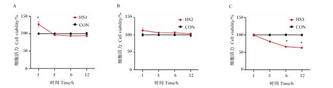
图1所示,处理1 h时HS1组细胞活力显著高于CON组(P<0.05),但是随着处理时间的增加,2组之间无显著差异(P>0.05);各处理时间时HS2组细胞活力与CON组之间均无显著差异(P>0.05),但其细胞活力为升高状态;随着处理时间的增加,HS3组细胞活力呈下降趋势,处理6 h时细胞活力降至临界值80%以下,与CON组差异显著(P<0.05)。
图1 不同处理温度及时间对细胞活力的影响

CON:CON组;HS1:HS1组;HS2:HS2组;HS3:HS3组。下图同。41和42 ℃下,各处理时间的细胞活力均高于90%;43 ℃下,处理6~12 h时细胞活力低于80%。“*”表示差异显著(P<0.05)。

Fig.1 Effects of different treatment temperatures and time on cell viability (n=3)

CON: CON group; HS1: HS1 group; HS2: HS2 group; HS3: HS3 group. The same as below. Under both 41 and 42 ℃, the cell viability remains above 90% at all time points. However, under 43 ℃, the cell viability drops below 80% at 6 and 12 h. “*” indicates a significant difference (P<0.05).

2.2 不同处理温度及时间对细胞内ROS水平的影响

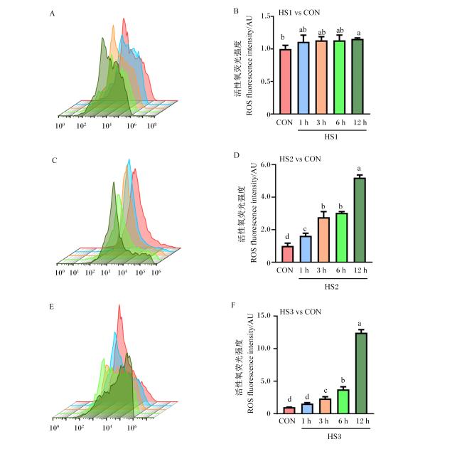
图2所示,在不同温度条件下体外培养细胞时,其ROS水平(通过荧光强度来反映)呈现出不同的变化情况:41 ℃下处理12 h时ROS水平与CON组相比显著升高(P<0.05);ROS水平随着处理时间的增加逐渐升高,42 ℃下处理1 h后即较CON组显著升高(P<0.05)。当培养温度提升到43 ℃时,处理3 h后ROS水平较CON组显著升高(P<0.05),且在12 h时ROS水平达到最高。
图2 不同处理温度及时间对细胞内ROS水平的影响

图A、C和E为不同温度下的流式细胞仪图,图B、D和F为ROS荧光强度统计分析图(流式图A、C和E中的荧光强度峰面积分别对应统计图B、D和F中相应颜色数据柱)。数据柱标相同小写字母表示差异不显著(P>0.05),不同小写字母表示差异显著(P<0.05)。

Fig.2 Effects of different treatment temperatures and time on ROS level in cells (n=3)

Figures A, C, and E are flow cytometry graphs under different temperatures. Figures B, D, and F are statistical analysis graphs of ROS fluorescence intensity (the fluorescence intensity peak area in the flow cytometry graphs A, C, and E corresponds to the respective color data column in the statistical graphs B, D, and F). The same lowercase letters on the data bars indicate no significant difference (P>0.05), while different lowercase letters indicate significant difference (P<0.05).

2.3 43 ℃下不同处理时间对细胞形态的影响

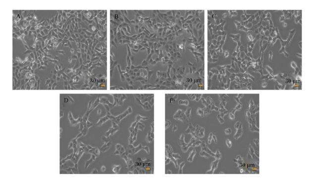
图3所示,未进行热处理的CON组细胞呈现出良好的贴壁状态,形态正常且分布均匀;与之相比,进行热处理的HS3组细胞呈现出随处理时间变化的特征:处理1 h时,细胞密度及形态无明显变化;处理3~6 h时,细胞密度明显降低,细胞量逐渐减少,细胞间隙变大;处理12 h时,细胞出现皱缩、空泡等凋亡现象。
图3 43 ℃下不同处理时间对细胞形态的影响

图A为未进行热应激处理(37 ℃,CON组)的细胞形态,图B为43 ℃下处理1 h的细胞形态,图C为43 ℃下处理3 h的细胞形态,图D为43 ℃下处理6 h的细胞形态,图E为43 ℃下处理12 h的细胞形态。细胞图像在显微镜下拍摄,比例尺为30 μm。

Fig.3 Effects of different treatment time on cell morphology at 43 ℃

Figure A shows the cell morphology without heat stress treatment (at 37 ℃, CON group), figure B shows the cell morphology after treatment at 43 ℃ for 1 h, figure C shows the cell morphology after treatment at 43 ℃ for 3 h, figure D shows the cell morphology after treatment at 43 ℃ for 6 h, and figure E shows the cell morphology after treatment at 43 ℃ for 12 h. The cell images were obtained under a microscope with a scale bar of 30 μm.

2.4 热应激对细胞内抗氧化指标的影响

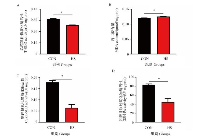
图4所示,与CON组相比,HS组在体外培养细胞6 h后,细胞内T-SOD、GSH-Px和CuZn-SOD的活性显著降低(P<0.05),而MDA含量则显著升高(P<0.05)。
图4 热应激对细胞内抗氧化指标的影响

CON组细胞维持在37 ℃,HS组细胞在43 ℃下处理6 h。*表示差异显著(P<0.05)。图5图7同。

Fig.4 Effects of heat stress on antioxidant indexes in cells (n=3)

Cells in the CON group were maintained at 37 ℃, while those in the HS group were treated at 43 ℃ for 6 h. “*” indicates a significant difference (P<0.05). The same as Fig.5 and Fig.7.

2.5 热应激对细胞内HSP70和HSP90含量的影响

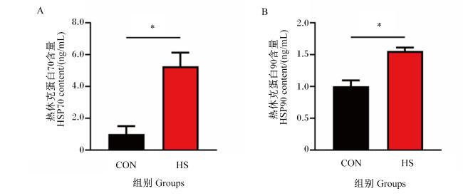
图5所示,HS组细胞内HSP70和HSP90含量显著高于CON组(P<0.05)。
图5 热应激对细胞内HSP70和HSP90含量的影响

Fig.5 Effects of heat stress on HSP70 and HSP90 contents in cells (n=3)

2.6 热应激对细胞内热应激相关基因表达的影响

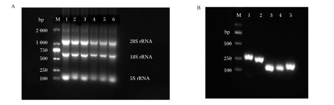
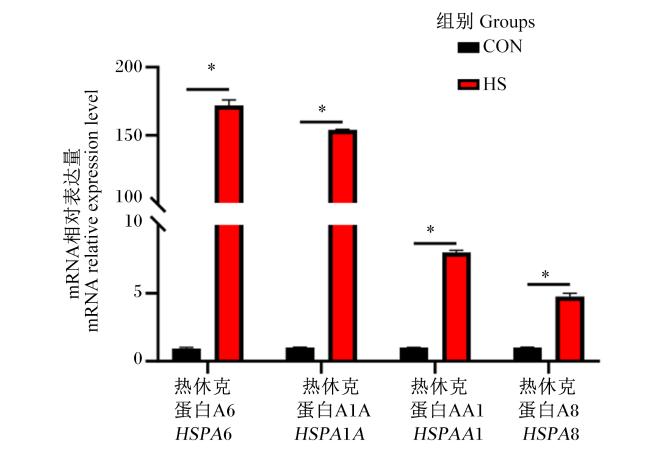
图6-A中,28S rRNA、18S rRNA和5S rRNA的条带清晰且无明显拖带现象,表明所提取的总RNA质量良好;图6-B中,扩增产物的条带清晰且单一,片段大小与预期结果完全一致;图7中,HS组细胞内4种热应激相关基因HSPA6、HSPA1AHSPAA1和HSPA8的mRNA相对表达量均显著高于CON组(P<0.05)。
图6 总RNA电泳图及实时荧光定量PCR扩增结果

图A中,泳道M为2000 DNA Marker,泳道1~3为CON组(37 ℃),泳道4~6为HS组(43 ℃,6 h);图B中,泳道M为2000 DNA Marker;泳道1为HSPA6,泳道2为HSPA8,泳道3为HSPAA1,泳道4为HSPA1A,泳道5为GAPDH

Fig.6 Total RNA electropherogram and RT-qPCR amplification results

In figure A, lane M is 2000 DNA Marker, lanes 1 to 3 stand for CON group (37 ℃), and lanes 4 to 6 stand for HS group (43 ℃, 6 h); In figure B, lane M stand for 2000 DNA Marker, lane 1 stand for HSPA6, lane 2 stand for HSPA8, lane 3 stand for HSPA1A, lane 4 stand for HSPA1A, and lane 5 stand for GAPDH.

图7 热应激对细胞内热应激相关基因mRNA相对表达量的影响

Fig.7 Effects of heat stress on mRNA relative expression levels of heat stress-related genes in cells (n=3)

3 讨论

我国南方地区夏季炎热潮湿,这种气候对畜禽的生产、繁殖、新陈代谢及免疫力均会产生不利影响。自19世纪末以来,全球变暖趋势不断加剧,进一步放大了这些季节性环境因素所带来的负面影响。炎热潮湿的气候易诱发动物的热应激反应,一旦动物出现热应激,其疾病发生的风险便会显著增加。目前,在细胞水平的热应激研究领域,大多以BMECs为模型开展相关研究。然而,至今仍缺乏系统的试验数据来明确揭示BMECs在特定热应激温度和时间条件下的具体状态以及各项指标的变化情况。因此,建立可靠的BMECs热应激模型显得尤为重要,这一模型的建立,有助于深入探究热应激的分子机制,同时对于研究开发缓解热应激的添加剂等也具有关键意义。
不同细胞建立热应激模型的条件存在显著差异,这可能与细胞本身的耐受性有关,不过所选用细胞在热应激下细胞活力大都处于50%~70%,细胞活力过高,意味着细胞所受的热应激不明显,而细胞活力过低,则表明细胞产生了不可逆损伤。尤其在乳腺上皮细胞中,过低的细胞活力可能会导致细胞凋亡[23-24]。有研究发现,在构建奶牛小肠上皮细胞热应激模型的过程中,当细胞经受43 ℃热处理4 h后,其细胞活力降至62.6%[25]。此外,在针对BMECs的相关研究中,也已明确证实高温会显著降低细胞活力,同时还会对氨基酸的转移以及乳蛋白的合成过程产生不利影响[26]
ROS作为生物有氧代谢过程中的副产物,活细胞具备有效平衡其形成的能力。然而,过量的ROS会对诸多生物分子造成损害。一旦ROS的产生量超出了抗氧化防御系统对其进行中和的能力,机体便会进入应激状态,进而在动物的健康、生长和发育等方面诱发不良后果[27-29]。大量研究表明,当细胞处在热应激状态时,ROS水平会有所升高;当细胞遭受不可逆的伤害时,ROS水平会急剧升高[30-34]。在研究沉默调节蛋白3(SIRT3)通过激活腺苷酸活化蛋白激酶(AMPK)信号通路对BMECs的保护作用时发现,当敲低SIRT3时,热应激对细胞的损伤加剧,抗氧化酶的生成受到抑制,进而导致BMECs中ROS水平显著升高[35]。通过细胞活力及ROS水平的测定结果,本研究初步确定了BMECs热应激模型的建模条件为43 ℃下处理6 h。
对初步建立的BMECs热应激模型进行氧化应激验证,数据显示43 ℃下处理6 h的热应激显著提升了细胞内脂质过氧化终产物MDA的含量,同时降低细胞内SOD和GSH-Px活性,这与前人关于热应激引发氧化应激的结论[36-39]一致。值得关注的是,本研究还发现热应激显著提升了细胞内CuZn-SOD活性,CuZn-SOD也可作为细胞热应激状态的生物标志物,这与p53-Sp1相互作用介导抗氧化基因抑制机制形成理论呼应,提示线粒体靶向抗氧化剂(如MitoQ10)可能通过调控此通路发挥保护作用[40]。在模型建立方面,本研究采用培养箱控温法在43 ℃下处理6 h成功构建了BMECs热应激模型,这与金钊等[25]构建奶牛小肠上皮细胞热应激模型的条件(43℃处理4 h)几乎一致,不同类型细胞差异可能影响细胞的应激阈值。值得注意的是,不同物种细胞耐热性呈现显著差异,如大鼠小肠上皮细胞(IEC-6)在43 ℃下处理1 h即建立有效热应激模型[41],而BMECs则需要更高时长处理,这提示建立标准化热应激模型需综合考虑细胞类型特异性。
从分子机制的角度来看,HSP70和HSP90的协同作用在热应激下尤为重要。HSP70能够迅速结合到变性蛋白质上,防止其聚集;而HSP90则进一步协助蛋白质的成熟和功能维持。在关于热应激对BMECs内HSP基因表达和合成的研究显示,热应激时细胞内HSP70和HSP90基因的转录水平和蛋白的表达水平显著升高[22],本试验研究结果与此一致。在研究热应激对水牛淋巴细胞内HSP70含量影响的试验中,与对照动物相比,直接热暴露对淋巴细胞内HSP70含量有显著影响(升高200倍);不过,在体外培养条件下,热处理导致淋巴细胞内HSP70含量轻度升高(升高2.5倍)[42]。Lee等[43]研究表明,在40 ℃热应激条件下,猪肌肉细胞内HSP90含量显著升高,HSP70 mRNA相对表达量无显著变化,这可能是由于物种与细胞不同导致的。热应激诱导的细胞内HSP70和HSP90含量增加以及基因表达量的上调,反映了细胞在热应激下通过上调HSP基因的表达来维持蛋白质稳态和细胞功能的适应性机制。
热应激会对细胞产生多种影响,其中HSP基因表达的变化是重要的响应机制。HSPA6、HSPA1A和HSPA8作为HSP70家族中的成员,在细胞应对热应激时发挥着关键作用。Banerjee等[44]研究表明,热应激会诱导HSPA8、HSPA6和HSPA1A基因表达上调。此外,热应激对奶牛生产性能及乳腺细胞蛋白质代谢基因表达的研究显示,热应激使得HSPA1A的丰度显著升高[45];同样,在杂交牛和热带适应型矮牛维丘尔牛(Vechur)和卡萨拉戈德牛(Kasaragod)HSPA1A基因表达谱及其与耐热性关系的研究中也表明,HSPA1A基因在不同品系和不同温湿指数(THI)下的表达均有显著差异[46]

4 结论

本研究通过系统评估BMECs对温度与时间变化的响应关系,确定43 ℃处理6 h可成功构建BMECs体外热应激模型;基于上述条件对BMECs进行热应激处理可显著抑制细胞活力,触发细胞内ROS及MDA异常积累,并且同步下调GSH-Px、T-SOD及CuZn-SOD等核心抗氧化酶的活性,同时上调HSP70和HSP90的含量以及相关基因的表达量。
[1]
WIJFFELS G, SULLIVAN M, GAUGHAN J. Methods to quantify heat stress in ruminants:current status and future prospects[J]. Methods, 2021, 186:3-13.

DOI

[2]
白慧. 热应激对奶牛乳腺上皮细胞功能的影响及牛磺酸的缓解作用[D]. 博士学位论文. 南京: 南京农业大学, 2022.

BAI H. Effects of heat stress on function of bovine mammary epithelial cells and the alleviative effects of taurine[D]. Ph.D.Thesis. Nanjing: Nanjing Agricultural University, 2022. (in Chinese)

[3]
VITALI A, FELICI A, LEES A M, et al. Heat load increases the risk of clinical mastitis in dairy cattle[J]. Journal of Dairy Science, 2020, 103(9):8378-8387.

DOI PMID

[4]
DAHL G E, TAO S, LAPORTA J. Heat stress impacts immune status in cows across the life cycle[J]. Frontiers in Veterinary Science, 2020, 7:116.

DOI PMID

[5]
OLIVEIRA C E A, TINÔCO I D F F, SOUSA F C D, et al. Health and thermal comfort of dairy cattle in compost-bedded pack barns and other types of housing:a comparative systematic review[J]. Agriengineering, 2024, 6(2):1395-1416.

DOI

[6]
HECK J M L, VAN VALENBERG H J F, DIJKSTRA J, et al. Seasonal variation in the Dutch bovine raw milk composition[J]. Journal of Dairy Science, 2009, 92(10):4745-4755.

DOI PMID

[7]
HAMMAMI H, VANDENPLAS J, VANROBAYS M L, et al. Genetic analysis of heat stress effects on yield traits,udder health,and fatty acids of Walloon Holstein cows[J]. Journal of Dairy Science, 2015, 98(7):4956-4968.

DOI

[8]
ZHAO S G, MIN L, ZHENG N, et al. Effect of heat stress on bacterial composition and metabolism in the rumen of lactating dairy cows[J]. Animals, 2019, 9(11):925.

DOI

[9]
LOUIE A P, ROWE J D, LOVE W J, et al. Effect of the environment on the risk of respiratory disease in preweaning dairy calves during summer months[J]. Journal of Dairy Science, 2018, 101(11):10230-10247.

DOI PMID

[10]
KHAN M Z, KHAN A, CHEN W T, et al. Advancements in genetic biomarkers and exogenous antioxidant supplementation for safeguarding mammalian cells against heat-induced oxidative stress and apoptosis[J]. Antioxidants, 2024, 13(3):258.

DOI

[11]
HUBER E, NOTARO U S, RECCE S, et al. Fetal programming in dairy cows:effect of heat stress on progeny fertility and associations with the hypothalamic-pituitary-adrenal axis functions[J]. Animal Reproduction Science, 2020, 216:106348.

DOI

[12]
DOVOLOU E, GIANNOULIS T, NANAS I, et al. Heat stress:a serious disruptor of the reproductive physiology of dairy cows[J]. Animals, 2023, 13(11):1846.

DOI

[13]
GOPINADHAN A, HUGHES J M, CONROY A L, et al. A human pluripotent stem cell-derived in vitro model of the blood-brain barrier in cerebral malaria[J]. Fluids and Barriers of the CNS, 2024, 21(1):38.

DOI

[14]
FU J Y, LI L, HUO D, et al. Astrocyte-derived TGFβ1 facilitates blood-brain barrier function via non-canonical hedgehog signaling in brain microvascular endothelial cells[J]. Brain Sciences, 2021, 11(1):77.

DOI

[15]
ZHANG H Y, YAMAGUCHI T, KAWABATA K. The maturation of iPS cell-derived brain microvascular endothelial cells by inducible-SOX18 expression[J]. Fluids and Barriers of the CNS, 2023, 20(1):10.

DOI PMID

[16]
KHAN A, KHAN M Z, DOU J H, et al. RNAi-mediated silencing of catalase gene promotes apoptosis and impairs proliferation of bovine granulosa cells under heat stress[J]. Animals, 2020, 10(6):1060.

DOI

[17]
HABASHY W S, MILFORT M C, REKAYA R, et al. Cellular antioxidant enzyme activity and biomarkers for oxidative stress are affected by heat stress[J]. International Journal of Biometeorology, 2019, 63(12):1569-1584.

DOI PMID

[18]
AL-JARYAN I L, AL-THUWAINI T M, AL-JEBORY H H. Heat shock protein 70 and its role in alleviating heat stress and improving livestock performance[J]. Reviews in Agricultural Science, 2023, 11:234-242.

DOI

[19]
RAKIB M R H, MESSINA V, GARGIULO J I, et al. Graduate student literature review:potential use of HSP70 as an indicator of heat stress in dairy cows—a review[J]. Journal of Dairy Science, 2024, 107(12):11597-11610.

DOI

[20]
JEYACHANDRAN S, CHELLAPANDIAN H, PARK K, et al. A review on the involvement of heat shock proteins (Extrinsic chaperones) in response to stress conditions in aquatic organisms[J]. Antioxidants, 2023, 12(7):1444.

DOI

[21]
BALAKRISHNAN K N, RAMIAH S K, ZULKIFLI I. Heat shock protein response to stress in poultry:a review[J]. Animals, 2023, 13(2):317.

DOI

[22]
HU H, ZHANG Y D, ZHENG N, et al. The effect of heat stress on gene expression and synthesis of heat-shock and milk proteins in bovine mammary epithelial cells[J]. Animal Science Journal, 2016, 87(1):84-91.

DOI PMID

[23]
ZENG H F, LI S J, CHANG H M, et al. Circ_002033 regulates proliferation,apoptosis,and oxidative damage of bovine mammary epithelial cells via the miR-199a-5p-MAP3K11 axis in heat stress[J]. Journal of Agricultural and Food Chemistry, 2024, 72(25):14386-14401.

DOI

[24]
GAI Z C, WANG Y J, WANG J, et al. Downregulation of CASTOR1 inhibits heat-stress-induced apoptosis and promotes casein and lipid synthesis in mammary epithelial cells[J]. Journal of Agricultural and Food Chemistry, 2022, 70(17):5386-5395.

DOI

[25]
金钊, 张艳芳, 周华, 等. 奶牛小肠上皮细胞热应激模型的建立[J]. 家畜生态学报, 2022, 43(12):64-68.

JIN Z, ZHANG Y F, ZHOU H, et al. Establishment of heat stress model in cow intestinal epithelial cells[J]. Acta Ecologae Animalis Domastici, 2022, 43(12):64-68. (in Chinese)

DOI

[26]
ZHOU J, YUE S, XUE B C, et al. Effect of hyperthermia on cell viability,amino acid transfer,and milk protein synthesis in bovine mammary epithelial cells[J]. Journal of Animal Science and Technology, 2022, 64(1):110-122.

DOI

[27]
SURAI P F, KOCHISH I I, FISININ V I, et al. Antioxidant defence systems and oxidative stress in poultry biology:an update[J]. Antioxidants, 2019, 8(7):235.

DOI

[28]
KOZLOV A V, JAVADOV S, SOMMER N. Cellular ROS and antioxidants:physiological and pathological role[J]. Antioxidants, 2024, 13(5):602.

DOI

[29]
SLIMEN I B, NAJAR T, GHRAM A, et al. Reactive oxygen species,heat stress and oxidative-induced mitochondrial damage.A review[J]. International Journal of Hyperthermia, 2014, 30(7):513-523.

DOI

[30]
BELHADJ SLIMEN I, NAJAR T, GHRAM A, et al. Heat stress effects on livestock:molecular,cellular and metabolic aspects,a review[J]. Journal of Animal Physiology and Animal Nutrition, 2016, 100(3):401-412.

DOI

[31]
WANG Y, WU J, XIA S W, et al. miR-27a-3p relieves heat stress-induced mitochondrial damage and aberrant milk protein synthesis through MEK/ERK pathway in BMECs[J]. Cell Stress and Chaperones, 2023, 28(3):265-274.

DOI

[32]
JIN X L, WANG K, LIU L, et al. Nuclear factor-like factor 2-antioxidant response element signaling activation by tert-butylhydroquinone attenuates acute heat stress in bovine mammary epithelial cells[J]. Journal of Dairy Science, 2016, 99(11):9094-9103.

DOI PMID

[33]
WANG Y, WANG H L, XING G D, et al. S-allyl cysteine ameliorates heat stress-induced oxidative stress by activating Nrf2/HO-1 signaling pathway in BMECs[J]. Toxicology and Applied Pharmacology, 2021, 416:115469.

DOI

[34]
SAMMAD A, AHMED T, ULLAH K, et al. Vitamin C alleviates the negative effects of heat stress on reproductive processes by regulating amino acid metabolism in granulosa cells[J]. Antioxidants, 2024, 13(6):653.

DOI

[35]
SUN X C, WANG Y, ZENG H F, et al. SIRT3 protects bovine mammary epithelial cells from heat stress damage by activating the AMPK signaling pathway[J]. Cell Death Discovery, 2021, 7(1):304.

DOI

[36]
YANG L H, HUANG H, WANG J J. Antioxidant responses of citrus red mite,Panonychus citri (McGregor) (Acari:Tetranychidae),exposed to thermal stress[J]. Journal of Insect Physiology, 2010, 56(12):1871-1876.

DOI

[37]
LI C M, WANG Y R, LI L, et al. Betaine protects against heat exposure-induced oxidative stress and apoptosis in bovine mammary epithelial cells via regulation of ROS production[J]. Cell Stress and Chaperones, 2019, 24(2):453-460.

DOI

[38]
LU Z, HE X F, MA B B, et al. Dietary taurine supplementation improves breast meat quality in chronic heat-stressed broilers via activating the Nrf2 pathway and protecting mitochondria from oxidative attack[J]. Journal of the Science of Food and Agriculture, 2019, 99(3):1066-1072.

DOI PMID

[39]
YANG W R, LI B B, HU Y, et al. Oxidative stress mediates heat-induced changes of tight junction proteins in porcine sertoli cells via inhibiting CaMKKβ-AMPK pathway[J]. Theriogenology, 2020, 142:104-113.

DOI

[40]
GONG J, SUN P P, LI L, et al. Heat stress suppresses MnSOD expression via p53-Sp1 interaction and induces oxidative stress damage in endothelial cells:protective effects of MitoQ10 and Pifithrin-α[J]. Heliyon, 2023, 9(12):e22805.

DOI

[41]
PAN Z G, HE X, SHAO Y, et al. ROS/JNK-mediated lysosomal injury in rat intestinal epithelial-6 cells during heat stress[J]. Journal of Thermal Biology, 2022, 109:103326.

DOI

[42]
MISHRA A, HOODA O K, SINGH G, et al. Influence of induced heat stress on HSP70 in buffalo lymphocytes[J]. Journal of Animal Physiology and Animal Nutrition, 2011, 95(4):540-544.

DOI PMID

[43]
LEE J, BELAL S A, LIN X, et al. Insect peptide CopA3 mitigates the effects of heat stress on porcine muscle satellite cells[J]. Animals, 2023, 13(20):3209.

DOI

[44]
BANERJEE D, UPADHYAY R C, CHAUDHARY U B, et al. Seasonal variation in expression pattern of genes under HSP70[J]. Cell Stress and Chaperones, 2014, 19(3):401-408.

DOI

[45]
CORAZZIN M, SACCÀ E, LIPPE G, et al. Effect of heat stress on dairy cow performance and on expression of protein metabolism genes in mammary cells[J]. Animals, 2020, 10(11):2124.

DOI

[46]
ELAYADETH-MEETHAL M, KEAMBOU TIAMBO C, POONKUZHI NASEEF P, et al. The profile of HSPA1A gene expression and its association with heat tolerance in crossbred cattle and the tropically adapted dwarf Vechur and Kasaragod[J]. Journal of Thermal Biology, 2023, 111:103426.

DOI

Outlines

/