REVIEW

Research Progress on Effects of Probiotics on Intestinal Epithelial Regeneration and Repair in Livestock and Poultry

  • CHEN Man , 1 ,
  • JU Xianghong 1 ,
  • YIN Hong 2 ,
  • LI Youquan 1, *
Expand
  • 1 College of Coastal Agricultural Sciences, Guangdong Ocean University, Zhanjiang 524088, China
  • 2 Lanzhou Veterinary Research Institute, Chinese Academy of Agricultural Sciences, Lanzhou 730046, China
* professor, E-mail: youquan-li@163.com

Received date: 2025-06-11

  Online published: 2026-01-13

Abstract

Intact intestinal mucosal epithelium can effectively prevent the invasion of harmful microorganisms and is extremely important for animal health. Probiotics have functions such as promoting the regeneration and repair of intestinal epithelium, improving intestinal epithelial permeability and maintaining intestinal homeostasis, inhibiting pathogenic bacteria, adjusting the structure of intestinal microbiota, alleviating intestinal inflammation, and enhancing the performance of livestock and poultry. This article reviewed the research progress on the regeneration and repair of intestinal epithelium, the regulatory mechanism of probiotics on it, and the application of probiotics in livestock breeding, aiming to provide a reference for studying the regulation of intestinal health in livestock and poultry by probiotics.

Cite this article

CHEN Man , JU Xianghong , YIN Hong , LI Youquan . Research Progress on Effects of Probiotics on Intestinal Epithelial Regeneration and Repair in Livestock and Poultry[J]. Chinese Journal of Animal Nutrition, 2026 , 38(1) : 172 -183 . DOI: 10.12418/CJAN2026.015

肠道是动物重要的消化器官,分为小肠和大肠2部分,其中小肠包括十二指肠、空肠和回肠,大肠包括盲肠、结肠和直肠。小肠和大肠是消化系统的核心组成部分,二者在结构和功能上紧密协作,共同完成食物的消化、吸收、排泄及免疫防御等生理过程。肠道作为畜禽生理机能的核心调控枢纽,其状态直接关联其生产性能、饲料转化率及疾病抵抗力等关键生产指标。而肠道上皮组织的再生与修复机制,在维系肠道内环境稳态过程中扮演着不可替代的生物学角色。肠道作为畜禽体内更新率最快的器官之一,其动态平衡主要依赖于隐窝底部的富含亮氨酸重复序列G蛋白偶联受体5(Lgr5)阳性(Lgr5+)干细胞的高效增殖和分化机制。肠道菌群作为宿主消化系统的“微生物器官”,在维持机体生理稳态中发挥多种生物学功能,其机制涉及屏障维护、代谢调控、病原防御及免疫调控等核心领域。某些特定的益生菌菌株能够通过上调Wnt/β-连环蛋白(β-catenin)信号通路、激活G蛋白偶联受体43(GPR43)受体等途径,推动肠道上皮细胞的再生与修复,进而维持畜禽肠道内环境的稳定。此外,这些益生菌产生的乙酸、丙酸、丁酸等短链脂肪酸(short chain fatty acids,SCFAs)及吲哚-3-丙酸等色氨酸代谢产物,同样可介导肠道上皮的再生与修复过程。益生菌作为抗生素的有效补充或替代品之一,具备绿色安全的显著特性,在畜牧业领域主要应用于疾病防控、促进生长及健康保健等方面。因此,在畜禽养殖过程中,依据实际需求适时且有针对性地补充益生菌,能够有效优化畜禽肠道微生物群落结构,这对于维持肠道内环境稳定、推动肠道上皮组织再生与修复以及提升畜禽生产性能等均具有重要意义。
本文围绕畜禽肠道上皮再生与修复、肠道上皮损伤诱因、益生菌对肠道上皮再生与修复的作用机制以及益生菌在畜牧业生产中的实际应用展开综述,旨在为后续动物肠道益生菌的深入研究与推广应用、以及益生菌调控肠道微生物群落稳态的相关研究提供参考。

1 肠道上皮再生与修复

1.1 肠道干细胞的增殖与分化

动物小肠上皮通过折叠形成隐窝和小肠绒毛的特殊结构。每个小肠绒毛呈指状突起,基部环绕多个隐窝。隐窝底部存在一类肠道干细胞,即隐窝基底柱状细胞[1]。该细胞的显著特征是表达Lgr5(又称G蛋白偶联受体49),Lgr5作为G蛋白偶联受体超家族成员,其分子结构包含18个富含亮氨酸的重复单位和7个跨膜区域[2]。因此,这类细胞又被称为Lgr5+肠道干细胞,通常位于+4标记保留干细胞下方,与潘氏细胞交替排列,构成肠道的主要干细胞[3]。当接近小肠绒毛顶端的上皮细胞发生凋亡时,隐窝基底柱状干细胞的子细胞从隐窝迁移至转运扩增区(TA区),经历短暂的增殖分化后,沿隐窝-绒毛轴向上迁移。离开TA区后,这些细胞分化为不同种类的肠道上皮细胞,继续上移填充绒毛区并执行其生理功能[4]。与此同时,终末分化后的潘氏细胞向下迁移至隐窝底部,与肠道间充质细胞共同构成维持Lgr5+肠道干细胞的生态位,提供Wnt、表皮生长因子和Notch等必需的信号分子[5]

1.2 肠道上皮再生与修复的调控

在正常生理状态下,肠道上皮细胞每3~5 d完成1次更新[6]。肠道上皮的这种高度有序的自我更新过程依赖于再生程序的精准调控,以维持上皮屏障的结构完整性和功能稳定性。肠道上皮再生与修复涉及Wnt、Notch、R-spondin、Hedgehog、骨形态发生蛋白(bone morphogenetic protein,BMP)和Hippo等多条信号通路,通过复杂的信号转导、细胞增殖及分化网络实现调控(图1)。其中,Wnt/β-catenin信号通路是驱动肠隐窝基部肠道干细胞增殖的核心调控途径[7]。而局部高浓度Wnt信号对维持肠道上皮自我更新至关重要,其信号来源包括潘氏细胞与肠道间充质细胞分泌的Wnt5a、β-catenin等配体[8-9]。肠道间充质细胞还可分泌R-spondin家族蛋白,作为Wnt信号的高效增强子[10]。Hippo信号通路的活性不仅受机械应变和细胞-细胞及细胞外基质黏附变化的调节,也受营养物质和其他细胞应激的影响,细胞间黏附受损会对Hippo信号通路活性产生强烈影响,并导致Yes相关蛋白(Yes-associated protein,YAP)/具有PDZ结合基序的转录共激活因子(transcriptional coactivator with PDZ-binding motif,TAZ)的激活或抑制[11]。YAP通过瞬时抑制Wnt信号传导和Lgr5+肠道干细胞群来保护肠道干细胞池,同时阻断肠道干细胞分化为潘氏细胞,并驱动依赖表皮生长因子受体信号诱导的促再生程序[12]。Notch信号通过“横向抑制”机制维持肠道干细胞未分化状态,其激活因子主要由潘氏细胞表达,对保持肠道干细胞多能性起决定性作用[13]。当隐窝受损时,肠内分泌细胞和簇状细胞可替代缺失的潘氏细胞,成为Notch信号的代偿来源;该通路抑制会诱导细胞向分泌型细胞(如杯状细胞)分化[14]。肠道上皮损伤后的修复过程遵循严格的时序调控,依次经历3个关键阶段:首先进入急性控制期,通过炎症应答抑制损伤扩散;随后进入爆发性增殖期(再生阶段),干细胞被激活并大量增殖;最终通过稳态重建期恢复上皮结构与功能平衡[15]。谱系追踪研究证实,当隐窝因损伤消融后,+4标记保留干细胞可感知生态位微环境信号,重新激活干细胞特性,通过增殖分化补充上皮细胞谱系,驱动肠道上皮组织的再生与修复[16]。在此过程中,Wnt/β-catenin和Hippo等信号通路通过精确调控相关基因的表达模式(如上调增殖基因、下调凋亡信号),确保再生程序的有序进行,避免细胞增殖失控或修复不足。
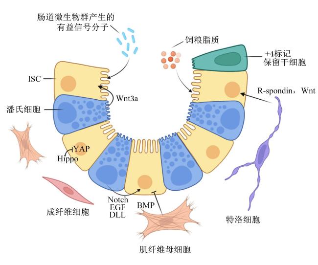
图1 肠道干细胞生态位中的经典信号通路

ISC:肠道干细胞 intestinal stem cell;YAP:Yes相关蛋白 Yes-associated protein;EGF:表皮生长因子 epidermal growth factor;DLL:delta样配体 delta-like ligand;BMP:骨形态发生蛋白 bone morphogenetic protein。

Fig.1 Classical signaling pathways in intestinal stem cell niche

1.3 免疫细胞对肠道上皮再生与修复的调节

免疫细胞与肠道上皮细胞的相互作用对肠道上皮细胞的再生与修复具有重要调节功能。在肠道固有层微环境中,集落刺激因子1受体(CSF1R)mRNA特异性表达于与隐窝上皮紧密相邻的巨噬细胞亚群。研究证实,通过阻断CSF1R信号通路诱导巨噬细胞耗竭后,Lgr5+肠道干细胞的数量呈现显著下降趋势,同时其向功能性肠道上皮细胞的分化进程亦受到明显干扰[17]。在结肠组织中,巨噬细胞与隐窝上皮细胞直接相邻,通过哺乳动物雷帕霉素靶蛋白复合体1(mTORC1)信号通路形成“代谢共生体”,为上皮细胞提供能量支持并驱动其自我更新,从而维持肠道上皮细胞稳态[18]。上皮内淋巴细胞是肠道上皮层中的主要免疫细胞,主要通过CD160与肿瘤坏死因子(TNF)受体超家族成员疱疹病毒入侵介质(herpesvirus entry mediator,HVEM)互作,进一步激活核因子-κB(NF-κB)信号传导来促进肠道再生修复[19]。调节性T细胞(regulatory T cells,Tregs)作为以Foxp3+、CD4+和CD25+为表型特征的T细胞亚群,负责调控免疫系统终止从而避免机体出现过度免疫反应,因而在抑制过度免疫反应中起核心作用[20]。研究证实,Tregs分泌的白细胞介素-10(IL-10)可通过信号转导与转录激活因子3(STAT3)信号通路激活隐窝基部肠道干细胞,显著增强肠道上皮细胞的增殖和修复能力[21-22]。此外,Tregs还能通过调控肠道基质细胞活性,促进关键再生信号分子(如Wnt10B)的分泌,进而维持隐窝微环境稳定。该过程通过多重机制实现肠道上皮细胞再生与修复的调控,包括抑制局部炎症反应、调节肠道菌群稳态以及增强上皮紧密连接蛋白表达,最终加速肠道屏障功能重建[23-24]。白细胞介素-22作为一种关键细胞因子,可由肠道微生物诱导免疫细胞产生,其主要功能包括诱导上皮细胞快速再生与分化,通过激活抗凋亡基因和DNA损伤反应通路提升上皮细胞存活率,并在急性肠道上皮损伤修复中发挥独特作用[25]

2 畜禽常见肠道上皮损伤诱因

肠道屏障是保障畜禽健康的关键生理结构,其稳态失衡将显著降低畜禽对营养物质的消化吸收效率,引发腹泻、病原体感染等临床症状,严重制约畜禽的产肉、产蛋等核心生产性能。病原体感染、氧化应激、断奶应激和抗生素滥用等因素,均会对动物肠道屏障功能造成损害,进而威胁畜禽健康。在病原体感染方面,多种病毒和细菌均可引发肠道损伤。流行性腹泻病毒、传染性胃肠炎病毒、轮状病毒和禽呼肠孤病毒等感染均可导致动物肠道组织损伤,诱发腹泻、坏死性肠炎等疾病,影响畜禽生产性能并造成经济损失[26-29]。沙门氏菌、产气荚膜梭菌和大肠杆菌等细菌感染,同样会破坏畜禽肠道屏障完整性,致使畜禽生长迟缓、淘汰率上升,严重影响畜牧生产效益[30-32]。值得注意的是,沙门氏菌病作为二类人畜共患传染病,鸡感染后不仅会出现肠道损伤、饲料转化率降低及生殖系统损伤,在肉鸡养殖中还会导致生长性能下降、死亡率升高;在蛋鸡养殖中则表现为产蛋率显著降低。此外,感染鸡群还可通过污染的鸡肉、鸡蛋及粪便传播病菌,对公共卫生安全构成潜在威胁[33-34]。应激因素对畜禽肠道健康的影响也不容忽视。在生猪养殖过程中,断奶应激和氧化应激均会导致生猪出现腹泻、免疫力下降等问题,严重影响其生产性能。其中,断奶应激作为生猪养殖中最为常见的应激源,由营养和环境等多重应激因素叠加而成,常引发仔猪腹泻、生长速度减缓及成活率降低等问题,对仔猪后续生长发育产生持续性负面影响,给养猪业带来严重的经济损失[35]。在国家“减抗禁抗”法规实施前,抗生素常被用作畜禽促生长药物。然而,长期滥用抗生素导致微生物抗生素抗性基因污染问题日益严峻,已演变为全球性公共卫生挑战[36]。更为严重的是,抗生素滥用会抑制畜禽肠道有益菌群生长,破坏肠道上皮细胞间紧密连接蛋白结构,扰乱肠道免疫屏障功能,影响肠道代谢,最终导致肠道上皮组织损伤,严重损害畜禽生产性能[37-39]。综上所述,健康的肠道微环境是畜禽绿色高效健康养殖的基础。在现代畜禽生产中,应高度重视肠道保健工作,同时采取有效措施避免各类因素对肠道上皮造成损伤,从而保障畜禽健康,提升养殖效益。

3 益生菌促进肠道上皮再生与修复的作用机制
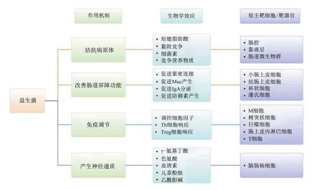
联合国粮农组织(FAO)、世界卫生组织(WHO)以及国际益生菌与益生元科学协会(ISAPP)将益生菌定义为:在摄入一定量时可给宿主带来健康作用的活性微生物[40]。乳杆菌、芽孢杆菌、双歧杆菌等多种细菌均可用作益生菌,不同种类的益生菌凭借独特的生物学特性,可定植在动物消化道的不同部位,给宿主带来不同的健康益处[41]。当肠道屏障因饲粮变更、抗生素滥用、病原体入侵等因素受损时,益生菌及其次级代谢产物便迅速“集结”,通过多种途径发挥拮抗病原体、改善肠道屏障、免疫调节以及产生神经递质等多种功能(图2)[42]
图2 益生菌主要作用机制示意图

Muc:黏蛋白 mucin;IgA:免疫球蛋白A immunoglobulin A。

Fig.2 Schematic diagram of main mechanisms of action of probiotics[42]

3.1 直接调控途径

益生菌可通过调控宿主代谢,加速肠道上皮再生与修复,从而矫正稳态失衡、恢复肠道稳态(图3)。其产生的SCFAs、色氨酸代谢物和胞外多糖等能通过多个途径直接介导肠道上皮的再生与修复。SCFAs主要包括乙酸、丙酸和丁酸等,作为肠道上皮细胞的主要能源,在维持肠道屏障完整、肠道稳态和结肠健康方面发挥着关键作用[43]。研究表明,丙酸盐可通过抑制Ⅰ类组蛋白去乙酰化酶(HDAC)活性、活化GPR43并激活STAT3,进而促进上皮细胞迁移,驱动肠道上皮细胞的再生与修复[44]。研究报道,SCFAs可在隐窝底部HDAC活性较低部位促进干细胞增殖;同时,SCFAs还能调节肠道上皮细胞中与能量代谢(如脂质代谢等)相关的基因表达,促进小鼠肠道类器官的发育,对成熟肠道上皮细胞维持正常细胞周期意义重大[45-46]。吲哚-3-甲醛(indole-3-aldehyde,3-IAld)作为重要的肠道微生物色氨酸代谢产物,已有研究证实其可增加断奶仔猪杯状细胞数量,上调紧密连接蛋白——闭合蛋白(Occludin)、密封蛋白(Claudin)1的表达;同时,能够显著提高猪肠道类器官的出芽效率和表面积,以及显著提高Lgr5和β-catenin的蛋白表达水平[47],这表明3-IAld可促进仔猪小肠干细胞的增殖。此外,3种色氨酸代谢物(吲哚-3-乙醇、吲哚-3-丙酮酸和3-IAld)在肠道炎症期间,可通过芳香烃受体(AhR)通路促进维持肠道屏障的完整性[48]。Zhou等[49]研究证实,植物乳杆菌NCU116分泌的胞外多糖116可通过显著提升肠道乳杆菌属的丰度,继而提高结肠隐窝微环境中乳酸的局部浓度,最终通过代谢微环境调控机制促进肠道干细胞的增殖与定向分化。
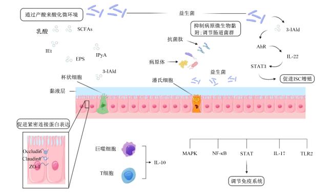
图3 益生菌调节肠道屏障、促进肠道上皮再生与修复的机制

SCFAs:短链脂肪酸 short-chain fatty acids;IEt:吲哚-3-乙醇 indole-3-ethanol;IPyA:吲哚-3-丙酮酸 indole-3-pyrubate;3-IAld:吲哚-3-甲醛 indole-3-aldehyde;EPS:胞外多糖 exopolysaccharides;AhR:芳香烃受体 aryl hydrocarbon receptor;IL-22:白细胞介素-22 interleukin-22;STAT:信号转导与转录激活因子 signal transducer and activator of transcription;ISC:肠道干细胞 intestinal stem cell;Occludin:闭合蛋白;Claudin 1:密封蛋白1;ZO-1:闭锁小带蛋白-1 zonula occludens-1;IL-10:白细胞介素-10 interleukin-10;MAPK:丝裂原活化蛋白激酶 mitogen-activated protein kinase;NF-κB:核因子-κB nuclear factor κB;IL-17:白细胞介素-17 interleukin-17;TLR2:Toll样受体2 Toll-like receptor 2。

Fig.3 Mechanism by which probiotics regulate intestinal barrier and promote regeneration and repair of intestinal epithelial cells

在信号通路调控层面,益生菌可通过介导Wnt/β-catenin信号通路促进肠道干细胞增殖分化,减轻肠道屏障损伤[50-51]。Qian等[52]从仔猪粪便中分离的发酵乳杆菌可激活Wnt/β-catenin信号通路,保护黏膜屏障完整性,刺激肠道上皮细胞增殖与分化,修复产肠毒素大肠杆菌(ETEC)K88诱导的损伤;同时,该菌能够降低促炎因子[TNF-α、白细胞介素-1β(IL-1β)、干扰素-γ(IFN-γ)]表达,促进抗炎因子[转化生长因子-β(TGF-β)、IL-10]表达,加速肠道炎症损伤修复并恢复肠道上皮稳态。Wang等[53-54]研究发现,解淀粉芽孢杆菌SC06可通过激活AhR/STAT3和Wnt/β-catenin双信号通路,促进肠道干细胞增殖并诱导其分化为杯状细胞,修复脂多糖诱导的肠道类器官损伤,改善断奶仔猪肠道炎症。Yang等[55]研究发现,植物乳杆菌可通过抑制Toll样受体4介导的丝裂原活化蛋白激酶(MAPK)信号通路的激活,以改善ETEC K88引起的先天免疫反应的过度激活,通过调节先天免疫来维持断奶仔猪的肠道健康。在肠道稳态调控中,鼠李糖乳杆菌GG可通过白细胞介素-17(IL-17)、视黄酸、NF-κB及MAPK信号通路介导肠道干细胞的增殖与自我更新[56]。其中,Th17细胞分泌的IL-17在肠道损伤修复过程中,能显著促进Lgr5+肠道干细胞中无调性BHLH转录因子1(Atoh1)基因的表达,进而诱导其向分泌型细胞谱系分化[57]。作为维生素A活性形式的视黄醇可代谢为视黄酸,二者通过提供早期信号启动级联调控网络,协同促进肠道干细胞向功能性肠道上皮细胞的定向分化[9]。此外,细菌在肠道的定植模式会影响Wnt激动剂(如Wnt5a、Wnt8b、Wnt11)的肠段特异性表达,而Wnt激动剂的差异表达可通过调控Wnt/β-catenin信号通路活性,影响肠道上皮细胞的再生修复过程[58]

3.2 间接调控途径

益生菌及其次级代谢物可影响畜禽肠道微生物群落的α和β多样性,降低肠道乙酸盐浓度,提高丙酸盐浓度,有效减少沙门氏菌和志贺氏菌的定植,抑制轮状病毒感染,维持肠道稳态,提高免疫力,减少疾病,减轻热应激等[59-62]。多种益生菌具有抑制肠道病原体、维持肠道微环境的功能。在肉鸡饲粮中添加枯草芽孢杆菌可显著提高其脏器指数,添加高剂量枯草芽孢杆菌还可清除回肠、盲肠和结肠中的沙门氏菌、产气荚膜梭菌和大肠杆菌,提高生产性能[63]。植物乳杆菌CCFM8661可通过改善绒毛长度和隐窝深度,抑制TNF-α、IL-1β和白细胞介素-6等促炎因子的表达,从而有效改善小鼠体重减轻和腹泻症状,减轻大肠杆菌带来的肠道损伤,改善小鼠肠道屏障功能[64]。Chamignon等[65]对3株乳杆菌促进肠道稳态的效果进行评价,发现三者均可增加肠道黏液的产生,恢复正常肠道上皮通透性,调节结肠超敏反应,并通过促进Claudin2和扣带蛋白(Cingulin)的表达来维持肠道稳态。益生菌亦可通过增强黏蛋白2(MUC2)、闭锁小带蛋白-1(ZO-1)和Occludin的表达来改善葡聚糖硫酸钠诱导的结肠屏障功能障碍[66]。罗伊氏乳杆菌产生的囊泡可被肠道上皮细胞摄取,通过上调β-N-乙酰己糖胺酶和α-N-乙酰氨基葡萄糖苷酶的表达,选择性地促进有益细菌的增殖,恢复肠道菌群平衡,并提高ZO-1和Occludin的表达,降低肠道通透性,对上皮紧密连接功能发挥保护作用[67]。此外,益生菌的次级代谢物如乙酰左旋肉碱、胆汁酸等可通过调节细胞黏附分子2(CADM2)表达、诱导宿主免疫细胞分化等方式来维持肠道屏障的稳态[68-69]

4 益生菌在畜牧业生产中的应用

在畜牧业生产中,病原微生物、抗营养因子、过敏原和毒素等均可能导致畜禽肠道屏障受损,诱发腹泻、肠道黏膜急性损伤以及肠道菌群结构改变等,并可能导致畜禽肠道功能紊乱和继发性损伤[70]。多年来,抗生素在畜禽养殖上多用于促生长和动物疫病的治疗,但抗生素的不科学使用或滥用导致微生物耐药基因的产生,诱发了大量生态、健康等问题。益生菌的高效、安全和无残留等特点,使其成为理想的减抗替抗产品之一,具有广阔的应用前景。在畜禽生产过程中适时、适量的补充益生菌可有效防治畜禽肠道损伤,提高畜禽生产性能。目前,益生菌在畜牧业(猪、家禽、反刍动物生产等)中已开始广泛应用。

4.1 益生菌在猪生产中的应用

研究表明,猪肠道菌群会随着猪的生长、环境变化等发生改变,肠道中定植的优势微生物种群也会发生变化,仔猪补充益生菌有助于其尽早建立健康的肠道菌群结构,有利于仔猪生长发育[71]。断奶是养殖过程中一个重要的时间节点。由于断奶仔猪的消化道尚未发育成熟,饲粮更换、环境应激均可造成仔猪腹泻,导致仔猪消化道功能减弱、营养物质吸收效率降低、生长发育缓慢甚至停滞,对养猪业造成严重影响[72]。研究证实,多种益生菌可缓解断奶仔猪腹泻症状,提高断奶仔猪生产性能。在无抗生素饲粮中添加马铃薯浓缩蛋白和布拉迪酵母菌可提高断奶仔猪空肠绒毛高度,上调Occludin表达,提高粪便乳杆菌属丰度,有助于在仔猪断奶后的关键阶段维持肠道健康[73]。阿根图拉特乳植杆菌(Lactiplantibacillus argentoratensis)AGMB00912可改善断奶仔猪的肠道菌群结构,降低螺杆菌属和弯曲杆菌属丰度,提高断奶仔猪生长性能并缓解腹泻,显著降低断奶仔猪的促炎因子TNF-α水平,并提高谷胱甘肽等抗氧化物质水平;同时,在饲粮中添加该菌可提高断奶仔猪空肠和回肠的绒毛高度与隐窝深度比值,增强肠道屏障完整性,稳定肠道结构并改善其发育[74]。罗伊氏乳杆菌可显著降低断奶仔猪肠道TNF-α水平,提高免疫球蛋白水平,并促进IL-10和Claudin1的表达,改善断奶仔猪肠道形态和屏障功能[75]。此外,在热应激生长猪饲粮中添加枯草芽孢杆菌可提高十二指肠和回肠绒毛高度,改善热应激猪的肠道完整性,有助于部分抵消热应激带来的负面影响[76]。在低蛋白质饲粮中添加益生菌可通过调节蛋白质降解和利用途径来调节肠道微生物群落的功能,促进肠道菌群代谢物的产生,提高育成猪平均日采食量和平均日增重,有利于提高经济效益[77]

4.2 益生菌在家禽生产中的应用

益生菌可通过改善肠道微生物群来促进肉鸡健康,预防坏死性肠炎等疾病。在肉鸡饲粮中添加枯草芽孢杆菌可提高链球菌属、丁酸球菌属和粪杆菌属等丰度,上调ZO-1、连接黏附分子2(JAM2)、IFN-γ、白细胞介素-12β(IL-12β)和TGF-β的表达,促进肠道屏障修复,减轻肠道炎症并恢复受损肠道的生理功能[78]。尽管亚临床坏死性肠炎(subclinical necrotic enteritis,SNE)在雏鸡中没有明显临床症状,但其已成为肉鸡养殖业中具有威胁性的问题之一[79]。约氏乳杆菌BS15可通过增强肉鸡肠道免疫力,改善细胞凋亡相关蛋白或mRNA的表达水平来影响回肠的抗氧化能力,并控制线粒体介导的细胞凋亡,从而预防SNE导致的生长性能降低[80]。在蛋鸡饲粮中添加益生菌可改善蛋鸡的生产性能和肠道发育,表现为体重、平均日增重、平均日采食量、绒毛高度、绒毛高度/隐窝深度值以及十二指肠、空肠和回肠的体重和长度的增加[81]。热应激是蛋鸡主要的应激源之一,给养鸡业带来了巨大的经济损失。植物乳杆菌4-2可显著提高热应激蛋鸡的生产性能,通过Kelch样ECH相关蛋白1(Keap1)-核因子E2相关因子2(Nrf2)信号通路增强抗氧化能力,减轻回肠屏障损伤,提高肠道紧密连接蛋白的表达,改善回肠绒毛高度,从而降低热应激对生产性能的影响[82]。沙门氏菌病目前已成为威胁全球家禽养殖业生产的重要疫病。益生菌可有效拮抗沙门氏菌,益生菌与中草药等联用或直接在饲粮中添加益生菌均可起到较好的抑菌效果,可对多重耐药的沙门氏菌起到抑制作用,有效维持家禽肠道稳态,预防沙门氏菌感染[83-85]。此外,在饲粮中添加益生菌还能有效提高动物福利和产品质量。枯草芽孢杆菌可降低肉鸡在屠宰前的应激反应,补充益生菌的肉鸡腿肉和胸肌颜色更亮、水分损失更小,肌肉风味更佳;副干酪乳杆菌NSMJ56可改善鸡蛋的哈夫单位,提高鸡蛋新鲜度[86-87]

4.3 益生菌在反刍动物生产中的应用

瘤胃菌群和肠道微生物在反刍动物生长发育过程中发挥重要作用。使用益生菌可以改善牛、羊的胃肠道菌群,提高瘤胃微生物群落丰富度,改变其粪便微生物群落组成,改善反刍动物健康[88-89]。饲粮中添加益生菌可提高奶牛的产奶量;益生菌也可与中草药组合使用,能够提高奶牛平均产奶量以及牛奶乳脂含量和非脂乳固体含量[90-91]。围产期对奶牛健康至关重要,该阶段的生理变化导致奶牛患病率增加,益生菌具有提高消化道益生菌丰度、增强消化功能、降低肠道pH以及增强黏膜免疫力的功能,在围产期奶牛控制炎症和提高生产效率方面发挥着关键作用[92]。在泌乳奶牛饲粮中添加益生菌复合物显著提高了奶牛肠道微生物群落的α和β多样性,并使得地中海杆菌属和梭菌属Ⅳ丰度提高,钙、铜、硒和锌的循环水平显著提高,泌乳奶牛肌酐水平下降,总胆固醇水平在正常范围内升高[93]。复合益生菌可提高新生犊牛肠道菌群丰富度,降低腹泻率[94]。发酵黏液乳杆菌可改善轮状病毒诱发腹泻导致的犊牛肠道菌群失调,提高厚壁菌门丰度,改善腹泻犊牛肠道菌群β多样性,降低犊牛腹泻对生产效益的影响[95]。反刍动物排放甲烷一直受到国际社会广泛关注,益生菌干预可显著降低牛瘤胃的甲烷排放量,并提高肉牛的生长性能,降低料重比[96]。在绵羊和山羊生产中,羔羊过早断奶常导致免疫力下降、肠道功能受损和对肠道疾病的易感性增加。约氏乳杆菌M5可显著降低羔羊腹泻率,提高免疫力和抗氧化能力,改善肠道形态并促进断奶羔羊的紧密连接蛋白表达[97]。益生菌可以作为有效的饲料添加剂改善肉的嫩度和风味。在饲粮中添加益生菌改变了苏尼特羊肉中壬醛、1-己醇和1-戊醇等挥发性风味化合物的组成,提高了肌肉总抗氧化能力和过氧化氢酶活性,同时降低了超氧化物歧化酶活性,改善了肉品质[98]

5 小结与展望

本文系统探讨了畜禽肠道上皮再生与修复机制、常见肠道上皮损伤诱因、益生菌对肠道健康的调控效应及其在畜牧生产中的应用实践,全面综述了益生菌在维护畜禽肠道健康中的关键作用,旨在为后续益生菌调控肠道上皮稳态的深入研究提供参考。不过,不同益生菌菌株对肠道上皮细胞稳态的调控效应存在显著差异,进而通过影响肠道微生态平衡对畜禽机体健康产生不同影响。通过益生菌干预调控肠道上皮再生与修复进程,同步改善肠道菌群结构、维持肠道微环境稳态、提升机体免疫功能及生产性能,是实现畜禽健康养殖的有效途径。未来研究应重点聚焦“全菌益生菌”及其次级代谢产物对肠道上皮细胞再生与修复的精准调控机制,深入解析益生菌/代谢产物-肠道上皮的交互作用网络,以期为畜禽健康养殖开拓新的营养调控和保健干预途径。
[1]
CAPDEVILA C, MILLER J, CHENG L, et al. Time-resolved fate mapping identifies the intestinal upper crypt zone as an origin of Lgr5+ crypt base columnar cells[J]. Cell, 2024, 187(12):3039-3055.e14.

DOI

[2]
BARKER N, VAN ES J H, KUIPERS J, et al. Identification of stem cells in small intestine and colon by marker gene Lgr5[J]. Nature, 2007, 449(7165):1003-1007.

DOI

[3]
DUBROVSKY G, DUNN J C Y. Mechanisms for intestinal regeneration[J]. Current Opinion in Pediatrics, 2018, 30(3):424-429.

DOI PMID

[4]
CLEVERS H. The intestinal crypt,a prototype stem cell compartment[J]. Cell, 2013, 154(2):274-284.

DOI

[5]
QUINTERO M, SAMUELSON L C. Paneth cells:dispensable yet irreplaceable for the intestinal stem cell niche[J]. Cellular and Molecular Gastroenterology and Hepatology, 2025, 19(4):101443.

DOI

[6]
LARUE A E M, ATLASI Y. The epigenetic landscape in intestinal stem cells and its deregulation in colorectal cancer[J]. Stem Cells, 2024, 42(6):509-525.

DOI PMID

[7]
OU W J, XU W M, WANG Y S, et al. Cooperation of Wnt/β-catenin and Dll1-mediated Notch pathway in Lgr5-positive intestinal stem cells regulates the mucosal injury and repair in DSS-induced colitis mice model[J]. Gastroenterology Report, 2024, 12:goae090.

[8]
FAIZO N L. The intestinal stem cell as a target:a review[J]. Medicine, 2024, 103(34):e39456.

DOI

[9]
LUKONIN I, SERRA D, CHALLET MEYLAN L, et al. Phenotypic landscape of intestinal organoid regeneration[J]. Nature, 2020, 586(7828):275-280.

DOI

[10]
HARNACK C, BERGER H, ANTANAVICIUTE A, et al. R-spondin 3 promotes stem cell recovery and epithelial regeneration in the colon[J]. Nature Communications, 2019, 10(1):4368.

DOI PMID

[11]
MOYA I M, HALDER G. Hippo-YAP/TAZ signalling in organ regeneration and regenerative medicine[J]. Nature Reviews Molecular Cell Biology, 2019, 20(4):211-226.

DOI PMID

[12]
GREGORIEFF A, LIU Y, INANLOU M R, et al. Yap-dependent reprogramming of Lgr5+ stem cells drives intestinal regeneration and cancer[J]. Nature, 2015, 526(7575):715-718.

DOI

[13]
CARULLI A J, KEELEY T M, DEMITRACK E S, et al. Notch receptor regulation of intestinal stem cell homeostasis and crypt regeneration[J]. Developmental Biology, 2015, 402(1):98-108.

DOI PMID

[14]
LI Y H, ZHANG M X, MA X R, et al. Tuft cells promote human intestinal epithelium regeneration as reserve stem cells after irradiation[J]. Cell Regeneration, 2024, 13(1):28.

DOI

[15]
MORRAL C, AYYAZ A, KUO H C, et al. p53 promotes revival stem cells in the regenerating intestine after severe radiation injury[J]. Nature Communications, 2024, 15(1):3018.

DOI PMID

[16]
BOHIN N, KEELEY T M, CARULLI A J, et al. Rapid crypt cell remodeling regenerates the intestinal stem cell niche after notch inhibition[J]. Stem Cell Reports, 2020, 15(1):156-170.

DOI PMID

[17]
SEHGAL A, DONALDSON D S, PRIDANS C, et al. The role of CSF1R-dependent macrophages in control of the intestinal stem-cell niche[J]. Nature Communications, 2018, 9(1):1272.

DOI PMID

[18]
FRITSCH S D, SUKHBAATAR N, GONZALES K, et al. Metabolic support by macrophages sustains colonic epithelial homeostasis[J]. Cell Metabolism, 2023, 35(11):1931-1943.e8.

DOI PMID

[19]
HUANG J Y, ZHANG X, XU H K, et al. Intraepithelial lymphocytes promote intestinal regeneration through CD160/HVEM signaling[J]. Mucosal Immunology, 2024, 17(2):257-271.

DOI PMID

[20]
JACOBSE J, LI J, RINGS E H H M, et al. Intestinal regulatory T cells as specialized tissue-restricted immune cells in intestinal immune homeostasis and disease[J]. Frontiers in Immunology, 2021, 12:716499.

[21]
LOFFREDO L F, SAVAGE T M, RINGHAM O R, et al. Treg-tissue cell interactions in repair and regeneration[J]. The Journal of Experimental Medicine, 2024, 221(6):e20231244.

DOI

[22]
LI D J, LIU L Y, DU X C, et al. MiRNA-374b-5p and miRNA-106a-5p are related to inflammatory bowel disease via regulating IL-10 and STAT3 signaling pathways[J]. BMC Gastroenterology, 2022, 22(1):492.

DOI PMID

[23]
TYAGI A M, YU M C, DARBY T M, et al. The microbial metabolite butyrate stimulates bone formation via T regulatory cell-mediated regulation of WNT10B expression[J]. Immunity, 2018, 49(6):1116-1131.e7.

DOI PMID

[24]
GU Y S, BARTOLOMÉ-CASADO R, XU C, et al. Immune microniches shape intestinal Treg function[J]. Nature, 2024, 628(8009):854-862.

DOI

[25]
KLOTSKOVA H B, KIDESS E, NADAL A L, et al. The role of interleukin-22 in mammalian intestinal homeostasis:friend and foe[J]. Immunity Inflammation and Disease, 2024, 12(2):e1144.

DOI

[26]
ZHENG H Q, QIU C Y, TIAN H L, et al. Host restriction factors against porcine epidemic diarrhea virus:a mini-review[J]. Veterinary Research, 2025, 56(1):67.

DOI

[27]
ANTAS M, OLECH M. First report of transmissible gastroenteritis virus (TGEV) and porcine respiratory coronavirus (PRCV) in pigs from Poland[J]. BMC Veterinary Research, 2024, 20(1):517.

DOI PMID

[28]
NEIRA V, MELGAREJO C, URZÚA-ENCINA C, et al. Identification and characterization of porcine rotavirus A in Chilean swine population[J]. Frontiers in Veterinary Science, 2023, 10:1240346.

[29]
DE FARIA V B, SILVA C C, DE PAULA DAMASO P, et al. Epidemiological insights into fowl adenovirus,astrovirus,and avian reovirus in Brazilian poultry flocks:a cross-sectional study[J]. Poultry Science, 2025, 104(5):104964.

DOI

[30]
LU J E, WU H, WU S B, et al. Salmonella:infection mechanism and control strategies[J]. Microbiological Research, 2025, 292:128013.

[31]
JOHNSON S, SKINNER A M, LOSTUTTER C, et al. Enteritis necroticans and Clostridium perfringens type C;epidemiological and pathological findings over the past 20 years[J]. PLoS Neglected Tropical Diseases, 2025, 19(2):e0012836.

DOI

[32]
SHA W L, BESHIR ATA E, YAN M, et al. Swine colibacillosis:analysis of the gut bacterial microbiome[J]. Microorganisms, 2024, 12(6):1233.

DOI

[33]
DE WITTE L, DE REU K, VAN IMMERSEEL F, et al. Research note:gut instincts:Salmonella contamination based on monitoring systems in the broiler sector[J]. Poultry Science, 2025, 104(3):104848.

DOI

[34]
KHOSRAVI V, MAHDAVI A H, HATAMZADE ESFAHANI N. Effects of zataria multiflora,salvia officinalis and probiotics on intestinal microbial colonization,immunocompetence,jejunal histology and performance of laying hens[J]. Veterinary Medicine and Science, 2025, 11(2):e70147.

DOI

[35]
ZHANG H Y, XIANG X, WANG C Y, et al. Different effects of acute and chronic oxidative stress on the intestinal flora and gut-liver axis in weaned piglets[J]. Frontiers in Microbiology, 2024, 15:1414486.

[36]
SUN J L, WANG X Q, HE Y J, et al. Environmental fate of antibiotic resistance genes in livestock farming[J]. Archives of Microbiology, 2025, 207(5):120.

DOI PMID

[37]
WANG C F, ZHAO L, XU J, et al. Trace antibiotic exposure affects murine gut microbiota and metabolism[J]. Science of the Total Environment, 2024, 955:177033.

[38]
ZHANG S F, TANG S L, LIU Z Q, et al. Baicalin restore intestinal damage after early-life antibiotic therapy:the role of the MAPK signaling pathway[J]. Pharmacological Research, 2024, 204:107194.

[39]
ABBAS W, BI R C, HUSSAIN D M, et al. Antibiotic cocktail effects on intestinal microbial community,barrier function,and immune function in early broiler chickens[J]. Antibiotics, 2024, 13(5):413.

DOI

[40]
HILL C, GUARNER F, REID G, et al. The international scientific association for probiotics and prebiotics consensus statement on the scope and appropriate use of the term probiotic[J]. Nature Reviews Gastroenterology & Hepatology, 2014, 11(8):506-514.

[41]
AZAD M A K, SARKER M, LI T J, et al. Probiotic species in the modulation of gut microbiota:an overview[J]. BioMed Research International, 2018, 2018:9478630.

[42]
SÁNCHEZ B, DELGADO S, BLANCO-MÍGUEZ A, et al. Probiotics,gut microbiota,and their influence on host health and disease[J]. Molecular Nutrition & Food Research, 2017, 61(1):1600240.

[43]
KUMAR S S, FATHIMA A, SRIHARI P, et al. Host-gut microbiota derived secondary metabolite mediated regulation of Wnt/β-catenin pathway:a potential therapeutic axis in IBD and CRC[J]. Frontiers in Oncology, 2024, 14:1392565.

[44]
BILOTTA A J, MA C Y, YANG W J, et al. Propionate enhances cell speed and persistence to promote intestinal epithelial turnover and repair[J]. Cellular and Molecular Gastroenterology and Hepatology, 2021, 11(4):1023-1044.

DOI

[45]
SINGH N, GURAV A, SIVAPRAKASAM S, et al. Activation of Gpr109a,receptor for niacin and the commensal metabolite butyrate,suppresses colonic inflammation and carcinogenesis[J]. Immunity, 2014, 40(1):128-139.

DOI

[46]
PARK J H, KOTANI T, KONNO T, et al. Promotion of intestinal epithelial cell turnover by commensal bacteria:role of short-chain fatty acids[J]. PLoS One, 2016, 11(5):e0156334.

DOI

[47]
ZHANG J Q, CHEN Y H, GUO X, et al. The gut microbial metabolite indole-3-aldehyde alleviates impaired intestinal development by promoting intestinal stem cell expansion in weaned piglets[J]. Journal of Animal Science and Biotechnology, 2024, 15(1):150.

DOI PMID

[48]
SCOTT S A, FU J J, CHANG P V. Microbial tryptophan metabolites regulate gut barrier function via the aryl hydrocarbon receptor[J]. Proceedings of the National Academy of Sciences of the United States of America, 2020, 117(32):19376-19387.

DOI PMID

[49]
ZHOU X T, ZHANG D D, QI W C, et al. Exopolysaccharides from Lactobacillus plantarum NCU116 facilitate intestinal homeostasis by modulating intestinal epithelial regeneration and microbiota[J]. Journal of Agricultural and Food Chemistry, 2021, 69(28):7863-7873.

DOI

[50]
ZHANG T, CHENG T T, GENG S, et al. Synbiotic combination between Lactobacillus paracasei VL8 and mannan-oligosaccharide repairs the intestinal barrier in the dextran sulfate sodium-induced colitis model by regulating the intestinal stem cell niche[J]. Journal of Agricultural and Food Chemistry, 2024, 72(4):2214-2228.

DOI

[51]
TONG D Q, LU Z J, ZENG N, et al. Dietary supplementation with probiotics increases growth performance,improves the intestinal mucosal barrier and activates the Wnt/β-catenin pathway activity in chicks[J]. Journal of the Science of Food and Agriculture, 2023, 103(9):4649-4659.

DOI

[52]
QIAN M Q, ZHOU X C, XU T T, et al. Evaluation of potential probiotic properties of Limosilactobacillus fermentum derived from piglet feces and influence on the healthy and E.coli-challenged porcine intestine[J]. Microorganisms, 2023, 11(4):1055.

DOI

[53]
WANG Q, WANG F, ZHOU Y H, et al. Bacillus amyloliquefaciens SC06 relieving intestinal inflammation by modulating intestinal stem cells proliferation and differentiation via AhR/STAT3 pathway in LPS-challenged piglets[J]. Journal of Agricultural and Food Chemistry, 2024, 72(12):6096-6109.

DOI

[54]
WANG Q, WANG F, TANG L, et al. Bacillus amyloliquefaciens SC06 alleviated intestinal damage induced by inflammatory via modulating intestinal microbiota and intestinal stem cell proliferation and differentiation[J]. International Immunopharmacology, 2024, 130:111675.

[55]
YANG K M, ZHU C, WANG L, et al. Early supplementation with Lactobacillus plantarum in liquid diet modulates intestinal innate immunity through Toll-like receptor 4-mediated mitogen-activated protein kinase signaling pathways in young piglets challenged with Escherichia coli K88[J]. Journal of Animal Science, 2021, 99(6):skab128.

[56]
CHEN L F, LI S M, PENG C T, et al. Lactobacillus rhamnosus GG promotes recovery of the colon barrier in septic mice through accelerating ISCs regeneration[J]. Nutrients, 2023, 15(3):672.

DOI

[57]
LIN X, GAUDINO S J, JANG K K, et al. IL-17RA-signaling in Lgr5+ intestinal stem cells induces expression of transcription factor ATOH1 to promote secretory cell lineage commitment[J]. Immunity, 2022, 55(2):237-253.e8.

DOI

[58]
NEUMANN P A, KOCH S, HILGARTH R S, et al. Gut commensal bacteria and regional Wnt gene expression in the proximal versus distal colon[J]. The American Journal of Pathology, 2014, 184(3):592-599.

DOI

[59]
GRANDMONT A, RHOUMA M, LÉTOURNEAU-MONTMINY M P, et al. Characterization of the effects of a novel probiotic on Salmonella colonization of a piglet-derived intestinal microbiota using improved bioreactor[J]. Animals, 2024, 14(5):787.

DOI

[60]
YANG H, FAN X Q, MAO X B, et al. The protective role of prebiotics and probiotics on diarrhea and gut damage in the rotavirus-infected piglets[J]. Journal of Animal Science and Biotechnology, 2024, 15(1):61.

DOI PMID

[61]
GUEVARRA R B, KIM E S, CHO J H, et al. Gut microbial shifts by synbiotic combination of Pediococcus acidilactici and lactulose in weaned piglets challenged with Shiga toxin-producing Escherichia coli[J]. Frontiers in Veterinary Science, 2022, 9:1101869.

[62]
HU R Q, LI S W, DIAO H, et al. The interaction between dietary fiber and gut microbiota,and its effect on pig intestinal health[J]. Frontiers in Immunology, 2023, 14:1095740.

[63]
KHAN S, CHAND N, HAFEEZ A, et al. Impact of Bacillus subtilis probiotic on growth,visceral and lymphatic organ weights,intestinal histomorphology,and pathogenic bacteria of broilers[J]. Annals of Animal Science, 2025, 25(2):661-668.

DOI

[64]
ZHANG Y, CHEN C, ZHAO J X, et al. Lactiplantibacillus plantarum CCFM8661 improves intestinal barrier function and regulates gut microbiota to alleviate enterotoxigenic Escherichia coli-induced diarrhea in mice[J]. Food Bioscience, 2025, 66:106229.

[65]
CHAMIGNON C, MALLARET G, RIVIÈRE J, et al. Beneficial effects of Lactobacilli species on intestinal homeostasis in low-grade inflammation and stress rodent models and their implication in the modulation of the adhesive junctional complex[J]. Biomolecules, 2023, 13(9):1295.

DOI

[66]
JIA D, LI Y Y, WANG Y J, et al. Probiotic Bacillus licheniformis ZW3 alleviates DSS-induced colitis and enhances gut homeostasis[J]. International Journal of Molecular Sciences, 2024, 25(1):561.

DOI

[67]
YUE N N, ZHAO H L, HU P, et al. Real-world of Limosilactobacillus reuteri in mitigation of acute experimental colitis[J]. Journal of Nanobiotechnology, 2025, 23(1):65.

DOI

[68]
LIN K, ZHENG W Y, GUO M Y, et al. The intestinal microbial metabolite acetyl L-carnitine improves gut inflammation and immune homeostasis via CADM2[J]. Biochimica et Biophysica Acta:Molecular Basis of Disease, 2024, 1870(4):167089.

DOI

[69]
PAIK D, YAO L N, ZHANG Y C, et al. Human gut bacteria produce TH17-modulating bile acid metabolites[J]. Nature, 2022, 603(7903):907-912.

DOI

[70]
KIM S W, DUARTE M E. Understanding intestinal health in nursery pigs and the relevant nutritional strategies[J]. Animal Bioscience, 2021, 34(3):338-344.

DOI

[71]
HE H, YANG M Z, LI W T, et al. Fecal microbial and metabolic characteristics of swine from birth to market[J]. Frontiers in Microbiology, 2023, 14:1191392.

[72]
HAN X B, HU X D, JIN W, et al. Dietary nutrition,intestinal microbiota dysbiosis and post-weaning diarrhea in piglets[J]. Animal Nutrition, 2024, 17:188-207.

DOI

[73]
DE SOUZA T C R, LANDÍN G M, CELIS U M, et al. Supplementation with potato protein concentrate and Saccharomyces boulardii to an antibiotic-free diet improves intestinal health in weaned piglets[J]. Animals, 2025, 15(7):985.

DOI

[74]
YOON K N, CHOI Y H, KEUM G B, et al. Lactiplantibacillus argentoratensis AGMB00912 alleviates diarrhea and promotes the growth performance of piglets during the weaning transition[J]. BMC Microbiology, 2024, 24(1):404.

DOI

[75]
XIE Q, YANG M, DUANMU Q, et al. Ningxiang pig-derived Lactobacillus reuteri improves the gut health of weaned piglets by regulating intestinal barrier function and cytokine profiles[J]. Scientific Reports, 2025, 15(1):3993.

DOI

[76]
GONZÁLEZ F, CERVANTES M, MORALES A, et al. Effect of supplementing a Bacillus subtilis-based probiotic on performance,intestinal integrity,and serum antioxidant capacity and metabolites concentrations of heat-stressed growing pigs[J]. Journal of Animal Science, 2024, 102:skae012.

[77]
VASQUEZ R, KIM S H, OH J K, et al. Multispecies probiotic supplementation in diet with reduced crude protein levels altered the composition and function of gut microbiome and restored microbiome-derived metabolites in growing pigs[J]. Frontiers in Microbiology, 2023, 14:1192249.

[78]
GHIMIRE S, SUBEDI K, ZHANG X W, et al. Efficacy of Bacillus subtilis probiotic in preventing necrotic enteritis in broilers:a systematic review and Meta-analysis[J]. Avian Pathology, 2024, 53(6):451-466.

DOI

[79]
SUN N, XUE Y, WEI S Y, et al. Compound probiotics improve body growth performance by enhancing intestinal development of broilers with subclinical necrotic enteritis[J]. Probiotics and Antimicrobial Proteins, 2023, 15(3):558-572.

DOI

[80]
WANG H S, NI X Q, QING X D, et al. Probiotic enhanced intestinal immunity in broilers against subclinical necrotic enteritis[J]. Frontiers in Immunology, 2017, 8:1592.

[81]
CUI Y M, LIU Y X, YANG J, et al. Microencapsulated Lactobacillus plantarum promotes intestinal development through gut colonization of layer chicks[J]. Animal Nutrition, 2024, 18:1-16.

DOI

[82]
XU L Z, GAO P C, WU H X, et al. Lactobacillus plantarum 4-2 alleviates cyclic heat stress-induced oxidative stress and damage in the ileum of laying hens via Keap1-Nrf2 pathway[J]. Journal of Thermal Biology, 2025, 127:104072.

[83]
WANG X, WU X J, CONG X Y, et al. The functional role of fecal microbiota transplantation on Salmonella enteritidis infection in chicks[J]. Veterinary Microbiology, 2022, 269:109449.

[84]
LAN D, XUN X Y, HU Y D, et al. Research on the effect of pediococcus pentosaceus on Salmonella enteritidis-infected chicken[J]. BioMed Research International, 2020, 2020(1):6416451.

DOI

[85]
SHEN J Q, LIU T G, QIAN Y L, et al. Therapeutic effect of probiotic-fermented herbal blend as antibiotic alternative on salmonellosis by multi-drug resistant Salmonella pullorum[J]. Food Bioscience, 2024, 57:103585.

[86]
MOHAMMED A A, MAHMOUD M A, ZAKI R S, et al. Effect of a probiotic supplement (Bacillus subtilis) on struggling behavior,immune response,and meat quality of shackled broiler chickens exposed to preslaughter stress[J]. Poultry Science, 2024, 103(10):104051.

DOI

[87]
KIM Y B, PARK J, LEE H G, et al. Dietary probiotic Lacticaseibacillus paracasei NSMJ56 modulates gut immunity and microbiota in laying hens[J]. Poultry Science, 2024, 103(4):103505.

DOI

[88]
PLAIZIER J C, DANESH MESGARAN M, DERAKHSHANI H, et al. Review:enhancing gastrointestinal health in dairy cows[J]. Animal, 2018, 12(Suppl.2):S399-S418.

[89]
WANG X Q, ZHANG Z C, WANG X P, et al. The impact of host genotype,intestinal sites and probiotics supplementation on the gut microbiota composition and diversity in sheep[J]. Biology, 2021, 10(8):769.

DOI

[90]
WANG X, XIE H J, LIU F, et al. Production performance,immunity,and heat stress resistance in Jersey cattle fed a concentrate fermented with probiotics in the presence of a Chinese herbal combination[J]. Animal Feed Science and Technology, 2017, 228:59-65.

DOI

[91]
NALLA K, MANDA N K, DHILLON H S, et al. Impact of probiotics on dairy production efficiency[J]. Frontiers in Microbiology, 2022, 13:805963.

[92]
MA Y, GAO X L, ELSABAGH M, et al. Anti-inflammatory potential of lactic acid bacteria for dairy cows during the periparturient period[J]. Animal Feed Science and Technology, 2025, 321:116234.

[93]
SIZOVA E A, YAUSHEVA E V, NECHITAILO K S, et al. Influence of the saccharomyces cerevisiae-based probiotic complex on gut microbiota,serum biochemistry,and circulating trace element and mineral levels in lactating dairy cows[J]. Veterinary World, 2024, 17(8):1864-1871.

[94]
LIU B, WANG C J, HUASAI S, et al. Compound probiotics improve the diarrhea rate and intestinal microbiota of newborn calves[J]. Animals, 2022, 12(3):322.

DOI

[95]
MURTAZA N, NAWAZ M, YAQUB T, et al. Impact of Limosilactobacillus fermentum probiotic treatment on gut microbiota composition in sahiwal calves with rotavirus diarrhea:a 16S metagenomic analysis study[J]. BMC Microbiology, 2024, 24(1):114.

DOI

[96]
PITTALUGA A M, YANG F, GAFFNEY J R, et al. Effect of supplementation with ruminal probiotics on growth performance,carcass characteristics,plasma metabolites,methane emissions,and the associated rumen microbiome changes in beef cattle[J]. Journal of Animal Science, 2023, 101:skac308.

[97]
WANG Z, ZHAO Y W, FAN D K, et al. Sheep-derived Lactobacillus johnsonii M5 enhances immunity and antioxidant capacity,alleviates diarrhea,and improves intestinal health in early-weaned lambs[J]. Microorganisms, 2025, 13(2):404.

DOI

[98]
LIU C, HOU Y R, SU R N, et al. Effect of dietary probiotics supplementation on meat quality,volatile flavor compounds,muscle fiber characteristics,and antioxidant capacity in lambs[J]. Food Science & Nutrition, 2022, 10(8):2646-2658.

DOI

Outlines

/