RESEARCH PAPER

Tea Polyphenols Activation of Silent Information Regulator 2-Related Enzyme 3 Alleviates Oxidative Stress and Inflammatory Damage in Bovine Mammary Epithelial Cells

  • JI Sitong , 1 ,
  • MA Xuehu 1 ,
  • AN Yanhao 2 ,
  • MA Min 1 ,
  • MA Yanfen , 1, **
Expand
  • 1 College of Animal Science and Technology, Ningxia University, Yinchuan 750021, China
  • 2 Ningxia Ruminant Germplasm Research Technology Co., Ltd., Yinchuan 750009, China
** professor, E-mail:

*Contributed equally

Received date: 2025-04-20

  Online published: 2026-01-13

Abstract

This experiment aimed to explore whether tea polyphenols could activate silent information regulator 2-related enzyme 3 (SIRT3) to alleviate the inflammatory response in bovine mammary epithelial cells (BMECs) induced by oxidative stress. By interfering with SIRT3, damaging BMECs with hydrogen peroxide (H2O2), and then intervening with tea polyphenols (TP), the effect of TP activating SIRT3 on the inflammatory response of oxidatively damaged BMECs was clarified. The results showed that after interfering with SIRT3 and treating with H2O2 lead to mitochondrial dysfunction, the mRNA relative expression levels of mitochondrial factors such as nuclear respiratory factor 1 (NRF1), peroxisome proliferator-activated receptor-gamma coactivator-1α (PGC-1α) and mitochondrial transcription factor A (TFAM) were significantly decreased (P<0.01), which in turn caused oxidative stress in cells, the content of malondialdehyde (MDA), an oxidative stress marker in BMECs, and the fluorescence intensity of reactive oxygen species (ROS) were significantly increased (P<0.01), the mRNA relative expression levels of the antioxidant pathway nuclear respiratory factor 2 (NRF2) and heme oxygenase-1 (HO-1) were significantly decreased (P<0.01), the contents of superoxide dismutase (SOD) and glutathione peroxidase (GSH-Px) in BMECs were significantly decreased (P<0.01), the mRNA relative expression levels of inflammatory factors interleukin-1β (IL-1β), tumor necrosis factor-α (TNF-α), interleukin-6 (IL-6), and interleukin-8 (IL-8) were significantly increased (P<0.01), finally, the mRNA relative expression levels of apoptosis factors such as B-cell lymphoma 2-associated X protein (BAX) and cysteine-containing aspartate-specific protease 3 (CASP3) were significantly increased (P<0.01), leading to the apoptosis of BMECs. However, the addition of TP could reverse this reaction, by increasing the contents of antioxidant factors and mitochondrial factors, and inhibiting the expression of inflammatory factors and apoptosis factors, alleviated the oxidative stress and inflammatory response of BMECs induced by H2O2. When SIRT3 was knocked out, this inhibitory effect disappeared, and mitochondrial dysfunction and oxidative damage were aggravated. In conclusion, this study reveals the effect and mechanism of TP in alleviating the mitochondrial dysfunction, oxidative stress and inflammatory response of BMECs induced by H2O2 through activating SIRT3, providing a solid theoretical and technical support for the popularization and application of TP in dairy cow production.

Cite this article

JI Sitong , MA Xuehu , AN Yanhao , MA Min , MA Yanfen . Tea Polyphenols Activation of Silent Information Regulator 2-Related Enzyme 3 Alleviates Oxidative Stress and Inflammatory Damage in Bovine Mammary Epithelial Cells[J]. Chinese Journal of Animal Nutrition, 2026 , 38(1) : 682 -692 . DOI: 10.12418/CJAN2026.052

奶牛乳腺炎是一种多因素炎性疾病,是奶牛围产期主要疾病之一,其主要特征是炎性细胞浸润和乳腺腺泡结构破坏,是导致产奶量和奶品质下降的主要因素之一[1]。围产期奶牛乳腺高代谢引起的乳腺抗氧化功能与活性氧(reactive oxygen species,ROS)生成之间的不平衡会增加乳腺炎的发病率。在泌乳早期,尤其是在高产奶牛中,大量乳汁的合成和分泌增加了乳腺组织的能量需求,导致大量ROS的积累,破坏细胞膜结构,继而引起乳腺组织的抗氧化功能障碍。虽然抗生素有助于治疗乳腺炎,但其易导致药物残留、细菌耐药性增强等问题[2]。因此,在当下畜牧业绿色可持续发展的迫切需求下,“替抗”进程刻不容缓,已然成为产业中亟待解决的关键问题。
沉默信息调节因子2相关酶3(silent information regulator 2-related enzyme 3,SIRT3)是一种调节能量和氧化还原平衡的线粒体去乙酰化酶,能拮抗过氧化氢(hydrogen peroxide,H2O2)对奶牛乳腺上皮细胞(bovine mammary epithelial cells,BMECs)的毒性作用[3]SIRT3可通过阻断炎性信号通路Toll样受体(Toll-like receptor,TLR)/核因子-κB(nuclear factor-kappa B,NF-κB)的激活发挥抗炎保护作用,缓解脂多糖在BMECs中引发的炎性反应[4]。研究表明,SIRT3敲除小鼠体内的NF-κBP65的表达显著增加,引起血管炎症加重,而过表达SIRT3则可降低血管内NF-κBP65的表达,减轻血管紧张素Ⅱ诱导的炎性反应[5]。此外,线粒体是细胞进行有氧呼吸的主要场所,与氧化应激关系密切。研究发现,激活SIRT3后可抑制线粒体功能障碍,主要通过介导线粒体转录因子A(mitochondrial transcription factor A,TFAM)脱乙酰化促进线粒体自噬和促进过氧化物酶体增殖物激活受体γ辅激活因子-1α(peroxisome proliferator-activated receptor γ coactivator-1α,PGC-1α)、核呼吸因子1(nuclear respiratory factor 1,NRF1)等重要线粒体因子的蛋白表达,进而发挥线粒体保护作用,抑制氧化应激和炎性反应的发生[6-7]
作为天然外源添加剂,茶多酚(tea polyphenol,TP)具有抗炎、抗氧化、抗癌和调节代谢等作用,在保护机体免受外部刺激和消除ROS方面发挥着重要作用[8]。研究表明,TP可通过靶向抑制NF-κB/NOD样受体蛋白3(NOD-like receptor protein 3,NLRP3)、转化生长因子-β1(transforming growth factor-β1,TGF-β1)/p38丝裂原活化蛋白激酶(p38 mitogen-activated protein kinase,p38MAPK)/c-Jun氨基末端激酶(c-Jun N-terminal kinase,JNK)轴和核因子E2相关因子2(nuclear factor erythroid 2-related factor 2,NRF2)等信号通路来缓解氧化应激和炎症的发生[9-10],但TP是否可通过靶向SIRT3来缓解H2O2诱导的奶牛乳腺上皮细胞的氧化应激和炎性反应尚不清楚。因此,本试验通过体外培养BMECs并敲除SIRT3后进行TP干预,观察TP对SIRT3的激活效应及对线粒体功能障碍和氧化损伤BMECs的缓解作用,为生产中防控奶牛乳腺发病提供技术支撑。

1 材料与方法

1.1 试剂与耗材

谷胱甘肽过氧化物酶(glutathione peroxidase,GSH-Px)、丙二醛(malondialdehyde,MDA)和烟酰胺腺嘌呤二核苷酸(nicotinamide adenine dinucleotide,NAD+)/还原型烟酰胺腺嘌呤二核苷酸(reduced nicotinamide adenine dinucleotide,NADH)比色法检测试剂盒购自泉州市睿信生物科技有限公司;PGC-1α检测试剂盒购自上海源桔生物科技中心;荧光定量PCR(qPCR)检测试剂盒(Q711)购自南京诺唯赞生物科技股份有限公司;DMEM高糖培养基购自美国Hvclone公司;胎牛血清(C04001)购自美国BI公司;0.25%胰蛋白酶(SH30042.018)购自大连美仑生物技术有限公司;细胞凋亡和ROS检测试剂盒购自上海碧云天生物技术有限公司;Trizol和反转录检测试剂盒购自宝生生物工程有限公司[3];TP购自金克隆(北京)生物技术有限公司。

1.2 试验设计

为探究TP能否通过激活SIRT3发挥缓解H2O2诱导BMECs的氧化应激和炎性反应,根据之前的试验筛选出TP添加剂量为5 μmol/L[3]。本试验先将培养好的BMECs转至六孔细胞培养板中干扰SIRT3后添加5 μmol/L TP培养12 h,再用配制好的600 μmol/L H2O2处理BMECs 6 h,分别构建si-NC+H2O2组、si-NC+TP+H2O2、si-SIRT3+H2O2和si-SIRT3+TP+H2O2组,每组3个重复,通过检测氧化指标、抗氧化指标、炎性因子和凋亡因子的表达,从而明确TP是否通过激活SIRT3发挥缓解H2O2诱导BMECs的氧化应激和炎性反应。

1.3 细胞复苏及培养

将细胞从液氮罐内取出,锅解冻后转入细胞间离心;弃上清液加入1 mL完全培养基混匀后接种在装有9 mL完全培养基直径为90 mm的细胞培养皿,摇匀后置于细胞培养箱(37 ℃、5% CO2)中培养[3]

1.4 氧化应激指标测定

MDA、SIRT3、GSH-Px、PGC-1α含量采用酶联免疫吸附测定(ELISA)法,试剂盒在室温放置120 min后与样本按1∶20比例配制洗涤液、底物溶液;每个试验组设置3个重复孔,每孔加入100 μL辣根过氧化物酶(HRP)标记的检测抗体和50 μL待测样本,在37 ℃恒温箱避光保存60 min去除液体并加入洗涤液洗涤;之后每孔中加入100 μL底物溶液放置恒温箱(37 ℃)避光15 min,最后每个孔加入50 μL终止液后在酶标仪上检测吸光度(OD)值[3]

1.5 ROS测定

用无血清培养基稀释2',7'-二氯荧光素二乙酸酯(DCFH-DA)使其终浓度10 μmol/L;弃培养液并洗涤后在每孔中加入1 mL稀释后的DCFH-DA;在细胞培养箱孵育20 min后用不含血清的培养基洗涤3次;置于荧光显微镜下记录ROS的荧光强度并用ImageJ分析图像[3]

1.6 引物设计

根据Primer 5.0软件设计引物。以β-肌动蛋白(β-actin)为内参基因,目的基因包括白细胞介素-6(IL-6)、白细胞介素-8(IL-8)、白细胞介素-1β(IL-1β)、肿瘤坏死因子-α(TNF-α)、B细胞淋巴瘤-2相关X蛋白(BAX)、B细胞淋巴瘤-2(BCL-2)、GSH-PxSODTFAM、半胱氨酸天冬氨酸蛋白酶3(CASP3)、PGC-1α、细胞色素C(CYTC)和血红素氧合酶-1(HO-1),引物序列见表1。引物由通用生物(安徽)有限公司合成,使用RNase Free Water溶解备用。
表1 引物序列

Table 1 Primer sequences

基因
Genes
引物序列
Primer sequences (5'—3')
基因
Genes
引物序列
Primer sequences (5'—3')
β-肌动蛋白
β-actin
F:GCTAACAGTCCGCCTAGAA
R:GCAGTCATCACCATCGGCAATGAG
B细胞淋巴瘤-2
BCL-2
F:ATGACCGAGTACCTGAAC
R:CATACAGCTCCACAAAGG
白细胞介素-6
IL-6
F:CTGGGTTCAATCAGGCGAT
R:CAGCAGGTCAGTGTTTGTGG
谷胱甘肽过氧化物酶
GSH-Px
F:TGCGAGGTGAATGGCGAGAA
R:GGGACCAGGTGATGAACTTAGGG
白细胞介素-8
IL-8
F:ACACATTCCACACCTTTCCAC
R:ACCTTCTGCACCCACTTTTC
超氧化物歧化酶
SOD
F:GACAAATCTGAGCCCTAA
R:AAGCAGCAATCTGTAAGC
白细胞介素-1β
IL-1β
F:CAACCGTACCTGAACCC
R:GACACCACCTGCCTGAA
线粒体转录因子A
TFAM
F:GCAGACTGGCAGGTATACAA
R:GCGAGGTCTTTTCGGTTT
肿瘤坏死因子-α
TNF-α
F:CTCACATACCCTGCCACAA
R:CAACCAGGAGGAAGGAGAA
半胱氨酸天冬氨酸
蛋白酶3
CASP3
F:AAGATTTAGTGCCGATGC
R:ACGCACCTCCCATTTCTC
B细胞淋巴瘤-2
相关X蛋白
BAX
F:GAGATGAATTGGACAGTAACA
R:TTGAAGTTGCCGTCAGAA
过氧化物酶体增殖物激
活受体γ辅激活因子-1α
PGC-1α
F:TGGACACGAGGAAAGGAAGGC
R:ACGGGTCGGAATCTGTGGAAG
细胞色素C
CYTC
F:GCACAAACACGCACCTCAAA
R:GTTAGCGGGAACTTCTCGGTC

1.7 RNA提取

将处理好的细胞每孔加入1 mL Trizol裂解5 min;加入200 μL氯仿,待溶液至乳白色反复剧烈振荡后室温静置5 min;4 ℃、8 049.6×g离心15 min,离心后取上清液转移至新的1.5 mL无酶离心管,加入500 μL异丙醇混匀室温静置10 min后;离心弃上清;加入1 mL 75%的无水乙醇洗涤沉淀,4 ℃、8 049.6×g离心5 min,弃上清后4 ℃、3 127.9×g离心5 min,吸弃残留液体,室温静置10 min后加入20 μL RNase-Free水溶解沉淀,取2 μL检测OD值,取3 μL分析RNA的完整性,剩余RNA转移至-80 ℃保存[3]

1.8 荧光定量PCR检测

反应程序如下:95 ℃ 3 min预变性,95 ℃ 10 s变性,60 ℃ 20 s退火温度,72 ℃ 30 s延伸;循环40次。反应体系为20 μL:cDNA 2 μL,无酶水6.4 μL,上游引物0.8 μL,下游引物0.8 μL,定量酶10 μL[3]

1.9 流式细胞术测定

采用流式细胞仪检测细胞凋亡情况。具体操作如下:吸出细胞培养液后用磷酸盐缓冲液(PBS)洗涤;室温加入胰酶消化后将细胞悬液转至1 mL离心管;55.9×g离心5 min,弃上清加PBS(冷)重悬;再次55.9×g离心5 min弃上清;加入195 μL膜联蛋白V-荧光素异硫氰酸酯(annexin V-fluorescein isothiocyanate,Annexin V-FITC)结合液重悬细胞,最后加入10 μL碘化丙啶(PI)染色液和5 μL Annexin V-FITC溶液;混匀20 min后用流式细胞仪检测,Flow jo软件分析数据[3]

1.10 细胞转染及筛选

利用荧光定量PCR检测干扰SIRT3转染BMECs后SIRT3的mRNA相对表达水平,筛选出SIRT3最佳干扰效率片段[3]

1.11 数据处理

利用2-ΔΔCt法分析荧光定量PCR的结果,数据以“平均值±标准误”表示。使用GraphPad Prism 9软件进行组间差异显著性检验,P<0.05表示差异显著,P<0.01表示差异极显著。

2 结果与分析

2.1 TP激活SIRT3缓解H2O2诱导的BMECs氧化应激

图1所示,与si-NC+H2O2组相比,si-SIRT3+H2O2组BMECs中ROS荧光强度极显著升高(P<0.01),MDA含量极显著升高(P<0.01),SOD、GSH-Px含量极显著降低(P<0.01)。与si-NC+H2O2组相比,si-NC+TP+H2O2组BMECs中ROS荧光强度极显著降低(P<0.01),MDA含量极显著降低(P<0.01),SOD、GSH-Px含量极显著升高(P<0.01)。与si-NC+TP+H2O2组相比,si-SIRT3+TP+H2O2组BMECs中ROS荧光强度及MDA、SOD、GSH-Px含量差异不显著(P>0.05)。以上结果说明,TP是依赖于SIRT3的高表达发挥抑制BMECs氧化应激的作用。
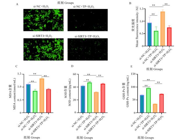
图1 TP激活SIRT3缓解H2O2诱导的BMECs氧化应激

*表示差异显著(P<0.05),**表示差异极显著(P<0.01),ns表示差异不显著(P>0.05)。下图同。

Fig.1 TP activation of SIRT3 alleviates H2O2-induced oxidative stress in BMECs

* indicated significant difference (P<0.05), ** indicated extremely significant difference (P<0.01), and ns indicated no significant difference (P>0.05). The same as below.

2.2 TP激活SIRT3抑制H2O2诱导的BMECs线粒体功能障碍

图2所示,与si-NC+H2O2组相比,si-SIRT3+H2O2组BMECs中NRF2、PGC-1αTFAMNRF1和HO-1的mRNA相对表达量极显著降低(P<0.01),NAD+/NADH比值极显著降低(P<0.01)。与si-NC+H2O2组相比,si-NC+TP+H2O2组BMECs中NRF2、PGC-1αTFAMNRF1和HO-1的mRNA相对表达量显著或极显著上升(P<0.05或P<0.01),NAD+/NADH比值极显著上升(P<0.01),ATP含量极显著上升(P<0.01)。与si-NC+TP+H2O2组相比,si-SIRT3+TP+H2O2组BMECs中PGC-1αTFAMNRF1等mRNA相对表达量差异不显著(P>0.05)。此外,与si-NC+H2O2组相比,si-SIRT3+H2O2组BMECs中SIRT3的mRNA相对表达量和含量显著降低(P<0.05)。与si-NC+TP+H2O2组相比,si-SIRT3+TP+H2O2组BMECs中SIRT3的mRNA相对表达量和含量差异不显著(P>0.05)。以上结果说明,TP依赖于SIRT3的高表达才能起到缓解H2O2诱导的BMECs的线粒体功能紊乱。
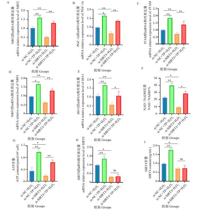
图2 TP激活SIRT3抑制H2O2诱导的BMECs线粒体功能障碍

Fig.2 TP activation of SIRT3 inhibits H2O2-induced mitochondrial mitochondrial dysfunction in BMECs

2.3 TP激活SIRT3抑制H2O2诱导的BMECs炎性反应

图3所示,与si-NC+H2O2组相比,si-SIRT3+H2O2IL-1βIL-6、IL-8和TNF-α的mRNA相对表达量极显著增加(P<0.01)。与si-NC+H2O2组相比,si-NC+TP+H2O2组BMECs中IL-1βIL-6和TNF-α的mRNA相对表达量显著或极显著降低(P<0.05或P<0.01)。与si-NC+TP+H2O2组相比,si-SIRT3+TP+H2O2组BMECs中IL-1βIL-6、IL-8和TNF-α的mRNA相对表达量差异不显著(P>0.05)。以上结果说明,TP依赖SIRT3的高表达起到对H2O2诱导的BMECs的抗炎作用。
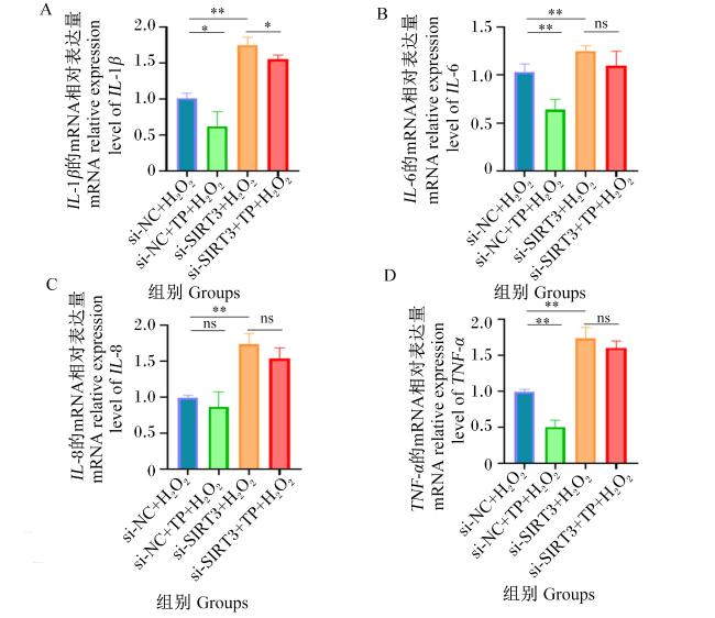
图3 TP激活SIRT3抑制H2O2诱导的BMECs炎性反应

Fig.3 TP activation of SIRT3 inhibits H2O2-induced inflammatory response in BMECs

2.4 TP激活SIRT3缓解H2O2诱导的BMECs凋亡

图4所示,与si-NC+H2O2组相比,si-SIRT3+H2O2组BMECs的细胞凋亡率极显著提高(P<0.01),BMECs中BAXCYTCCASP3的mRNA相对表达量极显著提高(P<0.01),BCL-2的mRNA相对表达量极显著降低(P<0.01),说明干扰SIRT3后加剧了H2O2诱导的BMECs凋亡。与si-NC+H2O2组相比,si-NC+TP+H2O2组BMECs的细胞凋亡率极显著降低(P<0.01),BMECs中BAXCYTCCASP3的mRNA相对表达量极显著降低(P<0.01),BCL-2的mRNA相对表达量极显著提高(P<0.01)。与si-NC+TP+H2O2组相比,si-SIRT3+TP+H2O2组BMECs的细胞凋亡率及BMECs中BAXBCL-2的mRNA相对表达量差异不显著(P>0.05),说明TP依赖SIRT3的高表达才能发挥抑制细胞凋亡的作用。以上结果说明,TP缓解BMECs氧化损伤的机制与线粒体转录因子SIRT3的高表达有关。
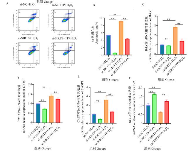
图4 TP激活SIRT3缓解H2O2诱导的BMECs凋亡

Fig.4 TP activation of SIRT3 alleviates H2O2-induced apoptosis in BMECs

3 讨论

奶牛乳腺炎会造成产奶量下降、乳品质降低、治疗成本增加、牛群更替成本增加等一系列危害,进而影响到牧场的经济效益。SIRT3在线粒体氧化代谢、能量代谢和氧化还原稳态以及细胞凋亡中发挥着不可或缺的作用[11-12]。研究表明,敲除SIRT3会导致ROS含量显著增加,线粒体内SIRT3/NRF1/TFAM信号通路受到抑制,最终导致严重的线粒体功能障碍和氧化应激,引发BMECs发生炎性反应[13],而SIRT3含量的增加可通过抑制ROS的生成,提升抗氧化酶SOD和GSH-Px的含量,改善线粒体损伤和氧化应激,进而缓解炎性反应,抑制细胞凋亡[14];线粒体功能障碍会导致动物体内产生大量氧自由基,进一步破坏线粒体结构和功能,从而使生物体处于氧化应激状态[15]。本课题组前期研究中发现,敲除SIRT3会上调BMECs内MDAROS的表达,诱发氧化应激,进而降低线粒体关键因子SIRT3、NRF1和TFAM的mRNA相对表达量,致使线粒体功能出现障碍,这进一步促使BMECs中炎性因子如TNF-αIL-1β的mRNA相对表达量增加,导致BMECs发生炎性损伤。与之相反,过表达SIRT3则能够有效抑制上述现象,维持BMECs内氧化还原稳态,保障线粒体功能正常,显著降低细胞炎性损伤程度,抑制细胞凋亡进程[3]。上述研究结果表明SIRT3在调控线粒体功能、氧化应激与炎性反应进程中发挥着核心作用,是缓解线粒体功能障碍、氧化应激损伤和减轻炎性症反应程度的有效干预靶点。适宜剂量的TP可以通过上调免疫反应来抵御免疫系统受到的刺激,不会对细胞产生负面效应[16]。研究表明,外源添加TP能够降低线粒体ROS的蛋白表达水平,提升SIRT3及线粒体复合物Ⅰ、复合物Ⅱ和复合物Ⅳ的蛋白表达水平,从而促进线粒体中三磷酸腺苷(adenosine triphosphate,ATP)的生成,缓解H2O2诱导的肝细胞和HEK293细胞氧化损伤[17-18]。本研究发现,外源添加TP后可以显著降低H2O2引发的BMECs中PGC-1αTFAMNRF1的mRNA相对表达量,改善线粒体功能障碍,进而恢复线粒体ATP含量。此外,当摄入或添加TP可有效激活SIRT3的表达,降低氧化因子MDA的表达,促进抗氧化因子SOD的表达,进而抑制机体和细胞的炎性损伤[19-20]。本研究发现,对BMECs进行TP干预后可以有效抑制由H2O2诱导的ROSMDA的表达,提高抗氧化因子GSH-Px和SOD含量,从而缓解氧化应激;而当敲除SIRT3后TP缓解H2O2诱导的氧化应激的作用消失,说明TP在发挥缓解BMECs线粒体功能障碍和氧化应激的作用中依赖SIRT3的高表达来实现。
氧化应激会增强炎性反应并导致乳腺的损伤[21]。NF-κB是炎性损伤的重要调节靶点,可调节几种炎性细胞因子如TNF-αIL-1βIL-6的表达参与不同的炎性疾病[22]。研究表明,通过外源添加剂TP可通过激活SIRT3上调抗氧化标致基因NRF2的表达,抑制NF-κB的表达,进而降低炎性因子TNF-αIL-1βIL-6的表达,缓解氧化应激引起的炎性反应[23-24]。本研究发现,外源添加TP后可显著激活NRF2/HO-1信号通路的表达,进而抑制炎性因子TNF-αIL-1βIL-6的表达,缓解H2O2诱导的BMECs氧化损伤;而敲除SIRT3后进行TP干预并未缓解氧化应激和炎性反应。此外,其他研究也得到了相似的结论,如外源添加齐墩果酸和柚皮苷等可以激活SIRT3的表达,从而降低NF-κB的表达,进而通过抑制p65和炎症小体NLRP3的蛋白表达来缓解细胞炎性损伤[25-26];褪黑素可以通过激活SIRT3的表达来降低线粒体氧化应激,进而增强NF-κB脱乙酰化,从而通过减少血清炎性因子TNF-α、IL-6和IL-10含量来缓解炎性反应[27]
当细胞发生炎性损伤后会激活CASP3触发细胞凋亡级联反应,导致细胞凋亡加剧[28]。研究表明,外源添加檀香醇后可通过激活SIRT3进而抑制PC12细胞中由血红素刺激激活CASP3触发的凋亡级联反应,减轻组织病理学损伤和细胞凋亡,并恢复细胞内ATP含量[29]。本研究发现,H2O2诱导可显著上调CASP3的表达,加入TP后则可显著抑制CASP3的表达,而敲除SIRT3后则抑制作用消失,CASP3依然处于激活状态,进而上调BMECs凋亡率,加速BMECs凋亡。此外,当线粒体发生功能障碍时,线粒体可以释放多种细胞凋亡诱导因子,激活半胱天冬酶级联反应,并促进细胞凋亡[30]CYTC是一种与细胞呼吸和细胞凋亡等生理过程密切相关的重要蛋白质,在细胞凋亡过程中,CYTC会从线粒体释放到细胞质中启动凋亡级联反应。研究表明,外源添加人参皂苷Rb1通过激活SIRT3信号通路抑制由高葡萄糖诱导激活的CASP3和CYTC的蛋白表达,进而抑制细胞凋亡的发生[31]。本研究中,经H2O2诱导后BMECs线粒体内凋亡因子CYTC的mRNA相对表达量显著上升,进而导致凋亡级联反应启动,BMECs中凋亡因子BAX的mRNA相对表达量显著上升,而凋亡抑制因子BCL-2的mRNA相对表达量显著下降,TP则可显著逆转这一反应,而敲除SIRT3后逆转作用消失。以上结果表明,TP可以通过激活SIRT3来抑制BMECs中凋亡的启动,从而抑制BMECs因炎性损伤导致的凋亡。

4 结论

TP可通过激活SIRT3下调炎性因子、氧化因子、促凋亡因子含量,上调抗氧化酶含量和线粒体关键调控因子表达,抑制凋亡因子表达,从而恢复线粒体功能,提高抗氧化能力,保护BMECs免受氧化应激与炎性损伤导致的细胞凋亡,进而缓解乳腺氧化损伤和炎性损伤。
[1]
ZHAO C J, WU K Y, BAO L J, et al. Kynurenic acid protects against mastitis in mice by ameliorating inflammatory responses and enhancing blood-milk barrier integrity[J]. Molecular Immunology, 2021, 137:134-144.

DOI PMID

[2]
ZHANG J, WANG J, JIN J, et al. Prevalence,antibiotic resistance,and enterotoxin genes of Staphylococcus aureus isolated from milk and dairy products worldwide:a systematic review and Meta-analysis[J]. Food Research International, 2022, 162(Pt A):111969.

[3]
马学虎, 冀思同, 曹佩佩, 等. 沉默信息调节因子2相关酶3通过提升线粒体功能改善奶牛乳腺氧化应激和炎性反应[J]. 动物营养学报, 2024, 36(12):8049-8061.

DOI

MA X H, JI S T, CAO P P, et al. Silent information regulator 2 related enzyme 3 ameliorated oxidative stress and inflammatory response of dairy cows by enhance mitochondrial function[J]. Chinese Journal of Animal Nutrition, 2024, 36(12):8049-8061. (in Chinese)

DOI

[4]
GAO J M, CHEN N N, LI N, et al. Neuroprotective effects of trilobatin,a novel naturally occurring SIRT3 agonist from Lithocarpus polystachyus Rehd.,mitigate cerebral ischemia/reperfusion injury:involvement of TLR4/NF-κB and Nrf2/keap-1 signaling[J]. Antioxidants & Redox Signaling, 2020, 33(2):117-143.

[5]
DIKALOVA A E, PANDEY A, XIAO L, et al. Mitochondrial deacetylase SIRT3 reduces vascular dysfunction and hypertension while SIRT3 depletion in essential hypertension is linked to vascular inflammation and oxidative stress[J]. Circulation Research, 2020, 126(4):439-452.

DOI PMID

[6]
DENG Z, HE M, HU H, et al. Melatonin attenuates sepsis-induced acute kidney injury by promoting mitophagy through SIRT3-mediated TFAM deacetylation[J]. Autophagy, 2024, 20(1):151-165.

DOI

[7]
DAI X Y, ZHAO Y, GE J, et al. Lycopene attenuates di (2-ethylhexyl) phthalate-induced mitophagy in spleen by regulating the sirtuin3-mediated pathway[J]. Food & Function, 2021, 12(10):4582-4590.

[8]
YANG J, GENG Y, ZHAO B, et al. Green tea polyphenols alleviate TBBPA-induced gastric inflammation and apoptosis by modulating the ROS-PERK/IRE-1/ATF6 pathway in mouse models[J]. Food & Function, 2024, 15(19):10179-10189.

[9]
XU R, ZHU M R, CAO J W, et al. Tea polyphenols protect the mammary gland of dairy cows by enhancing antioxidant capacity and regulating the TGF-β1/p38/JNK pathway[J]. Metabolites, 2022, 12(11):1009.

DOI

[10]
LIU W, MA C, LI H Y, et al. Tea polyphenols reduce inflammation of orbital fibroblasts in graves ophthalmopathy via the NF-κB/NLRP3 pathway[J]. Current Medical Science, 2023, 43(1):123-129.

DOI

[11]
LIU S W, SU Y, SUN B X, et al. Luteolin protects against CIRI,potentially via regulation of the SIRT3/AMPK/mTOR signaling pathway[J]. Neurochemical Research, 2020, 45(10):2499-2515.

DOI

[12]
ZHANG J, XIANG H G, LIU J, et al. Mitochondrial sirtuin 3:new emerging biological function and therapeutic target[J]. Theranostics, 2020, 10(18):8315-8342.

DOI

[13]
ZHENG J W, SHI L G, LIANG F, et al. SIRT3 ameliorates oxidative stress and mitochondrial dysfunction after intracerebral hemorrhage in diabetic rats[J]. Frontiers in Neuroscience, 2018, 12:414.

[14]
EID R A, BIN-MEFERIJ M M, EL-KOTT A F, et al. Exendin-4 protects against myocardial ischemia-reperfusion injury by upregulation of SIRT1 and SIRT3 and activation of AMPK[J]. Journal of Cardiovascular Translational Research, 2021, 14(4):619-635.

DOI

[15]
LEFORT K R, RUNGRATANAWANICH W, SONG B J. Contributing roles of mitochondrial dysfunction and hepatocyte apoptosis in liver diseases through oxidative stress,post-translational modifications, inflammation, and intestinal barrier dysfunction[J]. Cellular and Molecular Life Sciences, 2024, 81(1):34.

DOI

[16]
WAY T D, LIN H Y, KUO D H, et al. Pu-erh tea attenuates hyperlipogenesis and induces hepatoma cells growth arrest through activating AMP-activated protein kinase (AMPK) in human HepG2 cells[J]. Journal of Agricultural and Food Chemistry, 2009, 57(12):5257-5264.

DOI

[17]
QI G Y, WU W Q, MI Y S, et al. Tea polyphenols direct Bmal1-driven ameliorating of the redox imbalance and mitochondrial dysfunction in hepatocytes[J]. Food and Chemical Toxicology, 2018, 122:181-193.

DOI PMID

[18]
YI W, XIE X, DU M, et al. Green tea polyphenols ameliorate the early renal damage induced by a high-fat diet via ketogenesis/SIRT3 pathway[J]. Oxidative Medicine and Cellular Longevity, 2017(2017):9032792.

[19]
XIAO X T, HE S Q, WU N N, et al. Green tea polyphenols prevent early vascular aging induced by high-fat diet via promoting autophagy in young adult rats[J]. Current Medical Science, 2022, 42(5):981-990.

DOI

[20]
NIE C, LI T, FAN M, et al. Polyphenols in highland barley tea inhibit the production of advanced glycosylation end-products and alleviate the skeletal muscle damage[J]. Molecular Nutrition & Food Research, 2022, 66(18):e2200225.

[21]
BAE H, JEONG C H, CHENG W N, et al. Oxidative stress-induced inflammatory responses and effects of N-acetylcysteine in bovine mammary alveolar cells[J]. Journal of Dairy Research, 2017, 84(4):418-425.

DOI PMID

[22]
MALKO P, JIA X, WOOD I, et al. Piezo1 channel-mediated Ca2+ signaling inhibits lipopolysaccharide-induced activation of the NF-κB inflammatory signaling pathway and generation of TNF-α and IL-6 in microglial cells[J]. Glia, 2023, 71(4):848-865.

DOI

[23]
WANG D X, WANG T T, LI Z M, et al. Green tea polyphenols upregulate the Nrf2 signaling pathway and suppress oxidative stress and inflammation markers in D-galactose-induced liver aging in mice[J]. Frontiers in Nutrition, 2022, 9:836112.

[24]
NIE C, WANG B, FAN M, et al. Highland barley tea polyphenols extract alleviates skeletal muscle fibrosis in mice by reducing oxidative stress,inflammation,and cell senescence[J]. Journal of Agricultural and Food Chemistry, 2023, 71(1):739-748.

DOI

[25]
KURUNDKAR D, KURUNDKAR A R, BONE N B, et al. SIRT3 diminishes inflammation and mitigates endotoxin-induced acute lung injury[J]. JCI Insight, 2019, 4(1):e120722.

DOI

[26]
ZHAO W M, LI X L, ZHU Y Y, et al. Diosmin ameliorates renal fibrosis through inhibition of inflammation by regulating SIRT3-mediated NF-κB p65 nuclear translocation[J]. BMC Complementary Medicine and Therapies, 2024, 24(1):29.

DOI

[27]
XU S Q, LI L L, WU J, et al. Melatonin attenuates sepsis-induced small-intestine injury by upregulating SIRT3-mediated oxidative-stress inhibition,mitochondrial protection,and autophagy induction[J]. Frontiers in Immunology, 2021, 12:625627.

[28]
WANG J, HUANGFU M J, LI X J, et al. Osthole induces apoptosis and caspase-3/GSDME-dependent pyroptosis via NQO1-mediated ROS generation in HeLa cells[J]. Oxidative Medicine and Cellular Longevity, 2022, 2022:8585598.

[29]
ZHENG X C, GAO J L, ZHAO M M, et al. Honokiol attenuates mitochondrial fission and cell apoptosis by activating SIRT3 in intracerebral hemorrhage[J]. Chinese Medical Journal, 2023, 136(6):719-731.

DOI PMID

[30]
DORN G W N, KITSIS R N. The mitochondrial dynamism-mitophagy-cell death interactome:multiple roles performed by members of a mitochondrial molecular ensemble[J]. Circulation Research, 2015, 116(1):167-182.

DOI

[31]
KE S Y, YU S J, LIU D H, et al. Ginsenoside RB1 protects human umbilical vein endothelial cells against high glucose-induced mitochondria-related apoptosis through activating SIRT3 signalling pathway[J]. Chinese Journal of Integrative Medicine, 2021, 27(5):336-344.

DOI

Outlines

/