RESEARCH PAPER

Study on Dynamic Characteristics of Oxidative Stress and Inflammatory Response during Incremental Exercise in Yili Horses and Their Association with Exercise Performance

  • ZHANG Zihan , 1 ,
  • PENG Jiangfei 1 ,
  • WUSIMAN Adina 1 ,
  • BAO Yike 1 ,
  • CHU Hongzhong 2 ,
  • YAO Runchen 2 ,
  • ZENG Yaqi , 1, 3, *
Expand
  • 1 College of Animal Science, Xinjiang Agricultural University, Urumqi 830052, China
  • 2 Yili Prefecture Animal Husbandry Station, Yining 835099, China
  • 3 Xinjiang Key Laboratory of Equine Breeding and Exercise Physiology, Urumqi 830052, China
* associate professor, E-mail:

Received date: 2025-10-27

  Online published: 2026-02-12

Abstract

This experiment aimed to study the patterns of oxidative stress and inflammatory responses in horses during incremental exercise. Using an equine treadmill, an incremental exercise test was conducted with dynamic blood collection from seven Yili horses at different exercise load stages (3, 5, 7 and 9 m/s). Plasma antioxidant and immune parameters were measured to evaluate the levels of oxidative stress and inflammatory responses in the horses. The results showed that plasma total antioxidant capacity (T-AOC) was significantly higher at the 7 m/s stage compared with the 3 m/s stage (P<0.05), and plasma glutathione (GSH) content increased significantly when the exercise intensity increased from 5 to 7 m/s (P<0.05). In contrast, the activity of plasma glutathione peroxidase (GSH-Px) decreased significantly when the exercise intensity increased from 0 to 3 m/s (P<0.05). The activity of plasma catalase (CAT) showed a significant increase when the exercise intensity increased from 0 to 7 m/s (P<0.05). Furthermore, plasma interleukin-1β (IL-1β) content was significantly lower at the 7 m/s stage than that at the 3 m/s stage (P<0.05). The plasma T-AOC difference between the 3 and 5 m/s stages showed an extremely significant positive correlation with the competition time (P<0.01), the plasma GSH content difference between the 7 and 9 m/s stages showed a significant negative correlation with the competition time (P<0.05), and the plasma IL-1β content difference between the 5 and 7 m/s stages showed a significant positive correlation with the competition time (P<0.05). In conclusion, Yili horses dynamically adapt to treadmill incremental exercise at 3 to 5 m/s via total antioxidant system synergy, with 3 and 7 m/s identified as critical thresholds for significant inflammatory transition. This identifies a key window for oxidative-inflammatory management and training optimization.

Cite this article

ZHANG Zihan , PENG Jiangfei , WUSIMAN Adina , BAO Yike , CHU Hongzhong , YAO Runchen , ZENG Yaqi . Study on Dynamic Characteristics of Oxidative Stress and Inflammatory Response during Incremental Exercise in Yili Horses and Their Association with Exercise Performance[J]. Chinese Journal of Animal Nutrition, 2026 , 38(2) : 1277 -1285 . DOI: 10.12418/CJAN2026.102

跑步机增量运动在国外已成为评估马匹运动性能及诊断运动相关疾病的常用手段。例如,Mukai等[1]利用跑步机对纯血马进行高强度间歇训练,该研究在赛马模型中证实,“短时极量”间歇方案能在相同跑距下引发更强烈的急性代谢-缺氧应激,并同步激活线粒体生物发生与血管生成相关程序,从而为优化赛马的高强度训练周期提供了分子层面的依据。Lo Feudo等[2]通过递增跑台试验发现,马溃疡综合征(EGUS)会显著降低赛马的有氧阈值及酸中毒恢复能力。然而,目前利用跑步机增量运动对马匹进行运动负荷性能评价的研究仍较为有限。
活性氧(ROS)是细胞呼吸过程中产生的天然副产物,可通过破坏蛋白质、脂质和核酸等的细胞成分对机体产生毒性作用[3]。氧化应激是指机体因ROS生成与清除失衡,导致氧化水平升高,进而引发细胞成分氧化损伤的生物学过程[4]。研究表明,马匹在进行短时间急性运动时,机体能量消耗增加,新陈代谢加快,内环境发生改变,从而诱发运动性疲劳[5-7];而耐力运动同样会影响马匹的健康状况与运动表现[8-9]。这2种运动模式均会引发氧化应激和炎症反应,导致肌肉损伤、运动性能下降,并伴随心率加快、体温升高等生理变化[10]
伊犁马是我国自主培育的优良骑乘马种,以抗逆性强、耐力好著称,现已成为新疆地区主要的赛马品种之一[11-12]。本研究旨在探究伊犁马在不同负荷运动条件下,机体抗氧化与免疫系统在增量运动各阶段应对氧化应激的动态变化规律,并分析其与运动性能之间的关联,从而揭示运动训练对其生理机能的影响机制,评估马匹的氧化应激状态,为伊犁马的训练优化、性能提升、性能测定和健康管理提供理论基础与数据支撑。

1 材料与方法

1.1 试验设计

本研究方案已经新疆农业大学实验动物福利伦理委员会审核批准(批准编号:2023037)。
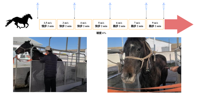
选取伊犁哈萨克自治州昭苏马场经相同调教训练、饲养管理、体型结构基本一致的7匹2岁伊犁马为研究对象(公马3匹、母马4匹)。马匹每天早、晚各饲喂2.5 kg精料(主要原料为熟化玉米压片、熟化大麦压片、熟化燕麦压片等)和4 kg干牧草,3个月后进行跑步机增量运动试验。试验前埋入留置针(图1),连接30 mL无菌注射器采集静态(0 m/s)血液后进行跑步机增量运动,程序设定为:坡度6%,1.5 m/s慢步3 min,3 m/s快步2 min,4 m/s快步2 min,5 m/s快步2 min,6 m/s跑步2 min,7 m/s跑步2 min,9 m/s跑步2 min,并分别在3、5、7、9 m/s这4个阶段结束后通过无菌注射器采集运动过程中的颈静脉动态血液样本12~15 mL[1],分装入乙二胺四乙酸(EDTA)(用于免疫指标测定)和肝素钠真空采血管(用于抗氧化指标测定)中,无菌注射器一次一换。血液样本经1 006×g离心10 min,吸取上清液置于2 mL的冻存管内,做好标记后放入液氮速冻,用于检测抗氧化和免疫指标。
图1 跑步机增量运动血液采集方案示意图

Fig.1 Schematic diagram of blood sampling protocol for treadmill graded exercise

增量运动试验结束24 h后开展1 200 m测试赛,由4名具有一定骑乘经验的骑师分2场(第1场4匹马,第2场3匹马,共7匹马),在相同的沙地跑道上进行比赛,记录比赛用时。

1.2 血浆抗氧化指标测定

所测血浆抗氧化指标包括超氧化物歧化酶(SOD)、谷胱甘肽过氧化物酶(GSH-Px)、过氧化氢酶(CAT)活性,总抗氧化能力(T-AOC)以及谷胱甘肽(GSH)、丙二醛(MDA)含量,使用南京建成生物工程研究所生产的试剂盒测定。

1.3 血浆免疫指标测定

所测血浆免疫指标包括免疫球蛋白G(IgG)、免疫球蛋白M(IgM)、白细胞介素-1β(IL-1β)、肿瘤坏死因子-α(TNF-α)、干扰素-γ(IFN-γ)和转化生长因子-β(TGF-β)含量,使用南京建成生物工程研究所生产的试剂盒测定。

1.4 数据统计与分析

获得的血浆抗氧化和免疫指标数据以及比赛用时使用Excel 2023进行整理,结果用平均值±标准差(mean±SD)表示。基于SPSS 27.0软件,对0、3、5、7、9 m/s 5个阶段马匹的血浆抗氧化和免疫指标进行单因素方差分析,并基于方差分析结果使用LSD法进行组间多重比较,P<0.05为差异显著。使用GraphPad Prism 10.1.2对统计结果进行图表绘制。
根据各阶段血浆抗氧化和免疫指标的数值,得到跑步机后一个运动阶段减去前一个阶段的差值(用3-0、5-3、7-5、9-7表示),随后通过迈维云平台相关性分析工具(https://cloud.metware.cn/#/home)将各阶段的数值和差值分别同比赛用时进行相关性分析,以反映比赛用时和氧化应激指标的相关性,再对抗氧化和免疫指标进行相关性分析,观察2种指标间的关联性。

2 结果与分析

2.1 2.1 1 200 m测试赛成绩

将7匹马的1 200 m测试赛的用时进行汇总后,计算出的平均用时为(88.70±5.35) s。

2.2 增量运动下血浆抗氧化指标差异分析

表1列出了不同速度阶段血浆抗氧化指标的具体数值。5个阶段血浆抗氧化指标的差异比较结果显示:随着速度的增加,T-AOC先降低后升高再降低,3和7 m/s阶段之间差异显著(P<0.05);GSH含量在由5 m/s阶段增加到7 m/s阶段时显著升高(P<0.05),随后在9 m/s阶段时又有所降低(P>0.05);GSH-Px活性在由0 m/s阶段增加到3 m/s阶段时显著降低(P<0.05),随后3个阶段均有变化但是不显著(P>0.05);CAT活性由0 m/s阶段增加到7 m/s阶段时显著升高(P<0.05);随着速度的增加,SOD活性先升高后降低随后重新升高最后再降低,MDA含量则先升高后降低,但各阶段之间均无显著差异(P>0.05)。
表1 增量运动对血浆抗氧化指标的影响

Table 1 Effects of progressive exercise on plasma antioxidant indexes

项目
Items
速度Speed/(m/s)
0 3 5 7 9
总抗氧化能力T-AOC/(mmol/L) 1.01±0.04ab 0.97±0.06b 1.03±0.07ab 1.04±0.07a 1.03±0.05ab
超氧化物歧化酶SOD/(U/mL) 24.93±4.40 26.58±6.54 25.87±7.09 29.93±7.35 27.43±8.37
谷胱甘肽GSH/(μmol/L) 38.62±6.93d 48.82±5.19cd 56.04±17.79bc 78.34±18.14a 67.35±11.61ab
谷胱甘肽过氧化物歧化酶
GSH-Px/(U/mL)
473.49±70.15a 374.97±60.02b 384.95±46.40b 384.09±57.40b 390.60±46.90b
过氧化氢酶CAT/(U/mL) 0.49±0.08b 0.48±0.08b 0.54±0.11ab 0.62±0.13a 0.57±0.03ab
丙二醛MDA/(mmol/L) 5.06±0.89 5.01±0.61 5.44±1.18 5.46±1.02 5.24±1.38

同行数据肩标不同小写字母表示差异显著(P<0.05),无字母或相同小写字母表示差异不显著(P>0.05)。下表同。

Values within the same row with different lowercase letter superscripts indicate significant difference (P<0.05), while with no letter or the same lowercase letter superscripts indicate no significant difference (P>0.05). The same as below.

2.3 增量运动下血浆免疫指标差异分析

表2列出了不同速度阶段血浆免疫指标的具体数值。5个阶段血浆免疫指标的差异比较结果显示:随着速度的增加,IgG和IL-1β含量先升高后降低最后升高,在3和7 m/s阶段差异显著(P<0.05);随着速度的增加,TNF-α含量先升高后降低,在3和9 m/s阶段差异显著(P<0.05);随着速度的增加,IFN-γ含量先升高后降低,在3 m/s阶段显著高于7、9 m/s阶段(P<0.05);随着速度的增加,IgM含量先升高后降低,TGF-β含量先升高后降低最后升高,各阶段之间差异均不显著(P>0.05)。
表2 增量运动对血浆免疫指标的影响

Table 2 Effects of progressive exercise on plasma immune indexes

项目
Items
速度Speed/(m/s)
0 3 5 7 9
免疫球蛋白G IgG/(mg/mL) 9.95±0.68ab 11.28±1.21a 10.53±2.17ab 9.20±1.26b 9.88±1.59ab
免疫球蛋白M IgM/(mg/mL) 0.86±0.15 1.08±0.23 0.98±0.27 0.85±0.22 0.85±0.15
白细胞介素-1β IL-1β/(ng/L) 27.17±2.61ab 28.97±2.76a 27.15±5.05ab 24.37±4.58b 25.61±3.65ab
肿瘤坏死因子-α TNF-α/(ng/L) 111.00±16.18ab 113.79±10.68a 109.25±19.98ab 99.17±15.86ab 96.69±10.20b
干扰素-γ INF-γ/(ng/L) 90.92±10.75ab 103.98±11.10a 97.70±19.28ab 87.34±14.57b 88.32±13.68b
转化生长因子-β TGF-β/(ng/L) 86.64±7.91 93.01±13.84 89.67±19.74 80.41±13.98 83.81±9.80

2.4 血浆抗氧化、免疫指标与比赛用时的相关性分析

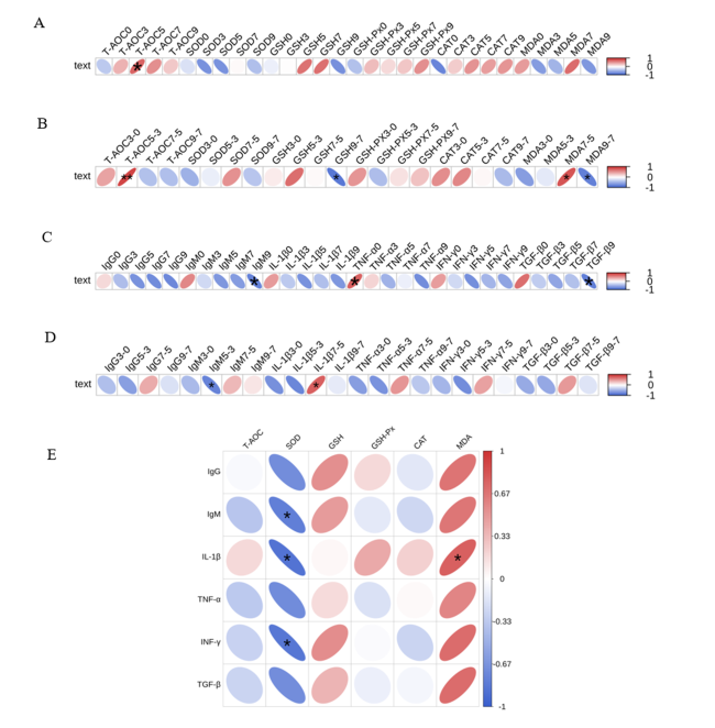
将0、3、5、7、9 m/s 5个阶段血浆抗氧化、免疫指标和前后2个阶段上述指标的差值分别同1 200 m测试的比赛用时进行相关性分析,并对血浆抗氧化与免疫指标进行相关性分析,结果如图2所示。相关性分析结果显示:比赛用时与5 m/s阶段T-AOC呈显著正相关(P<0.05);比赛用时与3~5 m/s阶段T-AOC差值极显著正相关(P<0.01),与5~7 m/s阶段MDA含量差值呈显著正相关(P<0.05),与7~9 m/s阶段GSH、MDA含量差值呈显著负相关(P<0.05);比赛用时与9 m/s阶段IgM、TGF-β含量呈显著负相关(P<0.05),与0 m/s阶段TGF-α含量呈显著正相关(P<0.05);比赛用时与3~5 m/s阶段IgM含量差值呈显著负相关(P<0.05),与5~7 m/s阶段IL-1β含量差值呈显著正相关(P<0.05);抗氧化指标SOD活性与免疫指标IgM、IL-1β和INF-γ含量呈显著负相关(P<0.05),MDA含量与IL-1β含量呈显著正相关(P<0.05)。
图2 血浆抗氧化、免疫指标和比赛用时的相关性分析

A:血浆抗氧化指标与比赛用时的相关性;B:血浆抗氧化指标差值与比赛用时的相关性;C:血浆免疫指标与比赛用时的相关性;D:血浆免疫指标差值与比赛用时的相关性;E:血浆抗氧化与免疫指标的相关性。T-AOC:总抗氧化能力;SOD:超氧化物歧化酶;GSH:谷胱甘肽;GSH-Px:谷胱甘肽过氧化物歧化酶;CAT:过氧化氢酶;MDA:丙二醛;IgG:免疫球蛋白G;IgM:免疫球蛋白M;IL-1β:白细胞介素-1β;TNF-α:肿瘤坏死因子-α;INF-γ:干扰素-γ;TGF-β:转化生长因子-β。0:0 m/s阶段;3:3 m/s阶段;5:5 m/s阶段;7:7 m/s阶段;9:9 m/s阶段;3-0:0~3 m/s阶段差值;5-3:3~5 m/s阶段差值;7-5:5~7 m/s阶段差值;9-7:7~9 m/s阶段差值。“*”表示显著相关(P<0.05),“**”表示极显著相关(P<0.01)。

Fig.2 Analysis of correlations among plasma antioxidant, immune indexes and competition time

A: correlation between plasma antioxidant indexes and competition time; B: correlation between plasma antioxidant index differences and competition time; C: correlation between plasma immune indexes and competition time; D: correlation between plasma immune index differences and competition time; E: correlation between plasma antioxidant and immune indexes. T-AOC: total antioxidant capacity; SOD: superoxide dismutase; GSH: glutathione; GSH-Px: glutathione peroxidase; CAT: catalase; MDA: malondialdehyde; IgG: immunoglobulin G; IgM: immunoglobulin M; IL-1β: interleukin-1β; TNF-α: tumor necrosis factor-α; INF-γ: interferon-γ; TGF-β: transforming growth factor-β. 0: 0 m/s stage; 3: 3 m/s stage; 5: 5 m/s stage; 7: 7 m/s stage; 9: 9 m/s stage; 3-0: difference between the 0 and 3 m/s stages; 5-3: difference between the 3 and 5 m/s stages; 7-5: difference between the 5 and 7 m/s stages; 9-7: difference between the 7 and 9 m/s stages. “*” indicates a significant correlation (P<0.05), and “**” indicates an extremely significant correlation (P<0.01).

3 讨论

3.1 运动过程中的抗氧化反应

T-AOC的变化可能证实马匹对氧化应激耐受性的高度适应,表现为抗氧化机制的加强,不仅有效防止氧化应激的形成,还有效防止ROS的有害影响[13]。本研究中,马匹血浆T-AOC表现为非线性变化规律,在增量运动3与7 m/s阶段具有显著差异,表明随着运动时间和运动负荷的增加,血浆T-AOC上升以应对马匹产生的氧化应激;值得注意的是,5 m/s阶段T-AOC与比赛用时呈显著正相关,特别是在3~5 m/s的加速阶段表现出极显著的正相关关系。这些结果表明总抗氧化系统的动态调节可能是马匹适应不同运动强度的重要生理机制[14],同时T-AOC可能是影响马匹运动表现的重要因素之一,通过监测血浆T-AOC的动态变化有助于评估马匹的运动适应状态。
作为机体抵御氧化应激的关键酶,GSH-Px的活性会在运动诱导的氧化胁迫下发生适应性上调,从而发挥细胞保护作用[15-16]。降低氧化应激反应可以增强机体的运动能力[17]。因此,在本研究中,增量运动3 m/s阶段血浆GSH-Px活性显著下降并在之后的3个阶段缓慢增长的原因可能是GSH-Px参与了抗氧化反应,随后在氧化胁迫下GSH-Px活性适应性上调使马匹应对氧化应激。GSH是所有生物体的抗氧化系统的重要组成部分[18],GSH-Px可以利用GSH还原多种过氧化物,将过氧化氢(H2O2)还原成水(H2O)[19],因此在本研究血浆GSH含量在增量运动过程中不断变化的原因可能是GSH-Px通过GSH进行了还原反应,引起马匹机体加速生成GSH,在7 m/s阶段血浆GSH含量达到最高,可能表明此阶段GSH正在充分参与抗氧化反应。有研究报道,高强度体力活动与高GSH-Px活性相关[20],而在7~9 m/s的高强度运动阶段,血浆GSH含量与比赛用时呈显著负相关,提示GSH的增加可能成为限制运动表现的重要因素。
细胞内的ROS水平可以通过SOD调节,将其转化为H2O2[21]。在本研究中,血浆SOD活性先升高后降低随后又升高再降低,这种波动可能反映了机体在不同阶段对ROS清除需求的动态调整。值得关注的是,本研究中发现血浆SOD活性与免疫指标IgM、IL-1β和INF-γ含量均呈现显著负相关,这一结果可能提示在增量运动下,抗氧化系统和免疫系统存在复杂的交互作用,过度氧化应激可能通过影响免疫调节因子而间接影响运动表现[22]
CAT是一种重要的酶促抗氧化剂,可有效地将H2O2分解成H2O和氧气(O2),从而减少氧化应激并防止细胞和分子崩解,它也是真核抗氧化系统的核心,这种酶在维持细胞氧化还原平衡方面起着关键作用[23-24]。SOD和CAT作为生物体内的ROS清除酶,可以进行级联反应去除超氧阴离子和H2O2,产生分子氧[25]。之前的报道显示,经过训练的马匹可以更好地应对氧化应激带来的影响[26]。本研究中,血浆CAT活性在增量运动7 m/s阶段达到最高,与0和3 m/s阶段具有显著差异,表明3 m/s运动阶段,马匹ROS水平开始增加,触发CAT代偿性活化,7 m/s时H2O2大量累积,此时CAT充分分解体内产生的H2O2,从而减轻氧化应激反应以维持氧化还原平衡;同时,肌肉的微创伤会使膜脂暴露于ROS[16],MDA作为脂质过氧化的副产物[27],在5 m/s和之后的负荷运动中的其含量均高于静息状态,显示马匹在5 m/s阶段开始产生氧化应激表现;在血浆MDA含量与比赛用时的相关性分析中也发现,MDA含量在5~7 m/s加速阶段与比赛用时呈显著正相关,此时MDA含量在这个阶段呈上升趋势,而在7~9 m/s加速跑阶段转为与比赛用时呈显著负相关,此阶段MDA含量呈下降趋势。这一变化提示:MDA含量的升高指示细胞膜损伤加剧,可能抑制肌肉收缩功能,导致比赛用时增加;而MDA含量的降低反映能量代谢加强,有助于提升马匹的运动表现。值得注意的是,血浆MDA含量与炎症因子IL-1β含量呈显著正相关。这表明,由MDA所代表的脂质过氧化损伤和由IL-1β介导的炎症反应可能存在协同作用,共同破坏组织完整性、加剧疼痛与疲劳感,并可能延缓恢复过程。高水平的MDA伴随高水平的IL-1β,共同构成对运动表现的抑制。该结果与之前发现的血浆SOD活性与IL-1β含量呈显著负相关相互印证,共同揭示了氧化应激-炎症轴的双向调控[28]

3.2 运动过程中的免疫反应

运动作为一种生理应激源,能够快速激活交感神经系统和下丘脑-垂体-肾上腺(HPA)轴,导致肾上腺素、去甲肾上腺素和皮质醇等应激激素的释放[29-30]。这些激素具有免疫调节作用,在运动初期可能刺激B细胞短暂性地增加抗体的合成与分泌,或促使储存的IgG从组织间隙进入血液循环,从而导致循环IgG含量的暂时性上升[31]。本研究中,增量运动3 m/s阶段血浆IgG含量的升高,可能是机体对运动应激产生的急性期反应,这可以视为马匹对运动挑战的一种即时免疫准备或动员状态;随后7 m/s阶段血浆IgG含量下降,则可能反映随着运动强度增大或时间延长,机体免疫系统开始承受更大的压力,提示运动强度可能是调节这种免疫应答的关键因素。
Young等[32]的研究发现,劳力性热应激对血浆内源性内毒素核心抗体(EndoCAb)浓度会产生影响,从而引起血浆IgM含量的改变。由于高温加速代谢,增加ROS的产生,热应激会加剧机体氧化应激[33]。本研究中,马匹在跑步机增量运动条件下,血浆IgM含量在运动早期(3 m/s阶段)即开始上升,这可能与运动诱导的多重应激交互作用有关。作为免疫应答中最早出现的抗体,IgM在多种应激叠加作用下变化最为显著,随着机体逐渐适应运动负荷,其含量随后下降并回落到正常水平。值得注意的是,血浆IgM含量在9 m/s阶段及3~5 m/s的加速阶段与比赛用时呈显著负相关,提示控制IgM含量的过度升高或许有助于优化马匹运动表现。因此,监测血浆IgM含量的动态变化有助于评估其免疫-代谢状态。
在长时间的剧烈运动中,白细胞亚群数量增加并引发炎症反应,此时IL-1β和TNF-α等炎症细胞因子被激活,同时免疫抑制机制启动,Th1细胞通过分泌INF-γ参与免疫调节[34-35]。本研究中,马匹血浆IL-1β和TNF-α含量自增量运动3 m/s阶段开始上升,但二者随运动强度增加呈现差异化响应:IL-1β含量在7 m/s阶段显著降低,而TNF-α含量则在9 m/s阶段显著下降,这可能反映了不同炎症因子对运动强度具有不同的敏感性[36]。此外,血浆INF-γ含量在3 m/s阶段升高后,自5 m/s阶段开始下降,并在7和9 m/s阶段显著降低。这一变化可能与高强度运动下IL-1β和TNF-α等炎症细胞因子的激活有关,其诱导INF-γ大量分泌以参与免疫调节,随后在7 m/s阶段因调节机制生效,炎症指标整体回落。进一步分析发现,血浆IL-1β含量在5~7 m/s加速阶段与比赛用时呈显著正相关。该结果与Nemet等[37]的研究结果一致,即剧烈运动可刺激炎症细胞因子释放,进而削弱肌肉力量、影响免疫反应并延长恢复时间。这表明血浆炎症细胞因子含量的升高可能导致马匹运动表现下降,比赛用时增加。
TGF-β在维持免疫稳态与诱导免疫耐受中起着关键作用,其主要通过抑制免疫系统中多种细胞的增殖与功能来实现[38]。氧化应激与炎症反应可诱导TGF-β的产生,进而通过抑制促炎信号通路来减轻组织损伤[39-40]。本研究中,血浆TGF-β含量在增量运动中的变化趋势与IL-1β和TNF-α等促炎细胞因子相似,均自3 m/s阶段开始上升,提示马匹在此强度下机体可能开始产生炎症反应,在Semenistaja等[41]的研究中也表示TGF-β含量的增加与更强的炎症有关联,此时机体通过分泌TGF-β来缓解炎症症状,增强机体的耐受力;随着运动强度增至7 m/s,炎症反应已显著减弱,同时血浆TGF-β含量在9 m/s阶段与比赛用时呈显著负相关,这进一步表明,降低机体的炎症反应与氧化应激水平对于缩短比赛用时具有关键作用。

3.3 研究存在的局限性

本研究仅选用7匹伊犁马完成增量运动试验,样本量有待提升,统计结果具有一定局限性。后续研究将扩大样本规模,通过制定不同增量运动程序进一步研究抗氧化和免疫系统的协同调节机制,以期获得更具普适性和深度的科学结论。

4 结论

本研究发现,3~5 m/s阶段是伊犁马抗氧化系统动态调节应对增量运动的关键区间;3和7 m/s为炎症反应的关键节点,血浆IL-1β和TGF-β含量可作为反映炎症状态的核心标志物。
[1]
MUKAI K, OHMURA H, TAKAHASHI Y, et al. Physiological and skeletal muscle responses to high-intensity interval exercise in thoroughbred horses[J]. Frontiers in Veterinary Science, 2023,10:1241266.

[2]
LO FEUDO C M, STUCCHI L, CONTURBA B, et al. Equine gastric ulcer syndrome affects fitness parameters in poorly performing Standardbred racehorses[J]. Frontiers in Veterinary Science, 2022,9:1014619.

[3]
PIETRUSZEWSKI M, NOWAK-KORNICKA J, ŻELAŹNIEWICZ A, et al. Muscle parameters in men and oxidative stress markers[J]. Journal of Physiological Anthropology, 2025, 44(1):2.

DOI PMID

[4]
WONG D, SAHOO D K, FAIVRE C, et al. Oxidative stress in critically ill neonatal foals[J]. Journal of Veterinary Internal Medicine, 2025, 39(1):e17297.

[5]
WHITE A, ESTRADA M, WALKER K, et al. Role of exercise and ascorbate on plasma antioxidant capacity in thoroughbred race horses[J]. Comparative Biochemistry and Physiology Part A:Molecular & Integrative Physiology, 2001, 128(1):99-104.

[6]
OTT E, CAVINDER C A, LEMLEY C O, et al. PSX-A-18 late-breaking:oxidative stress biomarkers in blood plasma of moderately exercised horses[J]. Journal of Animal Science, 2021, 99(S3):372.

[7]
SHONO S, GIN A, MINOWA F, et al. The oxidative stress markers of horses-the comparison with other animals and the influence of exercise and disease[J]. Animals, 2020, 10(4):617.

DOI

[8]
MARLIN D J, FENN K, SMITH N, et al. Changes in circulatory antioxidant status in horses during prolonged exercise[J]. The Journal of Nutrition, 2002, 132(6):1622S-1627S.

[9]
OTT E C, CAVINDER C A, WANG S S, et al. Oxidative stress biomarkers and free amino acid concentrations in the blood plasma of moderately exercised horses indicate adaptive response to prolonged exercise training[J]. Journal of Animal Science, 2022, 100(4):skac086.

[10]
SOUISSI W, BOUZID M A, FARJALLAH M A, et al. Effect of different running exercise modalities on post-exercise oxidative stress markers in trained athletes[J]. International Journal of Environmental Research and Public Health, 2020, 17(10):3729.

DOI

[11]
WANG C K, ZENG Y Q, WANG J W, et al. Estimation of genetic parameters of body conformation and racing performance traits in Yili horses[J]. Journal of Equine Veterinary Science, 2025,146:105378.

[12]
WANG T L, ZENG Y Q, MA C X, et al. Plasma non-targeted metabolomics analysis of Yili horses raced on tracks with different surface hardness[J]. Journal of Equine Veterinary Science, 2023,121:104197.

[13]
CICHOŃ-WOŹNIAK J, OSTAPIUK-KAROLCZUK J, CIEŚLICKA M, et al. Effect of 2 weeks rest-pause on oxidative stress and inflammation in female basketball players[J]. Scientific Reports, 2024, 14(1):14578.

DOI

[14]
HARIMANA Y, TANG X, XU P, et al. Effect of long-term moderate exercise on muscle cellularity and texture,antioxidant activities,tissue composition,freshness indicators and flavor characteristics in largemouth bass (Micropterus salmoides)[J]. Aquaculture, 2019,510:100-108.

[15]
ZHAO H Q, ZHANG R F, YAN X Y, et al. Superoxide dismutase nanozymes:an emerging star for anti-oxidation[J]. Journal of Materials Chemistry B, 2021, 9(35):6939-6957.

DOI

[16]
CARRERA-QUINTANAR L, FUNES L, HERRANZ-LÓPEZ M, et al. Acute antioxidant response to two types of exercises:2000 m run vs. Burpee test[J]. Antioxidants,2024, 13(2):144.

DOI

[17]
POZDNYAKOVA Y, MURZATAYEVA A. Neuroprotective potential of Stevia rebaudiana and Stachys sieboldii:effects on oxidative stress and locomotor activity in male rats fed a high-fat,high-sucrose diet[J]. Biology, 2025, 14(4):359.

DOI

[18]
FERREIRA M J, RODRIGUES T A, PEDROSA A G, et al. Glutathione and peroxisome redox homeostasis[J]. Redox Biology, 2023,67:102917.

[19]
LEE S H, TAKAHASHI K, HATAKAWA Y, et al. Lipid peroxidation-derived modification and its effect on the activity of glutathione peroxidase 1[J]. Free Radical Biology and Medicine, 2023,208:252-259.

[20]
COVAS M I, ELOSUA R, FITÓ M, et al. Relationship between physical activity and oxidative stress biomarkers in women[J]. Medicine and Science in Sports and Exercise, 2002, 34(5):814-819.

DOI

[21]
LIONTIS T, SENCHUK M M, ZHU S S, et al. Intestine-specific disruption of mitochondrial superoxide dismutase extends longevity[J]. Free Radical Biology and Medicine, 2025,229:195-205.

[22]
SINGH M, SAINI V P, MEENA L L. Heat stress induces oxidative stress and weakens the immune system in catfish Clarias magur:evidence from physiological,histological,and transcriptomic analyses[J]. Fish & Shellfish Immunology, 2025,161:110294.

[23]
ANWAR S, ALRUMAIHI F, SARWAR T, et al. Exploring therapeutic potential of catalase:strategies in disease prevention and management[J]. Biomolecules, 2024, 14(6):697.

DOI

[24]
BAKER A, LIN C C, LETT C, et al. Catalase:a critical node in the regulation of cell fate[J]. Free Radical Biology & Medicine, 2023,199:56-66.

[25]
KWON K, JUNG J, SAHU A, et al. Nanoreactor for cascade reaction between SOD and CAT and its tissue regeneration effect[J]. Journal of Controlled Release, 2022,344:160-172.

[26]
BUJOK J, PAVL’AK A, WALSKI T, et al. Changes in the blood redox status of horses subjected to combat training[J]. Research in Veterinary Science, 2024,171:105219.

[27]
THAKKAR H, CHATTERJEE S, VERMA A, et al. Malondialdehyde mediated alpha-synuclein aggregation:a plausible etiology of Parkinson’s disease in oxidative stress[J]. Chemical Research in Toxicology, 2025, 38(4):573-582.

DOI

[28]
THEOBALD D, SRIRAMULA S. Kinin B1 receptor mediates bidirectional interaction between neuroinflammation and oxidative stress[J]. Antioxidants, 2023, 12(1):150.

DOI

[29]
ZIEMBA A, ADAMCZYK J G, BARCZAK A, et al. Changes in the hormonal profile of athletes following a combat sports performance[J]. BioMed Research International, 2020, 2020(1):9684792.

DOI

[30]
JIANG Z Y, CHEN C, WEISS G L, et al. Stress-induced glucocorticoid desensitizes adrenoreceptors to gate the neuroendocrine response to somatic stress in male mice[J]. Cell Reports, 2022, 41(3):111509.

DOI

[31]
IGNATIUK V, SHAROVA V, ZAKHAROVA L. Prenatal inflammation reprograms hypothalamic-pituitary-gonadal axis development in female rats[J]. Inflammation, 2025, 48(5):2973-2985.

DOI

[32]
YOUNG P, RAUCH C, RUSSO I, et al. Plasma endogenous endotoxin core antibody response to exercise in endurance athletes[J]. International Journal of Sports Medicine, 2022, 43(12):1023-1032.

DOI

[33]
MUTWEDU V B, NYONGESA A W, ODUMA J A, et al. Thermal stress causes oxidative stress and physiological changes in female rabbits[J]. Journal of Thermal Biology, 2021,95:102780.

[34]
WENG P W, CHUNG Y C, LIN T C, et al. Enhancement of interferon-γ secretion by Lepidium meyenii extract supplementation after exhaustive endurance exercise in healthy men:a double-blind,placebo-controlled trial[J]. International Journal of Medical Sciences, 2025, 22(2):398-408.

DOI

[35]
DI PAOLO N C, SHAYAKHMETOV D M. Interleukin 1α and the inflammatory process[J]. Nature Immunology, 2016, 17(8):906-913.

DOI PMID

[36]
RYAN B J, SCHLEH M W, AHN C, et al. Moderate-intensity exercise and high-intensity interval training affect insulin sensitivity similarly in obese adults[J]. The Journal of Clinical Endocrinology and Metabolism, 2020, 105(8):e2941-e2959.

[37]
NEMET D, OH Y, KIM H S, et al. Effect of intense exercise on inflammatory cytokines and growth mediators in adolescent boys[J]. Pediatrics, 2002, 110(4):681-689.

PMID

[38]
WANG M Y, LIU W J, WU L Y, et al. The research progress in transforming growth factor-β2[J]. Cells, 2023, 12(23):2739.

DOI

[39]
HUANG Y P, LIU Z L, WANG X, et al. TGF-β3 protects neurons against intermittent hypoxia-induced oxidative stress and apoptosis through activation of the Nrf-2/KEAP1/HO-1 pathway via binding to TGF-βRI[J]. Neurochemical Research, 2023, 48(9):2808-2825.

DOI PMID

[40]
BOUCHET C, CARDOUAT G, DOUARD M, et al. Inflammation and oxidative stress induce NGF secretion by pulmonary arterial cells through a TGF-β1-dependent mechanism[J]. Cells, 2022, 11(18):2795.

DOI

[41]
SEMENISTAJA S, SOKOLOVSKA L, SVIRSKIS S, et al. Distinct late-stage osteoarthritis profiles identified through NF-κB,TNF-α,and TGF-β-driven synovial inflammation and pain[J]. Scientific Reports, 2025, 15(1):30288.

DOI

Outlines

/