RESEARCH PAPER

Effects of Different Concentrations of Microplastics on Growth Performance, Liver Oxidative Damage and Intestinal Microbiota of Yellow Catfish (Pelteobagrus fulvidraco)

  • ZHANG Fujia , 1 ,
  • KONG Qiuhong 2 ,
  • CHEN Dunxue 1 ,
  • ZHOU Xianjun , 1, * ,
  • WANG Julin 1 ,
  • LIU Qiao 1
Expand
  • 1 College of Animal Science, Guizhou University, Guiyang 550000, China
  • 2 Institute of Hydrobiology, Chinese Academy of Sciences, Wuhan 430072, China
* associate professor, E-mail:

Received date: 2025-07-08

  Online published: 2026-02-12

Abstract

This experiment was conducted to explore the effects of different concentrations of microplastics on the growth performance, liver oxidative damage and intestinal microbiota of yellow catfish (Pelteobagrus fulvidraco). A total of 360 healthy yellow catfish with a body weight of (2.63±0.03) g was selected and randomly divided into 4 groups, with 3 replicates in each group and 30 fish in each replicate. Each group was raised in water with microplastic (particle size 50 nm) concentrations of 0 [control group (CON group)], 100 (MP-100 group), 1 000 (MP-1000 group), and 10 000 μg/L (MP-10000 group), respectively, and fed the same basal diet. The experiment lasted for 21 days, and sampling was conducted on days 14 and 21 of the experiment, respectively. The results showed as follows: 1) after exposure to different concentrations of microplastics, the intestinal tract, gill filaments and liver of yellow catfish were damaged in varying degrees. 2) Compared with CON group, the weight gain rate (WGR) from days 1 to 14 in MP-100 group and MP-1000 group, as well as the WGR from days 1 to 21 in MP-1000 group, were significantly decreased (P<0.05), and the feed coefficient from days 1 to 21 in MP-1000 group and MP-10000 group was significantly increased (P<0.05). 3) Compared with CON group, on day 14, the reactive oxygen species (ROS) content in liver in MP-10000 group was significantly increased (P<0.05); on day 21, the activities of superoxide dismutase and glutathione peroxidase (GSH-Px) in liver in MP-10000 group were significantly increased (P<0.05), and the ROS content in liver in MP-10000 group was significantly increased (P<0.05). 4) Compared with CON group, on day 14, the tumor necrosis factor-α (TNF-α) content in liver in MP-1000 group and MP-10000 group was significantly increased (P<0.05), and the contents of interferon-γ (IFN-γ) and interleukin-10 (IL-10) in liver in MP-10000 group were significantly increased (P<0.05); on day 21, the contents of TNF-α and IFN-γ in liver in MP-10000 group were significantly increased (P<0.05), and the IL-10 content in liver in MP-100 group, MP-1000 group and MP-10000 group was all significantly increased (P<0.05). 5) At the phylum level, compared with CON group, on day 14, the Pseudomonadota relative abundance in intestine in MP-1000 group was significantly increased (P<0.05), while the Bacillota relative abundance in intestine in MP-10000 group was significantly decreased (P<0.05); on day 21, the Fusobacteriota relative abundance in intestine in MP-1000 group was significantly decreased (P<0.05). In conclusion, microplastics can induce the liver oxidative damage, inflammatory response and intestinal microbiota imbalance of yellow catfish, thereby delaying its growth.

Cite this article

ZHANG Fujia , KONG Qiuhong , CHEN Dunxue , ZHOU Xianjun , WANG Julin , LIU Qiao . Effects of Different Concentrations of Microplastics on Growth Performance, Liver Oxidative Damage and Intestinal Microbiota of Yellow Catfish (Pelteobagrus fulvidraco)[J]. Chinese Journal of Animal Nutrition, 2026 , 38(2) : 1286 -1296 . DOI: 10.12418/CJAN2026.103

塑料自发明以来,因其便利性和成本低,被广泛使用在日常生活中。随着使用量的增加,环境中的废弃塑料也随之增加,塑料受外力因素粉碎成小颗粒,粒径小于5 mm的塑料称之为微塑料[1-2]。同时,随着人类活动范围的扩大,微塑料污染范围也随之扩大,其因颗粒小、易扩散等特点[3-4],以及受自然因素的影响,在太平洋、大西洋(地中海)等海洋区域均已检测到微塑料的存在[5-7]。水环境中的微塑料会借助呼吸、摄食等途径侵入水生生物体内[8-9],持续在其体内富集,并随着食物链逐级传递、不断累积,不仅破坏生态系统的平衡稳定[10],还会对人类健康构成潜在威胁[11-12]
黄颡鱼(Pelteobagrus fulvidraco)属于鲶形目鲿科黄颡鱼属,在我国各大水系均有分布,是极具特色的重要淡水养殖品种[13]。据《2024中国渔业统计年鉴》数据显示,2023年我国黄颡鱼产量达62.2万t。黄颡鱼由于肉质鲜美且鱼刺较少,深受消费者喜爱。已有研究表明,微塑料对生物体造成的常见危害包括引发氧化损伤及损害肠道健康,具体表现为相关酶活性的改变和肠道菌群失衡[14-15],本研究聚焦于探究不同浓度微塑料对黄颡鱼造成生物损伤的潜在影响。

1 材料与方法

1.1 试验材料

试验所用微塑料为聚苯乙烯,购于拓渺生物科技(上海)有限公司;微塑料粒径为50 nm,该粒径的微塑料能在水环境中被广泛检测到,易被水生生物吞食[3]。试验中所使用的微塑料溶液使用紫外消毒过的蒸馏水加入微塑料原液进行配制。

1.2 试验饲料

根据黄颡鱼的营养需要[16]配制基础饲料,其组成及营养水平见表1。饲料配制完成后置于65 ℃烘箱烘干,于-18 ℃冰箱中保存备用。
表1 基础饲料组成及营养水平(风干基础)

Table 1 Composition and nutrient levels of the basal diet (air-dry basis) %

项目Items 含量Content
原料Ingredients
鱼粉Fish meal 35.00
豆粕Soybean meal 25.00
鸡肉粉Chicken meal 5.00
玉米蛋白粉Corn gluten meal 7.00
面粉Wheat flour 18.00
大豆油Soybean oil 5.00
磷酸二氢钙Ca(H2PO4)2 1.50
氯化胆碱Choline chloride 0.20
预混料Premix1) 1.00
玉米淀粉Corn starch 2.30
总计Total 100.00
营养水平Nutrient levels2)
水分Moisture 7.73
粗灰分Ash 10.36
粗蛋白质CP 43.28
粗脂肪EE 9.83

1)每千克预混料含有 One kilogram of the premix contained the following:VA 30 mg,VD 6 mg,VK 310 mg,VB1 25 mg,核黄素 riboflavin 45 mg,硫胺素 thiamine 25 mg,烟酸 niacin 200 mg,泛酸 pantothenic acid 60 mg,叶酸 folic acid 250 mg,Fe2(SO4)3 80 mg,ZnSO4 50 mg,NaCl 100 mg,NaF 2 mg,CoCl2 50 mg,CuSO4 10 mg,KI 0.8 mg,MgSO4 1 200 mg。

2)营养水平为实测值。Nutrient levels were measured values.

1.3 试验设计和饲养管理

试验所用黄颡鱼购于贵州镇远某渔业公司,运至贵州大学养殖室暂养14 d后开始试验。本试验经贵州大学实验动物伦理分委员会批准(批准编号:EAE-GZV-2025-T016)。选取360尾体重为(2.63±0.03) g的健康黄颡鱼,随机分成4组,每组3个重复(缸),每个重复30尾。各组饲养于微塑料浓度分别为0[对照组(CON组)]、100(MP-100组)、1 000(MP-1000组)和10 000 μg/L(MP-10000组)的玻璃养殖缸(长×宽×高=1.00 m×0.35 m×0.75 m)中,并均饲喂基础饲料。试验期21 d。
试验期间,24 h持续供氧,采用自然光照,并用加热棒保持水体恒温[(24±1) ℃];水体pH为(7.6±0.5),溶解氧浓度为(7.2±0.6) mg/L;每日投喂2次(09:00和17:00各1次),每次投喂量为黄颡鱼体重的3%;每天清除残余饲料和粪便,每24 h更换1/2含微塑料的水体,每48 h更换全部水体。

1.4 样品采集

分别于试验第14和21天进行解剖采样,采样前每尾黄颡鱼用蒸馏水进行冲洗。每个重复随机取2尾黄颡鱼用于收集肠道、鳃丝和肝脏组织,置于4%多聚甲醛,用于切片制备;每个重复随机取3尾黄颡鱼,分离肝脏,液氮速冻后置于-80 ℃保存,用于生化分析;每个重复随机取3尾黄颡鱼,收集肠道内容物,液氮速冻后置于-80 ℃保存,用于肠道菌群分析。

1.5 指标测定

1.5.1 饲料营养成分

参照AOAC(2006)[17]的方法测定饲料水分(930.15)、粗灰分(942.05)、粗蛋白质(984.13)和粗脂肪(920.39)含量。

1.5.2 生长性能

生长性能相关指标按照以下公式计算:
增重率(WGR,%)=100×(Wt-W0)/W0;
饲料系数(FCR)=F/(Wt-W0);
肝体比(HSI,%)=100×W/Wt
式中:W0为黄颡鱼初始体重(g);Wt为黄颡鱼终末体重(g);F为试验期间黄颡鱼摄入饲料总量(g);W为黄颡鱼肝脏重量(g)。

1.5.3 肝脏抗氧化指标和炎症因子含量

肝脏超氧化物歧化酶(SOD)、过氧化氢酶(CAT)、谷胱甘肽过氧化物酶(GSH-Px)活性以及活性氧(ROS)和炎症因子[肿瘤坏死因子-α(TNF-α)、干扰素-γ(IFN-γ)和白细胞介素-10(IL-10)]含量均使用试剂盒(南京建成生物工程研究所)进行测定。

1.5.4 肠道、鳃丝和肝脏组织形态

肠道、鳃丝和肝脏组织在4%多聚甲醛溶液中固定24 h后,进行脱水、包埋、切片、染色和封片等处理,采用光学显微镜对切片进行观察和扫描,使用CaseViewer 2.4软件分析组织形态。

1.5.5 肠道菌群

肠道内容物样品送至上海美吉生物医药科技有限公司进行肠道菌群测序分析,基于Illumina NovaSeq测序平台,利用双末端测序的方法,构建小片段文库进行测序。对reads进行拼接过滤、聚类或去噪,并进行物种注释及相对丰度分析。

1.6 数据统计分析

采用SPSS 22.0对试验数据进行单因素方差分析(one-way ANOVA)和Duncan氏法多重比较检验,显著性水平设为P<0.05;结果以“平均值±标准差(mean±SD)”形式表示。采用Origin 2024和Adobe Illustrator 2023对数据进行处理,并制作图表。

2 结果与分析

2.1 微塑料对黄颡鱼生长性能的影响

试验期间各组黄颡鱼摄食正常,由表2可知,与CON组相比,MP-100组和MP-1000组第14天体重以及MP-1000组第21天体重均显著降低(P<0.05);同时,MP-100组和MP-1000组第1~14天WGR以及MP-1000组第1~21天WGR均显著降低(P<0.05);此外,MP-1000组和MP-10000组第1~21天FCR显著提高(P<0.05)。
表2 微塑料对黄颡鱼生长性能的影响

Table 2 Effects of microplastics on growth performance of yellow catfish (Pelteobagrus fulvidraco)

项目
Items
组别Groups
CON MP-100 MP-1000 MP-10000
初始体重IBW/g 2.64±0.01 2.65±0.04 2.65±0.04 2.62±0.04
第14天体重BW on day 14/g 4.66±0.04b 4.26±0.22a 4.30±0.04a 4.42±0.26ab
第21天体重BW on day 21/g 5.20±0.12b 4.94±0.15ab 4.52±0.44a 4.82±0.21ab
第1~14天增重率WGR from days 1 to 14/% 76.54±0.90b 60.57±5.93a 62.54±1.37a 68.62±8.00ab
第1~21天增重率WGR from days 1 to 21/% 96.74±5.32b 86.52±3.07ab 70.62±15.98a 83.60±6.28ab
第1~14天饲料系数FCR from days 1 to 14 1.13±0.05 1.14±0.04 1.14±0.04 1.15±0.01
第1~21天饲料系数FCR from days 1 to 21 1.08±0.10a 1.08±0.09a 1.20±0.02b 1.21±0.02b
第14天肝体比HSI on day 14/% 1.68±0.06 1.68±0.06 1.74±0.01 1.70±0.02
第21天肝体比HSI on day 21/% 1.67±0.05 1.68±0.02 1.73±0.01 1.75±0.01

同行数据肩标无字母或相同字母表示差异不显著(P>0.05),不同字母表示差异显著(P<0.05)。下表同。

In the same row, values with no letter or the same letter superscripts indicated no significant difference (P>0.05), while with different letter superscripts indicated significant difference (P<0.05). The same as below.

2.2 微塑料对黄颡鱼肝脏抗氧化指标的影响

表3可知,第14天时,各组间黄颡鱼肝脏SOD、CAT和GSH-Px活性无显著差异(P>0.05);与CON组相比,MP-10000组肝脏ROS含量显著提高(P<0.05)。第21天时,与CON组相比,MP-10000组肝脏SOD和GSH-Px活性显著提高(P<0.05),MP-1000组肝脏CAT活性显著降低(P<0.05),MP-10000组肝脏ROS含量显著提高(P<0.05)。
表3 微塑料对黄颡鱼肝脏抗氧化指标的影响

Table 3 Effects of microplastics on liver antioxidant indices of yellow catfish (Pelteobagrus fulvidraco)

项目
Items
组别Groups
CON MP-100 MP-1000 MP-10000
第14天Day 14
超氧化物歧化酶SOD/(U/mg prot) 4.43±0.03 4.72±0.10 4.60±0.46 4.84±0.22
过氧化氢酶CAT/(U/mg prot) 1.03±0.01 1.19±0.13 1.13±0.15 1.10±0.02
谷胱甘肽过氧化物酶GSH-Px/(pmol/mg prot) 2.56±0.02 2.75±0.04 2.69±0.26 2.65±0.22
活性氧ROS/(U/mg prot) 38.43±1.18a 38.51±2.81ab 41.18±2.75ab 42.23±0.06b
第21天Day 21
超氧化物歧化酶SOD/(U/mg prot) 2.81±0.22a 2.91±0.45a 2.95±0.03a 3.56±0.08b
过氧化氢酶CAT/(U/mg prot) 0.85±0.06b 0.84±0.10b 0.71±0.00a 0.86±0.02b
谷胱甘肽过氧化物酶GSH-Px/(pmol/mg prot) 1.73±0.03a 1.83±0.13ab 1.90±0.23ab 2.06±0.04b
活性氧ROS/(U/mg prot) 45.60±1.16a 48.30±6.18ab 49.22±3.66ab 55.43±1.19b

2.3 微塑料对黄颡鱼肝脏炎症因子含量的影响

表4可知,第14天时,与CON组相比,MP-1000和MP-10000组黄颡鱼肝脏TNF-α含量显著提高(P<0.05),MP-10000组肝脏IFN-γ和IL-10含量显著提高(P<0.05)。第21天时,与CON组相比,MP-10000组肝脏TNF-α和IFN-γ含量显著提高(P<0.05),MP-100组、MP-1000和MP-10000组肝脏IL-10含量均显著提高(P<0.05)。
表4 微塑料对黄颡鱼肝脏炎症因子含量的影响

Table 4 Effects of microplastics on liver inflammatory factor contents of yellow catfish (Pelteobagrus fulvidraco) ng/g prot

项目
Items
组别Groups
CON MP-100 MP-1000 MP-10000
第14天Day 14
肿瘤坏死因子-α TNF-α 40.58±0.05a 46.00±3.05ab 47.05±5.55b 46.36±0.27b
干扰素-γ IFN-γ 8.04±0.10a 8.76±0.70ab 8.73±0.78ab 9.17±0.23b
白细胞介素-10 IL-10 19.59±0.25ab 20.75±1.45bc 18.61±1.19a 22.60±0.96c
第21天Day 21
肿瘤坏死因子-α TNF-α 46.99±1.99a 51.60±5.74ab 48.25±2.38ab 54.28±2.42b
干扰素-γ IFN-γ 9.53±0.26a 9.71±1.28a 10.06±0.42ab 11.41±0.49b
白细胞介素-10 IL-10 14.58±1.30a 17.43±0.82b 16.52±0.84b 17.64±0.15b

2.4 微塑料对黄颡鱼肠道、鳃丝和肝脏组织形态的影响

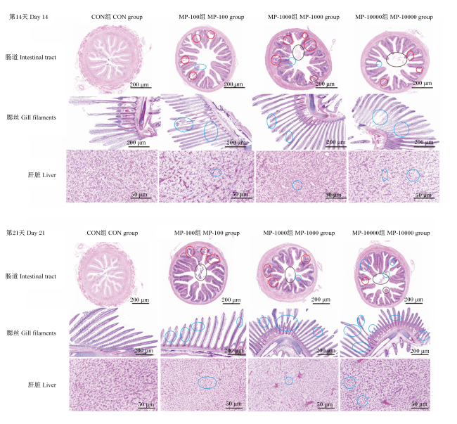
通过对黄颡鱼肠道、鳃丝和肝脏组织进行苏木精-伊红(HE)染色切片观察发现,微塑料暴露对其组织形态存在明显负面影响。如图1所示,第14天时,微塑料暴露组黄颡鱼肠道、鳃丝和肝脏组织均出现不同程度的病理损伤。持续至第21天时,微塑料暴露组肠道绒毛结构遭受严重破坏,出现明显溶解现象;鳃丝组织结构排列紊乱并断裂;肝脏组织则呈现出明显的空泡化病变。
图1 微塑料对黄颡鱼肠道、鳃丝和肝脏组织形态的影响

肠道红圈表示肠绒毛剥离并部分溶解,黑圈表示肠绒毛轻微脱落溶解,蓝圈表示肠绒毛尖端上皮细胞坏死;鳃丝蓝圈表示鳃丝排列紊乱、脱落、畸形和坏死;肝脏蓝圈表示肝细胞轻微空泡化。

Fig.1 Effects of microplastics on tissue morphology of intestinal tract, gill filaments and liver of yellow catfish (Pelteobagrus fulvidraco)

Red circle in intestinal tract indicated peeling and partial dissolution of intestinal villi, black circle indicated slight shedding and dissolution of intestinal villi, and blue circle indicated necrosis of epithelial cells at tips of intestinal villi. Blue circle in gill filaments indicated disordered arrangement, shedding, deformity and necrosis of gill filaments. Blue circle in liver indicated slight vacuolation of hepatocytes.

2.5 微塑料对黄颡鱼肠道菌群的影响

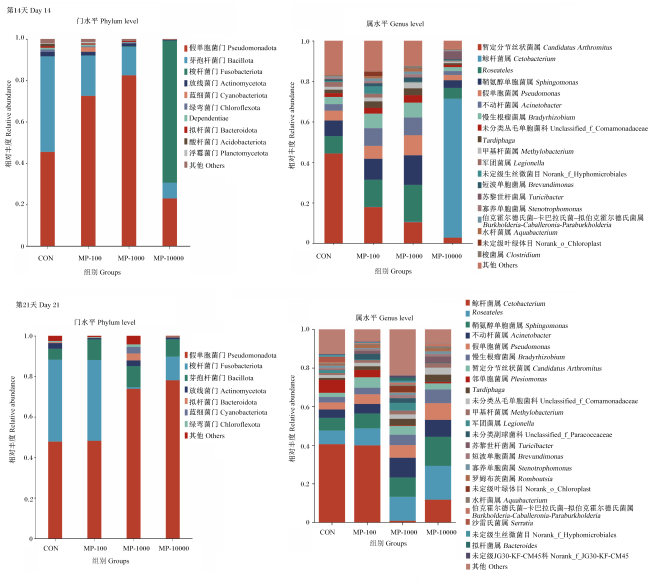
图2所示,在门水平上,黄颡鱼肠道主要由假单胞菌门(Pseudomonadota)、梭杆菌门(Fusobacteriota)和芽孢杆菌门(Bacillota)等组成,但随着暴露时间和微塑料浓度的变化,各菌门相对丰度发生变化。在属水平上,黄颡鱼肠道主要由暂定分节丝状菌属(Candidatus Arthromitus)、鲸杆菌属(Cetobacterium)、不动杆菌属(Acinetobacter)、鞘氨醇单胞菌属(Sphingomonas)、假单胞菌属(Pseudomonas)和Roseateles等组成。
图2 微塑料对黄颡鱼肠道菌群组成的影响

Fig.2 Effects of microplastics on gut microbiota composition of yellow catfish (Pelteobagrus fulvidraco)

表5可知,在门水平上,第14天时,与CON组相比,MP-1000组黄颡鱼肠道假单胞菌门相对丰度显著提高(P<0.05),MP-10000组肠道芽孢杆菌门相对丰度显著降低(P<0.05);第21天时,与CON组相比,MP-1000组肠道梭杆菌门相对丰度显著降低(P<0.05)。
表5 微塑料对黄颡鱼肠道菌群在门水平上相对丰度的影响

Table 5 Effects of microplastics on relative abundance of gut microbiota at phylum level of yellow catfish (Pelteobagrus fulvidraco)

项目
Items
组别Groups
CON MP-100 MP-1000 MP-10000
第14天Day 14
假单胞菌门Pseudomonadota 0.46±0.30ab 0.72±0.12bc 0.82±0.04c 0.23±0.16a
芽孢杆菌门Bacillota 0.46±0.34b 0.20±0.16ab 0.14±0.05ab 0.08±0.01a
梭杆菌门Fusobacteriota 0.04±0.09 0.00±0.00 0.00±0.00 0.69±0.16
拟杆菌门Bacteroidota 0.00±0.00 0.01±0.01 0.00±0.00 0.00±0.00
第21天Day 21
假单胞菌门Pseudomonadota 0.48±0.22 0.48±0.13 0.74±0.11 0.78±0.16
芽孢杆菌门Bacillota 0.05±0.06 0.10±0.03 0.10±0.06 0.09±0.05
梭杆菌门Fusobacteriota 0.40±0.31b 0.40±0.12b 0.01±0.00a 0.12±0.12ab
拟杆菌门Bacteroidota 0.00±0.00 0.01±0.01 0.03±0.04 0.00±0.00

3 讨论

3.1 微塑料对黄颡鱼肝脏氧化损伤和炎症因子含量的影响

氧化应激反应是识别微塑料颗粒暴露影响的重要生物标志,而微塑料在肝脏中富集会导致肝脏氧化损伤和代谢紊乱[18]。Matés[19]和Schülke等[20]研究表明,微塑料暴露会引起螃蟹体内ROS增加,刺激防御系统对抗氧化应激。本试验中,第14和21天时,MP-10000组组黄颡鱼肝脏ROS含量均显著高于CON组,表明在微塑料暴露刺激下,黄颡鱼氧化应激反应加剧,ROS生成增加。SOD和CAT是2种重要的抗氧化酶,已被证实是生物体内抗氧化剂防御的第一道防线[21];GSH-Px在保护生物体免受氧化损伤方面也起着重要作用[22]。在微塑料暴露试验中,红螯螯虾肝胰腺SOD活性显著升高[23],低浓度微塑料暴露使中华绒螯蟹肝脏SOD和GSH-Px活性升高[24]。本试验中,第14天时,各组间黄颡鱼肝脏SOD、CAT和GSH-Px活性无显著差异;第21天时,MP-10000组肝脏SOD活性显著高于CON组,这可能是在微塑料刺激下,机体通过提高SOD活性以应对持续的氧化反应;此外,第21天时,MP-10000组肝脏GSH-Px活性显著高于CON组,表明在持续的微塑料暴露下,机体可能通过提高GSH-Px活性来增强抗氧化防御能力,以此来应对持续的氧化应激。
潘春等[25]研究报道,5 μm微塑料可使小鼠前列腺组织炎症细胞大量浸润,炎症因子含量显著升高;聚苯乙烯暴露引起成年斑马鱼肠道IFN-γ蛋白表达水平变化[26]。本试验中,第14天时,与CON组相比,MP-1000和MP-10000组黄颡鱼肝脏TNF-α含量显著提高,MP-10000组肝脏IFN-γ和IL-10含量显著提高;第21天时,与CON组相比,各微塑料暴露组肝脏炎症因子含量均有所提高,由此推测,微塑料暴露可能会引起机体炎症反应。

3.2 微塑料对黄颡鱼生长性能和组织形态的影响

微塑料可通过主动进食等方式进入体内,并随着体循环转移至不同的组织。肝脏是鱼类最重要的解毒器官[5],微塑料颗粒可以穿过生物屏障进入体循环[15],在消化道内富集[18]。已有研究报道,微塑料暴露后富集在鱼类肠道中会导致消化道阻塞,诱发饱腹感,减少摄食,影响营养物质吸收,并最终影响鱼类生长[27-28]。研究表明,长期微塑料暴露会抑制雌性斑马鱼生长[29];投喂含有微塑料的饲料会抑制许氏平鲉的生长[30]。本试验中,经微塑料暴露后,各微塑料暴露组黄颡鱼终末体重和WGR均低于CON组,说明微塑料富集在黄颡鱼肠道影响了肠道对营养物质的吸收,从而减缓了生长。外环境微塑料的持续刺激会导致鳃丝持续分泌黏液,而黏液会进一步吸附微塑料颗粒,加重损伤。据报道,对斑马鱼进行21 d的微塑料摄入试验,在斑马鱼肝脏上发现微塑料颗粒[31];当日本青鳉摄入微塑料后会改变肌肉和肝脏的胆固醇分布,引起脂肪空泡的形成和单细胞坏死[32];幼年非洲鲶鱼暴露在聚乙烯环境中会加重肝脏组织损伤,并引起与生殖过程相关基因的表达发生变化[33];经微塑料暴露后的斑马鱼肠道和鳃丝出现损伤[11]。本试验中,各微塑料暴露组黄颡鱼肝脏都出现了不同程度的损伤,高浓度微塑料组(MP-10000组)黄颡鱼肝脏组织切片中出现明显空泡化,这可能是由于存在于环境中的微塑料通过体循环进入水生生物体内,刺激肝细胞导致细胞结构受损;同时,与第14天相比,第21天的肠道组织损伤程度进一步加剧,肠壁结构完整性受损,肠绒毛脱落、溶解等病理特征更为突出,表明长期高浓度微塑料暴露会对黄颡鱼肠道组织造成更为严重的损害;此外,各微塑料暴露组鳃丝出现了不同程度的损伤,且随着微塑料浓度的升高,鳃丝断裂和畸形情况加剧,这表明微塑料富集在一定程度上破坏了鳃丝的结构和功能。

3.3 微塑料对黄颡鱼肠道菌群的影响

肠道是鱼类营养物质消化吸收的重要场所,肠道健康是鱼类正常生长发育的前提,也是鱼类抵御病害的第一道防线[34]。肠道菌群影响肠道功能,稳定的菌群有利于鱼类的生长和健康[35];反之,肠道菌群的变化可能导致免疫系统紊乱,诱发多种疾病[36]。本试验中,在门水平上,黄颡鱼肠道主要由假单胞菌门、梭杆菌门和芽孢杆菌门等组成,这与陈严轩等[37]在黄颡鱼上的研究结果相似。研究表明,假单胞菌门(又名变形菌门)相对丰度被认为与诱导炎症有关[38-39];在水生生物体研究中,梭杆菌门能调节鱼类新陈代谢,被认为是有益菌[40];芽孢杆菌门(又名厚壁菌门)可以分泌消化酶,从而有利于分解蛋白质等营养物质[41]。本试验中,经过14 d微塑料暴露后,在门水平上,MP-100组和MP-1000组黄颡鱼肠道假单胞菌门相对丰度高于CON组,各微塑料暴露组肠道芽孢杆菌门相对丰度低于CON组;暴露至21 d时,MP-1000组和MP-10000组肠道假单胞菌门相对丰度高于CON组,肠道梭杆菌门相对丰度低于CON组。在属水平上,黄颡鱼肠道主要由暂定分节丝状菌属、鲸杆菌属和假单胞菌属等组成,这与陈严轩等[37]的研究结果相似。同时,经过21 d微塑料暴露后,MP-1000组和MP-10000组肠道鲸杆菌属相对丰度低于CON组。鲸杆菌属是鱼类肠道菌群的优势菌,也是鱼类肠道的“土著”菌,具有一定的抑制有害菌的作用,对鱼体健康起重要调控作用[11,42]。研究报道,鲸杆菌属具有增强肠道微生物与肠道屏障相互作用的能力,能够提高宿主抵抗力[43-44]

4 结论

综上所述,微塑料会诱发黄颡鱼肝脏氧化损伤、炎症反应以及肠道菌群失衡等,进而延缓其生长。
[1]
LACERDA A L D F, RODRIGUES L D S, VAN SEBILLE E, et al. Plastics in sea surface waters around the Antarctic Peninsula[J]. Scientific Reports, 2019, 9(1):3977.

DOI PMID

[2]
DI M X, WANG J. Microplastics in surface waters and sediments of the three gorges reservoir,China[J]. Science of the Total Environment, 2018,616/617:1620-1627.

[3]
LIU H F, YANG X M, LIU G B, et al. Response of soil dissolved organic matter to microplastic addition in Chinese loess soil[J]. Chemosphere, 2017,185:907-917.

[4]
XU S, MA J, JI R, et al. Microplastics in aquatic environments:occurrence,accumulation,and biological effects[J]. Science of the Total Environment, 2020,703:134699.

[5]
RHODES C J. Plastic pollution and potential solutions[J]. Science Progress, 2018, 101(3):207-260.

DOI PMID

[6]
ERIKSEN M, LEBRETON L C M, CARSON H S, et al. Plastic pollution in the world’s oceans:more than 5 trillion plastic pieces weighing over 250,000 tons afloat at sea[J]. PLoS One, 2014, 9(12):e111913.

DOI

[7]
TANG Y Q, LIU Y G, CHEN Y, et al. A review:research progress on microplastic pollutants in aquatic environments[J]. Science of the Total Environment, 2021,766:142572.

[8]
GRIGORAKIS S, MASON S A, DROUILLARD K G. Determination of the gut retention of plastic microbeads and microfibers in goldfish (Carassius auratus)[J]. Chemosphere, 2017,169:233-238.

[9]
AVIO C G, GORBI S, MILAN M, et al. Pollutants bioavailability and toxicological risk from microplastics to marine mussels[J]. Environmental Pollution, 2015,198:211-222.

[10]
SANA S S, DOGIPARTHI L K, GANGADHAR L, et al. Effects of microplastics and nanoplastics on marine environment and human health[J]. Environmental Science and Pollution Research, 2020,27:44743-44756.

[11]
PEI X, HENG X, CHU W H. Polystyrene nano/microplastics induce microbiota dysbiosis,oxidative damage,and innate immune disruption in zebrafish[J]. Microbial Pathogenesis, 2022,163:105387.

[12]
HUANG J N, WEN B, ZHU J G, et al. Exposure to microplastics impairs digestive performance,stimulates immune response and induces microbiota dysbiosis in the gut of juvenile guppy (Poecilia reticulata)[J]. Science of the Total Environment, 2020,733:138929.

[13]
WARING R H, HARRIS R M, MITCHELL S C. Plastic contamination of the food chain:a threat to human health?[J]. Maturitas, 2018,115:64-68.

[14]
ZHANG X, WEN K, DING D X, et al. Size-dependent adverse effects of microplastics on intestinal microbiota and metabolic homeostasis in the marine medaka (Oryzias melastigma)[J]. Environment International, 2021,151:106452.

[15]
FU L L, HAN D, YI J H, et al. Effects of dietary yeast hydrolysate on the growth performance,intestine health and digestion of juvenile yellow catfish (Pelteobagrus vachelli♂×Pelteobagrus fulvidraco♀)[J]. Aquaculture Reports, 2023,29:101496.

[16]
HAN Y K, XU Y C, LUO Z, et al. Fish meal replacement by mixed plant protein in the diets for juvenile yellow catfish Pelteobagrus fulvidraco:effects on growth performance and health status[J]. Aquaculture Nutrition, 2022, 2022(1):2677885.

[17]
AOAC. Official methods of analysis of AOAC international[S]. 18th ed. Gaithersburg: Association of Official Analytical Chemists, 2006.

[18]
JEONG C B, WON E J, KANG H M, et al. Microplastic size-dependent toxicity,oxidative stress induction,and p-JNK and p-p38 activation in the monogonont rotifer (Brachionus koreanus)[J]. Environmental Science & Technology, 2016, 50(16):8849-8857.

DOI

[19]
MATÉS J M. Effects of antioxidant enzymes in the molecular control of reactive oxygen species toxicology[J]. Toxicology, 2000, 153(1/2/3):83-104.

DOI

[20]
SCHÜLKE S, DREIDAX D, MALIK A, et al. Living with stress:regulation of antioxidant defense genes in the subterranean,hypoxia-tolerant mole rat,Spalax[J]. Gene, 2012, 500(2):199-206.

DOI

[21]
WRIGHT S L, THOMPSON R C, GALLOWAY T S. The physical impacts of microplastics on marine organisms:a review[J]. Environmental Pollution, 2013,178:483-492.

[22]
VELMA V, TCHOUNWOU P B. Chromium-induced biochemical,genotoxic and histopathologic effects in liver and kidney of goldfish,Carassius auratus[J]. Mutation Research, 2010, 698(1/2):43-51.

[23]
CHEN Q, LV W W, JIAO Y, et al. Effects of exposure to waterborne polystyrene microspheres on lipid metabolism in the hepatopancreas of juvenile redclaw crayfish,Cherax quadricarinatus[J]. Aquatic Toxicology, 2020,224:105497.

[24]
YU P, LIU Z Q, WU D L, et al. Accumulation of polystyrene microplastics in juvenile Eriocheir sinensis and oxidative stress effects in the liver[J]. Aquatic Toxicology, 2018,200:28-36.

[25]
潘春, 范臻成, 洪润洋, 等. 聚苯乙烯微塑料对雄性小鼠前列腺的影响及机制[J]. 中国组织工程研究, 2025, 29(34):7353-7361.

PAN C, FAN Z C, HONG R Y, et al. Effect and mechanism of polystyrene microplastics on prostate in male mice[J]. Chinese Journal of Tissue Engineering Research, 2025, 29(34):7353-7361. (in Chinese)

[26]
JIN Y X, XIA J Z, PAN Z H, et al. Polystyrene microplastics induce microbiota dysbiosis and inflammation in the gut of adult zebrafish[J]. Environmental Pollution, 2018,235:322-329.

[27]
MATTSSON K, EKVALL M T, HANSSON L A, et al. Altered behavior,physiology,and metabolism in fish exposed to polystyrene nanoparticles[J]. Environmental Science & Technology, 2015, 49(1):553-561.

DOI

[28]
MAZURAIS D, ERNANDE B, QUAZUGUEL P, et al. Evaluation of the impact of polyethylene microbeads ingestion in European sea bass (Dicentrarchus labrax) larvae[J]. Marine Environmental Research, 2015, 112(Pt A):78-85.

DOI PMID

[29]
孔娟. 环境浓度氧四环素和聚苯乙烯微塑料复合暴露对斑马鱼生长及肠道健康的影响[D]. 硕士学位论文. 武汉: 华中农业大学, 2022.

KONG J. Effects of environmental concentrations of oxytetracycline and polystyrene microplastics on growth and intestinal health of zebrafish[D]. Master’s Thesis. Wuhan: Huazhong Agricultural University, 2022. (in Chinese)

[30]
赵美静. 聚氯乙烯微塑料和重金属铜对许氏平鲉的联合毒性效应[D]. 硕士学位论文. 天津: 天津农学院, 2021.

ZHAO M J. Combined toxic effects of polyvinyl chloride microplastics and copper on Sebastes schlegelii[D]. Master’s Thesis.Tianjin: Tianjin Agricultural University, 2021. (in Chinese)

[31]
RAINIERI S, CONLLEDO N, LARSEN B K, et al. Combined effects of microplastics and chemical contaminants on the organ toxicity of zebrafish (Danio rerio)[J]. Environmental Research, 2018,162:135-143.

[32]
ROCHMAN C M, HOH E, KUROBE T, et al. Ingested plastic transfers hazardous chemicals to fish and induces hepatic stress[J]. Scientific Reports, 2013, 3(1):3263.

DOI

[33]
KARAMI A, ROMANO N, GALLOWAY T, et al. Virgin microplastics cause toxicity and modulate the impacts of phenanthrene on biomarker responses in African catfish (Clarias gariepinus)[J]. Environmental Research, 2016,151:58-70.

[34]
艾业睿, 胡毅, 张俊智, 等. 低鱼粉饲料中添加丁酸钠对黄颡鱼生长性能、血清免疫及肠道健康的影响[J]. 水生生物学报, 2025, 49(11):102509.

AI Y R, HU Y, ZHANG J Z, et al. Sodium butyrate addition to low fishmeal diets on growth performance,serum immunity,and intestinal health of catfish Pelteobagrus fulvidraco[J]. Acta Hydrobiologica Sinica, 2025, 49(11):102509. (in Chinese)

[35]
QIAO R X, SHENG C, LU Y F, et al. Microplastics induce intestinal inflammation,oxidative stress,and disorders of metabolome and microbiome in zebrafish[J]. Science of the Total Environment, 2019,662:246-253.

[36]
MIRPURI J, RAETZ M, STURGE C R, et al. Proteobacteria-specific IgA regulates maturation of the intestinal microbiota[J]. Gut Microbes, 2014, 5(1):28-39.

DOI PMID

[37]
陈严轩, 王国霞, 邱建强, 等. 姜黄素改善氧化鱼油对黄颡鱼肠道的不利影响[J]. 动物营养学报, 2024, 36(8):5297-5307.

DOI

CHEN Y X, WANG G X, QIU J Q, et al. Curcumin improves adverse effects of oxidized fish oil on intestine of Pelteobagrus fulvidraco[J]. Chinese Journal of Animal Nutrition, 2024, 36(8):5297-5307.

[38]
WANG Y, QI W T, SONG G, et al. High-fructose diet increases inflammatory cytokines and alters gut microbiota composition in rats[J]. Mediators of Inflammation, 2020, 2020(1):6672636.

[39]
LITVAK Y, BYNDLOSS M X, TSOLIS R M, et al. Dysbiotic Proteobacteria expansion:a microbial signature of epithelial dysfunction[J]. Current Opinion in Microbiology, 2017,39:1-6.

[40]
WU X X, HAO Q, TEAME T, et al. Gut microbiota induced by dietary GWF contributes to growth promotion,immune regulation and disease resistance in hybrid sturgeon (Acipenserbaerii×Acipenserschrenckii):insights from a germ-free zebrafish model[J]. Aquaculture, 2020,520:734966.

[41]
KIMURA I, OZAWA K, INOUE D, et al. The gut microbiota suppresses insulin-mediated fat accumulation via the short-chain fatty acid receptor GPR43[J]. Nature Communications, 2013,4:1829.

[42]
VAN KESSEL M A, DUTILH B E, NEVELING K, et al. Pyrosequencing of 16S rRNA gene amplicons to study the microbiota in the gastrointestinal tract of carp (Cyprinus carpio L.)[J]. AMB Express, 2011, 1(1):41.

DOI

[43]
LARSEN A M, MOHAMMED H H, ARIAS C R. Characterization of the gut microbiota of three commercially valuable warmwater fish species[J]. Journal of Applied Microbiology, 2014, 116(6):1396-1404.

DOI PMID

[44]
QI X Z, ZHANG Y, ZHANG Y L, et al. Vitamin B12 produced by Cetobacterium somerae improves host resistance against pathogen infection through strengthening the interactions within gut microbiota[J]. Microbiome, 2023, 11(1):135.

DOI

Outlines

/