

1 材料与方法
1.1 试验材料
1.2 试验方法
1.2.1 溶液配制
1.2.1.1 AFB1工作液的配制
1.2.1.2 POP工作液配制
1.2.2 细胞培养
1.2.3 细胞分组与处理
1.2.3.1 POP作用浓度、预处理时间和AFB1作用浓度的筛选
1.2.3.2 POP对AFB1诱导的细胞凋亡和氧化应激的影响
1.2.4 细胞活力检测
1.2.5 细胞内线粒体膜电位检测
1.2.6 细胞内ROS相对含量测定
1.2.7 细胞内抗氧化酶活性测定
1.2.8 细胞内ATP相对水平检测
1.2.9 细胞内凋亡相关蛋白和线粒体动力学相关蛋白相对表达量检测
1.3 数据分析
2 结果与分析
2.1 POP作用浓度、预处理时间和AFB1作用浓度的确定
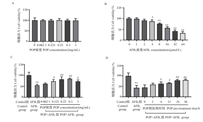
图1 POP和AFB1对IPEC-J2细胞活力的影响POP:平菇多糖;AFB1:黄曲霉毒素B1。*和**分别表示与0 μmol/L AFB1(图B)或Control组(图C和D)相比有显著(P<0.05)或极显著(P<0.01)差异;#和##分别表示与AFB1组(图C和D)相比有显著(P<0.05)或极显著(P<0.01)差异。下图同。 Fig.1 Effects of POP and AFB1 on viability of IPEC-J2 cells POP: Pleurotus ostreatus polysaccharides; AFB1: aflatoxin B1. * and ** indicate significant (P<0.05) or extremely significant (P<0.01) differences compared with the 0 μmol/L AFB1(figure B) or the Control group (figures C and D), respectively; # and ## indicate significant (P<0.05) or extremely significant (P<0.01) differences compared with the AFB1 group (figures C and D), respectively. The same applies to the figures below. |
2.2 POP和AFB1对IPEC-J2细胞内凋亡相关蛋白表达的影响
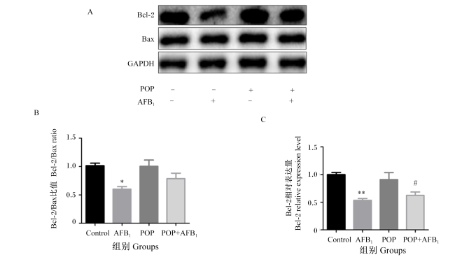
2.3 POP和AFB1对IPEC-J2细胞内ROS含量、ATP相对水平和线粒体膜电位的影响
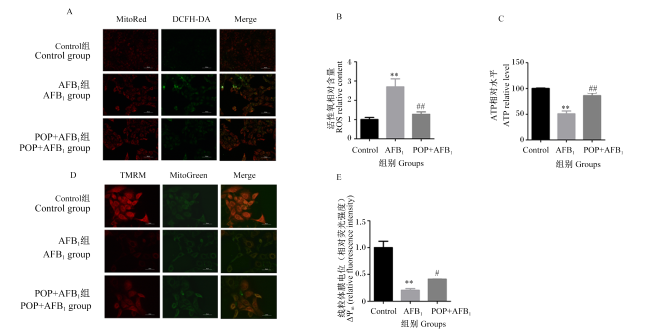
图3 POP和AFB1对IPEC-J2细胞内ROS相对含量、ATP相对水平和线粒体膜电位的影响MitoRed:线粒体红色荧光探针 mito-tracker red;TMRM:四甲基罗丹明甲酯 tetramethylrhodamine methyl ester;MitoGreen:线粒体绿色荧光探针 mito-tracker green;DCFH-DA:2',7'-二氯荧光素二乙酸酯 2',7'-dichlorodihydrofluorescein diacetate;Merge:合并。 Fig.3 Effects of POP and AFB1 on ROS relative content, ATP relative level and ΔΨm in IPEC-J2 cells |
2.4 POP和AFB1对IPEC-J2细胞内抗氧化酶活性的影响
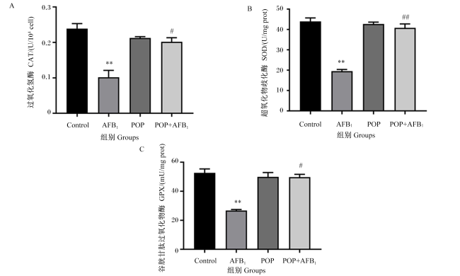
2.5 POP和AFB1诱导的IPEC-J2细胞内线粒体动力学相关蛋白表达的影响
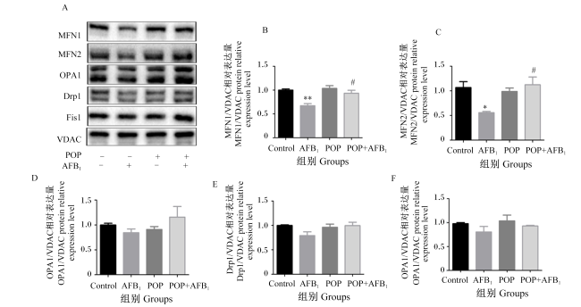


图5 POP和AFB1对IPEC-J2细胞内线粒体动力学相关蛋白表达的影响VDAC:电压依赖性阴离子通道 voltage-dependent anion channel;MFN1:线粒体融合蛋白1 mitofusin 1;MFN2:线粒体融合蛋白2 mitofusin 2;Fis1:线粒体分裂蛋白1 mitochondrial fission 1 protein;OPA1:视神经萎缩蛋白1 optic atrophy 1;Drp1:动力蛋白相关蛋白1 dynamin-related protein 1。 Fig.5 POP and AFB1 on expression of mitochondrial dynamics-related proteins in IPEC-J2 cells |