RESEARCH PAPER

Arginine Stimulating Proliferation and Milk Fat Fatty Acid Synthesis of Bovine Mammary Epithelial Cells through GPRC6A/Akt-mTOR Signaling Pathway

  • LANG Jiaojiao , 1 ,
  • WU Qi 2 ,
  • XUE Changjian 1 ,
  • ZHANG Zhenyu 1 ,
  • YIN Wenya 1 ,
  • XIA Chengqiang 1 ,
  • ZHANG Jing , 1, * ,
  • LIU Qiang , 1, *
Expand
  • 1 College of Animal Science, Shanxi Agricultural University, Taigu 030801, China
  • 2 Wenshui County Animal Husbandry and Veterinary Service Center, Wenshui 032100, China
*ZHANG Jing, associate professor, E-mail: ;
LIU Qiang, professor, E-mail:

Received date: 2025-07-25

  Online published: 2026-03-16

Abstract

This study aimed to investigate the effects of arginine (Arg) on proliferation and milk fat fatty acid synthesis of bovine mammary epithelial cells (BMECs) through G protein-coupled receptor family C, group 6 and subtype A (GPRC6A)/protein kinase B (Akt)-mammalian target of rapamycin (mTOR) signaling pathway, and systematically evaluate its regulatory effects and molecular mechanisms on the proliferation activity, apoptosis level and milk fat synthesis ability of BMECs. Using BMECs as a model, the cell proliferation was assessed by CCK-8, and the expression of proteins related to proliferation, apoptosis and fatty acid synthesis was detected by Western blot. The cells were treated with the Akt inhibitor (AKT-IN-1), mTOR inhibitor (rapamycin), AMP-activated protein kinase (AMPK) activator (EX229) and GPRC6A mall interfering RNA (siRNA), to verify the signaling pathway dependency of Arg’s effects. The results showed that 1.00 μmol/L Arg significantly promoted BMECs proliferation (P<0.05), significant or extremely significant up-regulated the protein expression levels of proliferation marker proteins such as proliferating cell nuclear antigen (PCNA), cyclin D1 (Cyclin D1) and B-cell lymphoma 2 (BCL2) (P<0.05 or P<0.01), and significant or extremely significant down-regulated the protein expression levels of pro-apoptotic proteins such as BCL2 associated X protein (BAX), cysteine aspartic acid specific protease-3 (Caspase-3) and cysteine aspartic acid specific protease-9 (Caspase-9) (P<0.05 or P<0.01). Arg activated GPRC6A and its downstream Akt-mTOR signaling pathway, and the proliferative and anti-apoptotic effects were reversed by GPRC6A siRNA or Akt/mTOR inhibitors. In addition, 1.00 μmol/L Arg significant or extremely significant up-regulated the protein expression levels of fatty acid synthesis-related proteins such as peroxisome proliferator-activated receptor gamma (PPARγ), sterol regulatory element-binding protein 1 (SREBP1), fatty acid synthase (FASN) and stearoyl-CoA desaturase 1 (SCD1) (P<0.05 or P<0.01), and the AMPK activation or GPRC6A siRNA could blocked the fatty acid synthesis-promoting effect of Arg. Collectively, the Arg promotes BMECs proliferation and inhibits apoptosis via the GPRC6A receptor mediated activation of the Akt-mTOR signaling pathway, and enhances the milk fat fatty acid synthesis through the GPRC6A/AMPK axis, providing a theoretical basis for targeted regulation of milk component synthesis.

Cite this article

LANG Jiaojiao , WU Qi , XUE Changjian , ZHANG Zhenyu , YIN Wenya , XIA Chengqiang , ZHANG Jing , LIU Qiang . Arginine Stimulating Proliferation and Milk Fat Fatty Acid Synthesis of Bovine Mammary Epithelial Cells through GPRC6A/Akt-mTOR Signaling Pathway[J]. Chinese Journal of Animal Nutrition, 2026 , 38(3) : 2173 -2183 . DOI: 10.12418/CJAN2026.174

乳脂和乳蛋白作为牛乳中关键的经济性状指标,其总和占据乳固形物比例超过八成。研究表明,若这2项指标提升0.1个百分点,单头泌乳牛的日收益可增加1.2~1.5元[1]。然而,围产期至泌乳早期奶牛处于能量负平衡、氧化应激和免疫抑制的“三重困境”,乳腺组织虽快速增生,但是乳脂合成效率低[2]。因此,挖掘安全、高效、可产业化的乳成分调控营养素已成为奶业提质增效的关键技术瓶颈。精氨酸(Arg)作为一种条件性必需氨基酸,除了参与蛋白质合成外,还是多胺、一氧化氮(NO)、谷氨酸、肌酸等生物活性物质的前体[3]。Arg通过调控血管舒张、血流量、激素分泌、哺乳动物雷帕霉素靶蛋白(mTOR)及单磷酸腺苷活化蛋白激酶(AMPK)信号通路,直接调控乳腺上皮细胞的增殖、凋亡及脂质代谢[4]。因此,阐明Arg调控乳成分合成的分子机制,对制定奶牛精准营养策略、提升乳品质和产量具有重要现实意义。
已有奶牛试验指出,通过静脉或瘤胃后途径补充1.0~1.5 mmol/kg BW的Arg,可使乳脂率提升0.12%~0.28%,乳蛋白率增加0.08%~0.15%,日产奶量提高1.5~3.2 kg/d[5]。这是Arg调控乳腺发育和能量代谢所致[6-7]。Arg与N-氨甲酰谷氨酸协同可降低血浆非酯化脂肪酸(NEFA)和β-羟基丁酸(BHBA)含量,缓解能量负平衡[8]。细胞试验结果显示,在小鼠乳腺上皮细胞中,0.8 mmol/L Arg通过G蛋白偶联受体家族C第6组A亚型(GPRC6A)/磷脂酰肌醇3-激酶(PI3K)/蛋白激酶B(Akt)-mTOR信号通路促进细胞增殖[9];在奶牛乳腺上皮细胞(BEMCs)中,Arg上调甾醇调节元件结合蛋白1(SREBP1)、脂肪酸合酶(FASN)表达,但其上游受体及能量感受机制尚不清楚[10]。另有研究表明,GPRC6A是感应碱性氨基酸的G蛋白偶联受体,可激活PI3K-Akt-雷帕霉素靶蛋白复合物1(mTORC1)信号通路,促进蛋白质翻译[11]。AMPK是细胞能量感受器,AMPKα-苏氨酸(Thr)172磷酸化抑制乙酰辅酶A羧化酶α(ACACA)-丝氨酸(Ser)79磷酸化,从而抑制脂质合成[12]。尽管已有体内试验证明Arg可提高乳脂率,但相关作用机制仍存在未明确之处:1)Arg是否通过GPRC6A介导的信号通路发挥作用;2)该信号通路是否同时调控细胞增殖与脂质合成;3)AMPK在Arg促脂合成中的确切作用。因此,本研究拟确定Arg促进BMECs增殖与抑制凋亡的最适剂量与受体依赖机制,解析GPRC6A/Akt-mTOR信号通路在细胞增殖中的作用顺序,阐明GPRC6A/AMPK信号通路在乳脂脂肪酸合成中的调控作用,从而为奶牛饲粮中Arg的精准补充提供细胞水平的潜在分子机制和靶点。

1 材料与方法

1.1 试验材料

1.1.1 细胞与培养

所有动物试验均已获得山西农业大学实验动物伦理委员会批准(批准文号:SXAU-EAW-2022C.FU.00301402)。本研究所用BMECs系依据Zhao等[13]报道的分离方法,从3头处于泌乳中期的中国荷斯坦奶牛[平均体重(646.2±12.3) kg、(39.1±3.4)月龄、泌乳天数(29.3±1.6) d]乳腺组织中分离获得,经角蛋白-18免疫荧光鉴定,该细胞纯度>95%。细胞培养液为杜尔贝科改良伊格尔培养基(DMEM)/F12+10%胎牛血清(FBS)+1%青霉素-链霉素,37 ℃、5%二氧化碳(CO2)培养。按1.5×10⁶个在25 cm2培养瓶进行接种后,在37 ℃、5% CO2条件下的培养箱中进行培养传代,本次试验所用细胞为第4代细胞。

1.1.2 主要试剂

L-Arg(纯度≥99%,Sigma-Aldrich Corporation,美国);细胞计数试剂盒-8(CCK-8)(Dojindo Laboratories Co., Ltd.,日本);Akt抑制剂-1(AKT-IN-1)、mTOR抑制剂雷帕霉素(Rap)、AMPK激活剂EX229(MedChemExpress LLC,美国);GPRC6A小干扰RNA(siRNA)及阴性对照siRNA(苏州吉玛基因股份有限公司);Lipofectamine 3000转染试剂(Invitrogen Corporation,美国);二喹啉甲酸(BCA)蛋白定量试剂盒、放射免疫沉淀分析缓冲液(RIPA)裂解液、苯甲基磺酰氟(PMSF)及蛋白酶/磷酸酶抑制剂(上海碧云天生物技术股份有限公司);兔抗增殖细胞核抗原(PCNA)、细胞周期蛋白D1(cyclin D1)、B细胞淋巴瘤2(BCL2)、B细胞淋巴瘤2相关X蛋白(BAX)、半胱氨酸天冬氨酸蛋白酶-3(Caspase-3)、半胱氨酸天冬氨酸蛋白酶-9(Caspase-9)、GPRC6A、Akt、磷酸化(p)-Akt(Ser473)、mTOR、p-mTOR(Ser2448)、过氧化物酶体增殖物激活受体γ(PPARγ)、SREBP1、ACACA、p-ACACA(Ser79)、FASN、硬脂酰辅酶A去饱和酶1(SCD1)、AMPK、p-AMPK(Thr172)及β-肌动蛋白(β-actin)抗体(Cell Signaling Technology, Inc.,美国);辣根过氧化物酶(HRP)标记山羊抗兔免疫球蛋白G(IgG)(上海碧云天生物技术股份有限公司)。

1.2 试验设计

1.2.1 剂量-效应试验

参照Zhang等[14]报道的方法,按1×10³个/孔将BMECs接种于96孔板,待细胞贴壁以后分别用含0、0.50、0.75、1.00、1.50、3.00 μmol/L Arg的无血清DMEM/F12处理,在37 ℃、5% CO2条件下培养72 h,每组设8个复孔,重复3次。然后,每孔加入10 μL不含FBS的DMEM-F12和CCK-8检测液,继续孵育1 h。最后,用酶标仪(Infinite® 200 PRO,Tecan Group Ltd.,瑞士)测定450 nm处吸光度值(OD450 nm)[15],筛选最适Arg浓度。

1.2.2 信号通路阻断与受体沉默试验

以最适浓度1.00 μmol/L Arg为基础,分为4组:对照组(0 μmol/L Arg)、Arg组(1.00 μmol/L Arg)、AKT-IN-1组(30 nmol/L AKT-IN-1)或Rap组(50 pmol/LRap)或EX229组(150 μmol/L EX229)、Arg+AKT-IN-1组(1.00 μmol/L Arg+30 nmol/L AKT-IN-1)或Arg+Rap组(1.00 μmol/L Arg+50 pmol/LRap)或Arg+EX229组(1.00 μmol/L Arg+150 μmol/L EX229)。参照Zhang等[14]的方法进行细胞培养,抑制剂/激活剂于Arg加入前2 h预孵育。AKT-IN-1、Rap和EX229的用量是基于其有效浓度范围、预试验结果、细胞模型的特异性以及试验设计的合理性综合考虑确定的。
GPRC6A沉默试验设4组:对照组(0 μmol/L Arg)、Arg组(1.00 μmol/L Arg)、Arg+NC-siRNA组(阴性对照siRNA)、Arg+GPRC6A-siRNA组。siRNA转染按Lipofectamine 3000说明书进行,转染后24 h换液,再加入Arg处理72 h。

1.3 检测指标与方法

1.3.1 细胞增殖

采用CCK-8法,每孔加入10 μL CCK-8溶液,37 ℃孵育2 h,酶标仪(Infinite® 200 PRO,Tecan Group Ltd.,瑞士)测定OD450 nm,计算细胞增殖率。

1.3.2 蛋白表达

采用Western Blot对目标蛋白进行定量分析。细胞样本经RIPA裂解液充分裂解后提取总蛋白,使用BCA试剂盒测定浓度并统一上样量为20 μg。蛋白经10%或12%十二烷基硫酸钠-聚丙烯酰胺凝胶电泳(SDS-PAGE)分离后,湿转至聚偏二氟乙烯(PVDF)膜,随后以5%脱脂奶粉室温封闭2 h。封闭结束后,加入稀释比例为1∶2 000的一抗,置于4 ℃条件下过夜孵育。次日,用Tris-缓冲盐溶液-吐温20(TBST)缓冲液充分洗涤后,加入1∶5 000稀释的HRP标记二抗,于室温反应2 h。再次用TBST清洗,最终使用ChampChemi成像系统(ChampChemi 910 Plus,北京森西赛智科技有限公司)采集图像,并以β-actin为内参,通过ImageJ软件(National Institutes of Health,美国)对各蛋白条带灰度值进行定量分析[16]

1.4 数据处理

所有试验独立重复3次,数据以“平均值±标准误”表示。采用SigmaPlot 12.5软件进行统计分析,对不同水平Arg的处理效应进行线性和二次曲线分析,对其他数据以t检验进行生物统计。当P<0.01时表示差异极显著,当P<0.05时表示差异显著。文中所有数据制图均采用SigmaPlot 12.5统计软件完成。

2 结果与分析

2.1 Arg对BMECs增殖和凋亡的影响

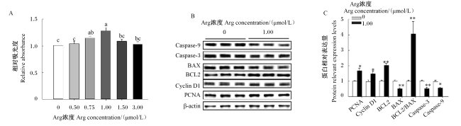
图1可见,CCK-8试验结果表明,Arg对BMECs的促增殖作用呈浓度依赖性,其中1.00 μmol/L Arg组的细胞增殖率显著高于对照组(P<0.05)。Western blot结果表明,1.00 μmol/L Arg显著或极显著上调增殖标志蛋白PCNA、Cyclin D1和BCL2的蛋白表达水平(P<0.05或P<0.01),显著或极显著下调促凋亡蛋白BAX、Caspase-3和Caspase-9的蛋白表达水平(P<0.05或P<0.01),导致BCL2/BAX比值极显著升高(P<0.01)。
图1 Arg对BMECs增殖和凋亡的影响

PCNA:增殖细胞核抗原 proliferating cell nuclear antigen;Cyclin D1:细胞周期蛋白D1 cyclin D1;BCL2:B淋巴细胞瘤2 B-cell lymphoma 2;BAX:B淋巴细胞瘤2相关X蛋白 B-cell lymphoma 2 associated X protein;Caspase-3:半胱氨酸天冬氨酸蛋白酶-3 cysteine aspartic acid specific protease-3;Caspase-9:半胱氨酸天冬氨酸蛋白酶-9 cysteine aspartic acid specific protease-9;Arg:精氨酸 arginine;β-actin:β-肌动蛋白。

数据柱标相同小写字母表示差异不显著(P>0.05),不同小写字母表示差异显著(P<0.05)。Value columns with the same small letter mean no significant difference (P>0.05), while with different small letters mean significant difference (P<0.05).

*表示差异显著(P<0.05),**表示差异极显著(P<0.01)。图2图6同。* indicated significant difference (P<0.05), and ** indicated extremely significant difference (P<0.01). The same as Fig.2 and Fig.6.

Fig.1 Effects of Arg on proliferation and apoptosis of BMECs

2.2 Arg激活GPRC6A/Akt-mTOR信号通路

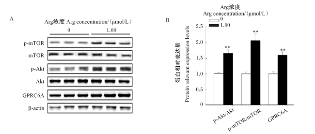
图2可见,1.00 μmol/L Arg极显著上调GPRC6A的蛋白表达水平(P<0.01),并极显著提高p-Akt/Akt和p-mTOR/mTOR比值(P<0.01),提示Arg通过GPRC6A受体激活下游Akt-mTOR信号通路。
图2 Arg对BMECs中Akt-mTOR信号通路的影响

GPRC6A:G蛋白偶联受体家族C第6组A亚型 G protein-coupled receptor family C, group 6 and subtype A;Akt:蛋白激酶B protein kinase B;p-Akt:磷酸化蛋白激酶B phosphorylated protein kinase B;mTOR:哺乳动物雷帕霉素靶蛋白 mammalian target of rapamycin;p-mTOR:磷酸化哺乳动物雷帕霉素靶蛋白 phosphorylated mammalian target of rapamycin;Arg:精氨酸 arginine;β-actin:β-肌动蛋白。

Fig.2 Effects of Arg on Akt-mTOR signal pathway in BMECs

2.3 阻断Akt信号通路逆转Arg的促增殖效应

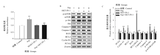
图3可见,与Arg组相比,Arg+AKT-IN-1组的细胞增殖率极显著降低(P<0.01),PCNA、Cyclin D1和BCL2的蛋白表达水平显著下调(P<0.05),BAX、Caspase-3、Caspase-9的蛋白表达水平显著上调(P<0.05);此外,Arg+AKT-IN-1组的p-mTOR/mTOR比值显著下调(P<0.05),初步表明Akt是mTOR的上游调控节点。
图3 阻断Akt信号通路逆转了Arg对BMECs中增殖相关蛋白表达的影响

Arg:精氨酸 arginine;AKT-IN-1:AKT抑制剂;Cyclin D1:细胞周期蛋白D1;PCNA:增殖细胞核抗原 proliferating cell nuclear antigen;BAX:B淋巴细胞瘤2相关X蛋白 BCL2 associated X protein;BCL2:B淋巴细胞瘤2 B-cell lymphoma 2;Caspase-3:半胱氨酸天冬氨酸蛋白酶-3 cysteine aspartic acid specific protease-3;Caspase-9:半胱氨酸天冬氨酸蛋白酶-9 cysteine aspartic acid specific protease-9;Akt:蛋白激酶B protein kinase B;p-Akt:磷酸化蛋白激酶B phosphorylated protein kinase B;mTOR:哺乳动物雷帕霉素靶蛋白 mammalian target of rapamycin;p-mTOR:磷酸化哺乳动物雷帕霉素靶蛋白 phosphorylated mammalian target of rapamycin;β-actin:β-肌动蛋白。图4图5同 the same as Fig.4 and Fig.5

*和**分别表示与对照组相比差异显著(P<0.05)和极显著(P<0.01),#和##分别表示与Arg组相比差异显著(P<0.05)和极显著(P<0.01)。图4图5图7图8同。* and ** indicated significant difference (P<0.05) and extremely significant difference (P<0.01) compared with the control group, and # and ## indicated significant difference (P<0.05) and extremely significant difference (P<0.01) compared with the Arg group. The same as Fig.4, Fig.5, Fig.7 and Fig.8.

Fig.3 Suppression of Akt signal pathway reversed effects of Arg on expression of proliferation-related proteins in BMECs

2.4 阻断mTOR信号通路抑制Arg的抗凋亡效应

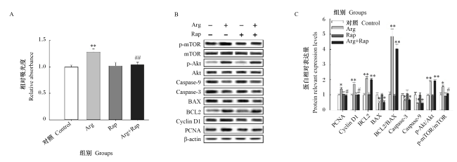
图4可见,与Arg组相比,Rap组的细胞增殖率极显著降低(P<0.01),PCNA、Cyclin D1和BCL2的蛋白表达水平显著或极显著下调(P<0.05或P<0.01),BAX、Caspase-3、Caspase-9的蛋白表达水平显著上调(P<0.05);此外,p-mTOR/mTOR比值显著下调(P<0.05),表明mTOR是Arg调控细胞增殖与凋亡的关键下游靶点。
图4 阻断mTOR信号通路逆转了Arg对BMECs中增殖相关蛋白表达的影响

Rap:雷帕霉素 rapamycin。

Fig.4 Suppression of mTOR signal pathway reversed effects of Arg on expression of proliferation-related proteins in BMECs

2.5 GPRC6A介导Arg的生物学效应

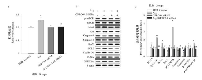
图5可见,与Arg组相比,Arg+GPRC6A-siRNA组的细胞增殖率显著降低(P<0.05),PCNA、Cyclin D1、BCL2的蛋白表达水平显著或极显著下调(P<0.05或P<0.01),BAX、Caspase-3、Caspase-9的蛋白表达水平显著上调(P<0.05);此外,p-Akt/Akt和p-mTOR/mTOR比值显著下调(P<0.05),表明GPRC6A是Arg激活Akt-mTOR信号通路的必需受体。
图5 GPRC6A siRNA逆转了Arg对BMECs中增殖相关蛋白表达的影响

GPRC6A:G蛋白偶联受体家族C第6组A亚型 G protein-coupled receptor family C, group 6, and subtype A;siRNA:小干扰RNA small interfering RNA。

Fig.5 GPRC6A siRNA reversed effects of Arg on expression of proliferation-related proteins in BMECs

2.6 Arg促进BMECs中脂肪酸合成相关蛋白表达

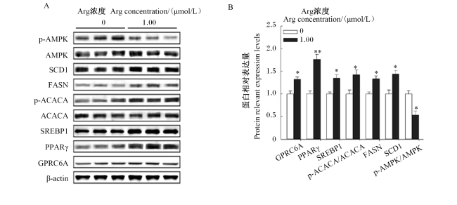
图6可见,1.00 μmol/L Arg显著或极显著上调PPARγ、SREBP1、FASN和SCD1的蛋白表达水平(P<0.05或P<0.01),显著提高p-ACACA/ACACA比值(P<0.05),显著降低p-AMPK/AMPK比值(P<0.05),提示其通过抑制AMPK活性促进脂质合成。
图6 Arg对BMECs中脂肪酸合成相关蛋白表达的影响

p-AMPK:磷酸化单磷酸腺苷活化的蛋白激酶 phosphorylated AMP-activated protein kinase;AMPK:单磷酸腺苷活化的蛋白激酶 AMP-activated protein kinase;SCD1:硬脂酰辅酶A去饱和酶1 stearoyl-CoA desaturase 1;FASN:脂肪酸合酶 fatty acid synthase;p-ACACA:磷酸化乙酰辅酶A羧化酶α phosphorylated acetyl- coenzyme A carboxylase α;ACACA:乙酰辅酶A羧化酶α acetyl-coenzyme A carboxylase α;SREBP1:甾醇调节元件结合蛋白1 sterol regulatory element-binding protein 1;PPARγ:过氧化酶增殖因子活化受体γ peroxisome proliferator-activated receptor gamma;GPRC6A:G蛋白偶联受体家族C第6组A亚型 G protein-coupled receptor family C, group 6, and subtype A;β-actin:β-肌动蛋白;Arg:精氨酸 arginine。下图同 the same as below。

Fig.6 Effects of Arg on expression of proteins related to fatty acid synthesis in BMECs

2.7 AMPK激活逆转Arg的促脂肪酸合成效应

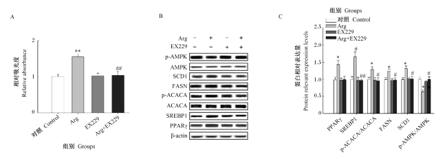
图7可见,与Arg组相比,EX229组的细胞增殖率极显著降低(P<0.01),SREBP1、FASN和SCD1的蛋白表达水平显著或极显著下调(P<0.05或P<0.01),p-AMPK/AMPK比值显著上调(P<0.05)。
图7 EX229逆转了Arg对BMECs中脂肪酸合成相关蛋白表达的影响

Fig.7 EX229 reversed effects of Arg on expression of proteins related to fatty acid synthesis in BMECs

2.8 GPRC6A调控Arg诱导的脂肪酸合成

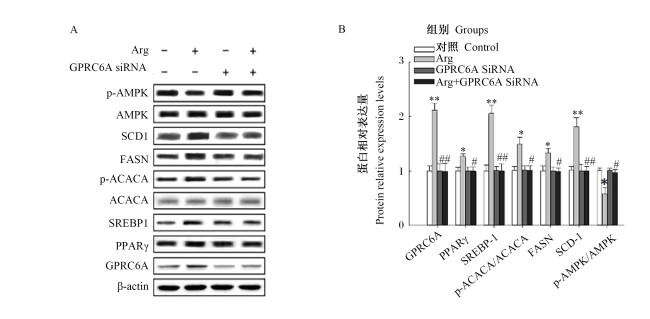
图8可见,与Arg组相比,Arg+GPRC6A-siRNA组的PPARγ、SREBP1、FASN和SCD1的蛋白表达水平显著或极显著下调(P<0.05或P<0.01),p-AMPK/AMPK比值显著上调(P<0.05),表明GPRC6A通过AMPK依赖性途径介导Arg对脂肪酸合成的调控。
图8 GPRC6A siRNA逆转了Arg对BMECs中脂肪酸合成相关蛋白表达的影响

Fig.8 GPRC6A siRNA reversed effects of Arg on expression of proteins related to fatty acid synthesis in BMECs

3 讨论

3.1 Arg调控BMECs增殖的分子机制

BMECs的持续增殖与适度凋亡是维持奶牛高产稳产的生物学基础。研究证实,Arg经过GPRC6A-Akt-mTOR信号通路,促进细胞增殖,有效抑制线粒体介导的凋亡,形成一个高度协同的增殖-抗凋亡网络,从而显著促进乳腺发育与乳成分合成[17]
研究发现,Arg可优先与GPRC6A受体结合,从而迅速启动Akt-mTOR级联磷酸化反应。本研究观察到1.0 μmol/L Arg使p-Akt/Akt和p-mTOR/mTOR比值分别提高1.8倍和2.1倍,与Ge等[6]在小鼠上的研究结果一致。单细胞测序证实,GPRC6A在小鼠乳腺基底细胞高表达,经0.8 mmol/L Arg处理后,处于S期的细胞占比由8.7%显著增至15.4%,并伴随p-Akt/Akt和p-mTOR/mTOR比值的同步上调[6],提示GPRC6A-Akt-mTOR轴在细胞周期启动中发挥关键调控作用。从分子机制看,激活后的mTORC1可催化真核翻译起始因子4E结合蛋白(4EBP1)和核糖体蛋白S6激酶1(S6K1)发生磷酸化修饰[18],显著提高Cyclin D1、和PCNA等的翻译效率[19-20]。Cyclin D1主要介导G1/S期转换,而PCNA作为DNA滑动夹蛋白,直接参与复制叉的延伸与修复,三者共同构成BMECs增殖的分子基础[21]。尽管0.75 μmol/L和1.00 μmol/L组的细胞增殖率没有显著差异,但1.00 μmol/L组的细胞增殖率在所有测试浓度中是最高的。这表明1.00 μmol/L可能更接近最佳浓度。另外,有研究发现,1.0~1.5 mmol/kg BW的Arg补充在动物模型中已经显示出显著的乳脂率和乳蛋白率提升效果[5]。因此,1.00 μmol/L的浓度在细胞水平上可能更接近生理相关浓度,能够更好地模拟体内环境。
本研究进一步研究发现,Arg通过同一信号通路对细胞凋亡进行负向调控。BCL2蛋白家族是线粒体凋亡通路的核心开关,其中BCL2具有抗凋亡功能,而BAX则具有促凋亡功能[22]。本研究中,Arg通过提升BCL2蛋白表达水平并抑制BAX蛋白表达水平,提高BCL2/BAX比值,进而减弱线粒体外膜通透性,阻断细胞色素C外逸。与此同时,Arg明显抑制Caspase-9与Caspase-3活性,截断凋亡信号下游的级联放大[23]。值得注意的是,功能验证进一步表明,Akt抑制剂AKT-IN-1可完全逆转Arg的促增殖与抗凋亡效应,而Rap仅阻断mTOR下游,证实Arg-GPRC6A信号流向为GPRC6A→Akt→mTOR。另外,在人前列腺癌细胞中,GPRC6A介导的Akt/mTOR磷酸化在氧化应激条件下仍能维持,提示该信号通路在炎症微环境中具有稳定性[24]。虽然目前尚无奶牛氧化应激模型下的直接证据,但基于BMEC炎症模型及GPRC6A-mTOR信号通路的保守性,可推测在奶牛氧化应激或脂多糖(LPS)低度炎症条件下,该信号通路仍可能维持磷酸化激活,未来可通过奶牛体内或原代BMEC-LPS模型进一步验证。
综上所述,Arg通过GPRC6A-Akt-mTOR信号通路,一方面加速Cyclin D1和PCNA的翻译,推动细胞周期运转;另一方面通过提升BCL2/BAX比值、遏制半胱氨酸天冬氨酸蛋白酶链式激活进程,阻断线粒体凋亡通路。这种增殖与抗凋亡的双重调控,使乳腺组织在能量负平衡与氧化应激环境下仍能维持结构完整与功能高效,为奶牛饲粮中精准补充Arg提供了坚实的细胞与分子依据,也为未来靶向乳腺营养的调控策略奠定了理论基础。

3.2 Arg调控BMECs脂肪酸合成的机制

Arg对BMECs脂质生成的调控并非单一通路,而是GPRC6A-AMPK与PPARγ-SREBP1两大模块协同作用的整体网络。首先,Arg通过GPRC6A介导的AMPK信号通路解除能量闸门。本研究显示,Arg显著降低p-AMPK/AMPK比值,使AMPK对ACACA的抑制解除;ACACA磷酸化水平下降后,丙二酰辅酶A生成增加,该代谢信号迅速触发SREBP1的成熟与核转位,进而激活FASNSCD1和甘油-3-磷酸酰基转移酶(GPAT)等靶基因,促使十六烷酸(C16∶0)经去饱和反应转化为油酸(C18∶1),并加速甘油三酯(TAG)与甘油二酯(DAG)的组装。脂质组学结果证实,Arg处理24 h即可使小鼠乳腺上皮细胞中TAG和DAG含量分别增加2.3倍和1.7倍[6]。当使用AMPK激活剂EX229时,上述脂质合成信号被阻断,同时SREBP1的核转位也被逆转[25],说明AMPK是Arg促脂通路中的“主开关”。
其次,PPARγ与SREBP1作为乳腺脂质生成的核心转录开关[26],可进一步放大Arg的效应。PPARγ与SREBP1不仅上调ACACA和FASN的蛋白表达,从而增强以乙酸和β-羟丁酸为底物的新生脂肪酸(DNFA)合成及其分泌[27],还通过SCD催化饱和脂肪酸(SFA)向单不饱和脂肪酸的转化[28]。在本研究中,Arg显著上调PPARγ、SREBP1、FASN和SCD1的蛋白表达水平及p-ACACA/ACACA比值。Ding等[5]进一步通过颈静脉灌注37.6 g/d Arg,发现奶牛乳腺组织中PPARγACACASCD基因表达显著升高,DNFA产量随之增加。虽然本研究没有直接测量脂滴合成量,但通过检测脂肪酸合成的关键调控因子(如PPARγ、SREBP1、FASN、SCD1等)的蛋白表达,已经提供了脂肪合成活性增加的有力间接证据。未来的研究将补充直接测量脂肪合成量的试验,以进一步完善研究结果。
值得注意的是,AMPK作为能量感应器,在细胞处于高葡萄糖及高Arg浓度的正向能量平衡时被抑制[5,29]。本研究观察到Arg显著抑制AMPK磷酸化,与Ding等[5]报道的Arg灌注下调AMP-活化蛋白激酶β1基因表达及Zhang等[17]报道的过瘤胃Arg显著抑制AMPK磷酸化相符。此外,Arg代谢产生的多胺(腐胺、精胺)可通过鸟氨酸脱羧酶(ODC)途径进一步上调SREBP1表达,形成“Arg-多胺-脂质”次级调控环[30]。综上所述,Arg通过GPRC6A-AMPK轴解除ACACA抑制,激活SREBP1/PPARγ主开关,协同提升DNFA合成与SFA去饱和,从而提高脂质生成效率。
Arg、谷氨酰胺与脯氨酸构成“谷氨酰胺-Arg-脯氨酸代谢轴”,吡咯啉-5-羧酸(P5C)作为核心中间体将三者动态耦合[31]。该轴不仅决定细胞内Arg供给,还通过还原型烟酰胺腺嘌呤二核苷酸磷酸(NADPH)再生维持脂质合成还原力[31]。Arg补充可在脂肪组织中同时触发抗氧化与促氧化效应:一方面通过一氧化氮(NO)抑制活性氧(ROS)产生,减少丙二醛(MDA)含量;另一方面通过增强线粒体呼吸链活性产生超氧阴离子[32]。然而,在泌乳奶牛乳腺中,由于高血流量和丰富抗氧化酶体系,Arg主要表现为抗氧化,从而保护脂质合成酶活性[1,5]。今后可结合GPRC6A基因多态性筛选高响应群体,并开发包被Arg或GPRC6A激活剂,实现乳腺靶向营养。

4 结论

1.00 μmol/L Arg可增强BMECs的增殖能力并抑制其凋亡过程,其作用机制涉及GPRC6A介导的Akt-mTOR信号通路调控细胞周期,并通过GPRC6A/AMPK轴促进乳脂脂肪酸的合成。该结果为奶牛精准饲养提供了新的分子靶点和理论依据。
[1]
ZHAO F F, WU T Y, WANG H R, et al. Jugular arginine infusion relieves lipopolysaccharide-triggered inflammatory stress and improves immunity status of lactating dairy cows[J]. Journal of Dairy Science, 2018, 101(7):5961-5970.

DOI PMID

[2]
LAPIERRE H, PACHECO D, BERTHIAUME R, et al. What is the true supply of amino acids for a dairy cow?[J]. Journal of Dairy Science, 2006, 89(S1):E1- E14.

[3]
WU G Y. Amino acids:metabolism,functions,and nutrition[J]. Amino Acids, 2009, 37(1):1-17.

[4]
ZHANG J L, WANG S J, GUO X M, et al. Arginine supplementation targeting tumor-killing immune cells reconstructs the tumor microenvironment and enhances the antitumor immune response[J]. ACS Nano, 2022, 16(8):12964-12978.

DOI PMID

[5]
DING L, SHEN Y, JAWAD M, et al. Effect of arginine supplementation on the production of milk fat in dairy cows[J]. Journal of Dairy Science, 2022, 105(10):8115-8129.

DOI PMID

[6]
GE Y S, LI F, HE Y, et al. L-arginine stimulates the proliferation of mouse mammary epithelial cells and the development of mammary gland in pubertal mice by activating the GPRC6A/PI3K/AKT/mTOR signalling pathway[J]. Journal of Animal Physiology and Animal Nutrition, 2022, 106(6):1383-1395.

DOI

[7]
HARDIE D G. AMPK:positive and negative regulation,and its role in whole-body energy homeostasis[J]. Current Opinion in Cell Biology, 2015, 33:1-7.

DOI

[8]
WANG H R, ZHU W J, WANG X Y. Mechanism of inhibition of arginine kinase by flavonoids consistent with thermodynamics of docking simulation[J]. International Journal of Biological Macromolecules, 2011, 49(5):985-991.

DOI

[9]
BURLINA A, ARDISSONE A, BATTINI R, et al. Arginase 1 deficiency:a treatable form of spastic paraplegia[J]. Neurological Sciences, 2025, 46(9):4219-4228.

DOI

[10]
MAGNUSON B, EKIM B, FINGAR D C. Regulation and function of ribosomal protein S 6 kinase (S6K) within mTOR signalling networks[J]. Biochemical Journal, 2012, 441(1):1-21.

DOI

[11]
SAXTON R A, SABATINI D M. mTOR signaling in growth,metabolism,and disease[J]. Cell, 2017, 168(6):960-976.

DOI

[12]
ZHOU G, SOMASUNDARAM T, BLANC E, et al. Transition state structure of arginine kinase:implications for catalysis of bimolecular reactions[J]. Proceedings of the National Academy of Sciences of the United States of America, 1998, 95(15):8449-8454.

[13]
ZHAO F F, HOU X P, ZHU Y H, et al. Establishment and characterization of bovine mammary epithelial cells[J]. In Vitro Cellular & Developmental Biology-Animal, 2016, 52(5):548-556.

[14]
张静, 卜丽君, 郎姣姣, 等. 丁酸钠通过G蛋白偶联受体41介导蛋白激酶B/哺乳动物雷帕霉素靶蛋白信号通路刺激牛乳腺上皮细胞增殖[J]. 动物营养学报, 2024, 36(3):1878-1891.

DOI

ZHANG J, BU L J, LANG J J, et al. Sodium butyrate through G protein-coupled receptor 41 mediated protein kinase B/mammalian target of rapamycin signaling pathway to promote proliferation of bovine mammary epithelial cells[J]. Chinese Journal of Animal Nutrition, 2024, 36(3):1878-1891. (in Chinese)

DOI

[15]
ZHANG J, LIU Y P, BU L J, et al. Effects of dietary folic acid supplementation on lactation performance and mammary epithelial cell development of dairy cows and its regulatory mechanism[J]. Animal Biotechnology, 2023, 34(8):3796-3807.

[16]
张静. 外源性硫化氢对哺乳动物初情期乳腺发育的影响及机制[D].博士学位论文. 广州: 华南农业大学, 2018.

ZHANG J. Effects of exogenous H2S on puberty mammary gland development of mammal and its mechanisms[D].Ph.D.Thesis. Guangzhou: South China Agricultural University, 2018. (in Chinese)

[17]
ZHANG J, LANG J J, BU L J, et al. Impacts of dietary arginine supplementation on performance,nutrient digestion and expression of proteins related to milk fatty acid and casein synthesis in early lactating dairy cows[J]. Animal Nutrition, 2025, 21:267-278.

DOI

[18]
VIDAL C M, OUYANG C, QI Y, et al. Arginine regulates HSPA5/BiP translation through ribosome pausing in triple-negative breast cancer cells[J]. British Journal of Cancer, 2023, 129(3):444-454.

DOI

[19]
LENTS N H, PISZCZATOWSKI R T. Cyclins,cyclin-dependent kinases,and cyclin-dependent kinase inhibitors[J]. Encyclopedia of Cell Biology, 2023, 5:224-234.

[20]
WANG Y C, KELSO A A, KARAMAFROOZ A, et al. Arginine shortage induces replication stress and confers genotoxic resistance by inhibiting histone H4 translation and promoting PCNA ubiquitination[J]. Cell Reports, 2023, 42(4):112296.

DOI

[21]
ZHANG J, BU L J, LIU Y P, et al. Dietary supplementation of sodium butyrate enhances lactation performance by promoting nutrient digestion and mammary gland development in dairy cows[J]. Animal Nutrition, 2023, 15:137-148.

DOI PMID

[22]
VAN DELFT M F, DEWSON G. The BCL-2 family proteins:insights into their mechanism of action and therapeutic potential[J]. Encyclopedia of Cell Biology, 2023, 5:184-198.

[23]
TAKAC P, KELLO M, PILATOVA M B, et al. New chalcone derivative exhibits antiproliferative potential by inducing G2/M cell cycle arrest,mitochondrial-mediated apoptosis and modulation of MAPK signalling pathway[J]. Chemico-Biological Interactions, 2018, 292:37-49.

DOI

[24]
YE R S, PI M, NOOH M M, et al. Human GPRC6A mediates testosterone-induced mitogen-activated protein kinases and mTORC1 signaling in prostate cancer cells[J]. Molecular Pharmacology, 2019, 95(5):563-572.

DOI PMID

[25]
SU F T, KOEBERLE A. Regulation and targeting of SREBP-1 in hepatocellular carcinoma[J]. Cancer and Metastasis Reviews, 2024, 43(2):673-708.

DOI

[26]
ABDELATTY A M, IWANIUK M E, GARCIA M, et al. Effect of short-term feed restriction on temporal changes in milk components and mammary lipogenic gene expression in mid-lactation Holstein dairy cows[J]. Journal of Dairy Science, 2017, 100(5):4000-4013.

DOI PMID

[27]
TIAN Z C, ZHANG Y Y, ZHANG H M, et al. Transcriptional regulation of milk fat synthesis in dairy cattle[J]. Journal of Functional Foods, 2022, 96:105208.

DOI

[28]
CONTE G, MELE M, CHESSA S, et al. Diacylglycerol acyltransferase 1,stearoyl-CoA desaturase 1,and sterol regulatory element binding protein 1 gene polymorphisms and milk fatty acid composition in Italian brown cattle[J]. Journal of Dairy Science, 2010, 93(2):753-763.

DOI

[29]
HUANG J, GUESTHIER M A, BURGOS S A. AMP-activated protein kinase controls lipid and lactose synthesis in bovine mammary epithelial cells[J]. Journal of Dairy Science, 2020, 103(1):340-351.

DOI PMID

[30]
梁飞腾, 邓琼, 王铸, 等. DDAH1参与调控精氨酸代谢介导肿瘤发展的研究进展[J]. 生命的化学, 2023, 43(5):728-734.

LIANG F T, DENG Q, WANG Z, et al. Progress of DDAH1 involvement in arginine metabolism in tumors[J]. Chemistry of Life, 2023, 43(5):728-734. (in Chinese)

[31]
PHANG J M. Proline metabolism in cell regulation and cancer biology:recent advances and hypotheses[J]. Antioxidants & Redox Signaling, 2019, 30(4):635-649.

[32]
KURHALUK N. The effectiveness of L-arginine in clinical conditions associated with hypoxia[J]. International Journal of Molecular Sciences, 2023, 24(9):8205.

DOI

Outlines

/