RESEARCH PAPER

Effects of High-Sugar Diet and Chronic Hypoxic Stress on Growth Performance, Liver Histomorphology and Antioxidant Capacity of Largemouth Bass (Micropterus salmoides)

  • TANG Zeming , 1, 2 ,
  • DOU Shilong 1, 2 ,
  • LI Hongye 1, 2 ,
  • XU Yihuan 1, 2 ,
  • ZHANG Lihan 1, 2 ,
  • XIA Hui 1, 2 ,
  • GUO Ran , 1, 2, *
Expand
  • 1 Ocean College, Hebei Agricultural University, Qinhuangdao 066000, China
  • 2 Key Laboratory of Nutritional Regulation and Disease Control in Aquaculture of Hebei Province, Qinhuangdao 066000, China
* professor, E-mail:

Received date: 2025-12-03

  Online published: 2026-07-13

Abstract

This experiment aimed to investigate the effects of a high-sugar diet and chronic hypoxic stress on the growth performance, liver histomorphology and antioxidant capacity of largemouth bass (Micropterus salmoides). A 2×2 two-factor randomized crossover experimental design was employed. The two factors were dietary sugar level (normal-sugar diet: digestible sugar level was 10%; high-sugar diet: digestible sugar level was 15%) and water dissolved oxygen (DO) concentration [normoxic water: DO concentration was (7.0±0.5) mg/L; hypoxic water: DO concentration was (3.0±0.5) mg/L]. A total of 240 juvenile largemouth bass with an initial average body weight of (11.50±0.03) g were randomly assigned to four groups. Each group had three replicates (aquariums), with 20 fish per aquarium. The feeding trial lasted for 8 weeks. The results showed as follows: 1) chronic hypoxic stress significantly reduced final body weight, weight gain rate, feed intake and specific growth rate (P<0.05). 2) Dietary sugar level significantly affected whole-body crude protein, ether extract and crude ash contents, as well as the hepatosomatic index and viscerosomatic index (P<0.05). Water DO concentration significantly affected whole-body moisture, crude protein and ether extract contents, as well as the viscerosomatic index (P<0.05). 3) Largemouth bass fed the high-sugar diet exhibited hepatocyte swelling, nuclear displacement, and pronounced vacuolation. 4) The interaction between dietary sugar level and water DO concentration significantly affected liver triglyceride content, as well as serum triglyceride and high-density lipoprotein cholesterol contents (P<0.05). 5) The interaction between dietary sugar level and water DO concentration significantly affected liver total antioxidant capacity, as well as the activities of peroxidase, superoxide dismutase and glutathione peroxidase (P<0.05). Therefore, chronic hypoxic stress reduces the growth performance of largemouth bass, impairs nutrient absorption and deposition, and induces oxidative stress. High-sugar diet leads to hepatic lipid accumulation and oxidative stress in largemouth bass, thereby compromising liver health. Compared with a single stressor, the combination of high-sugar diet and chronic hypoxic stress may exert a synergistic effect, exacerbating the negative impacts on largemouth bass.

Cite this article

TANG Zeming , DOU Shilong , LI Hongye , XU Yihuan , ZHANG Lihan , XIA Hui , GUO Ran . Effects of High-Sugar Diet and Chronic Hypoxic Stress on Growth Performance, Liver Histomorphology and Antioxidant Capacity of Largemouth Bass (Micropterus salmoides)[J]. Chinese Journal of Animal Nutrition, 2026 , 38(7) : 5295 -5305 . DOI: 10.12418/CJAN2026.424

水环境中溶解氧(DO)的浓度会因多种因素的影响在时间和空间上发生变化,对水环境中的不同生物产生不同影响[1]。在水产养殖中,水体溶解氧浓度介于0.28~4.00 mg/L被定义为缺氧[2]。大口黑鲈(Micropterus salmoides)养殖水体含氧量要求高于4.00 mg/L,其最低耐氧量为1.20 mg/L[3-4]。面对缺氧胁迫,鱼类会通过增加呼吸频率和血红蛋白水平来增强氧气摄取能力;同时,它们通过减少摄食量,降低与消化吸收相关的代谢需求,从而减轻对氧气的依赖。研究表明,缺氧可诱导鱼类产生活性氧(ROS)并引发氧化应激;为应对ROS的积累,其抗氧化防御系统通过级联反应发挥关键的调控作用[5]。参与该防御系统的酶主要包括超氧化物歧化酶(SOD)、过氧化氢酶(CAT)和谷胱甘肽过氧化物酶(GSH-Px)等[6]
碳水化合物作为一种价格低廉的能量来源,在饲料加工中常被用作稳定剂和膨胀剂,广泛用于各类鱼饲料中[7-8]。传统的大口黑鲈膨化浮球饲料通常需要不低于20%的饲料糖水平,以获得足够的膨胀和浮力特性[9]。然而,鱼类(尤其是肉食性鱼类)利用复合饲料中碳水化合物的能力较低。过量摄入碳水化合物可引发多种代谢紊乱,包括血糖升高、肝糖原蓄积增加及肝脏脂质沉积加重,进而损害肝脏功能,最终导致生长迟缓和代谢功能障碍[10-11]。先前研究表明,大口黑鲈饲料中适宜的碳水化合物水平约为10%,超过14.4%时则表现出生长抑制和氧化应激[7,12]
大口黑鲈是我国重要的经济肉食性鱼类,其养殖业发展迅速,截至2024年底,全国年总产量已达938 509 t[13]。然而,高密度养殖导致的缺氧问题以及高糖商业饲料的使用,共同制约着大口黑鲈养殖业的健康发展。目前,大多数研究集中于缺氧或高糖等单一因素对大口黑鲈的影响,而关于长期缺氧与高糖共同作用对大口黑鲈综合影响的研究相对较少。因此,本试验选择大口黑鲈作为试验对象,探究饲喂高糖饲喂条件下慢性缺氧胁迫对大口黑鲈生长性能、肝脏组织形态及抗氧化能力的影响,探究高糖与缺氧是否存在协同或拮抗效应,以期为优化大口黑鲈的营养策略与养殖模式提供新思路。

1 材料与方法

1.1 伦理声明

本次试验动物已通过河北农业大学试验动物伦理委员会审核(批准编号:2024100)。

1.2 试验设计

将购自河北省唐山市滦南县赵嘉庄水产养殖有限公司的大口黑鲈鱼苗在淡水中进行驯化,期间投喂商业饲料(营养成分含量:粗蛋白质51%,粗脂肪8%,粗纤维5%,粗灰分13%,水分10%)。驯化2周后,选择240条健康的大口黑鲈幼鱼[初始平均体重(11.50±0.03) g],随机分为4组,每组设3个重复(玻璃缸),每个玻璃缸放养20尾。配制2种糖水平饲料(常糖饲料:可消化糖水平10%;高糖饲料:可消化糖水平15%),并设置2种DO浓度水体[常氧水体:DO浓度为(7.0±0.5) mg/L;缺氧水体:DO浓度为(3.0±0.5) mg/L]。其中,2组试验鱼在常氧条件下分别饲喂常糖饲料和高糖饲料,并分别命名为常氧常糖组(N组)、常氧高糖组(S组);另外2组试验鱼在缺氧条件下分别饲喂常糖饲料和高糖饲料,并分别命名为缺氧常糖组(H组)、缺氧高糖组(HS组)。养殖试验持续8周。
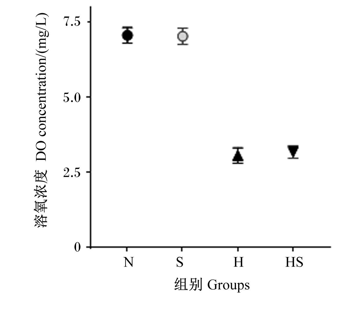
试验饲料组成及营养水平如表1所示。所有饲料原料经粉碎后过80目筛,充分混匀后采用单螺杆膨化机制成颗粒饲料,于-20 ℃保存备用。水体DO浓度参照Sun等[4]的方法设置:在玻璃缸上方放置同尺寸的玻璃罩,充气石从玻璃罩中央的1 cm2小孔通过,通过控制氧气输入量保持水体中DO浓度。在试验第1天,每隔2 h使用便携式氧气计(YSI,ProODO,美国)测量1次DO浓度,连续测6次;此后每周验证1次所设定的DO浓度。1 d内常氧与低氧水体的DO浓度如图1所示。
表1 试验饲料组成及营养水平(干物质基础)

Table 1 Composition and nutrient levels of experimental diets (DM basis) %

项目
Items
常糖饲料
Normal
sugar diet
高糖饲料
High sugar
diet
原料Ingredients
鱼粉Fish meal 40.00 40.00
豆粕Soybean meal 15.00 15.00
血粉Blood meal 7.00 7.00
面粉Wheat flour 14.50 14.50
小麦淀粉Wheat starch 5.40
鱼油Fish oil 3.00 3.00
豆油Soybean oil 5.50 5.50
卵磷脂Lecithin 1.00 1.00
氯化胆碱Choline chloride 1.00 1.00
黏合剂Binder 2.00 2.00
磷酸二氢钙Ca(H2PO4)2 1.00 1.00
预混料Premix1) 2.50 2.50
精氨酸Arg 1.40 1.40
蛋氨酸Met 0.20 0.20
微晶纤维素
Microcrystalline cellulose
5.90 0.50
合计Total 100.00 100.00
营养水平Nutrient levels2)
粗脂肪EE 9.62 9.09
粗蛋白质CP 47.53 47.37
粗灰分Ash 12.00 11.91
粗纤维CF 11.45 6.49
无氮浸出物NFE 10.66 15.38

1)预混料为每千克饲料提供The premix provided the following per kg of diets:VA 10 mg,VB1 6 mg,VB2 5 mg,VB6 7.5 mg,VB12 (1%) 4 mg,烟酰胺 nicotinic acid 50 mg,抗坏血酸 ascorbic acid (35%) 500 mg,泛酸钙 calcium pantothenate 20 mg,生物素 biotin (2%) 2.5 mg,叶酸 folic acid 5 mg,VE (50%) 200 mg,VK3 10 mg,VD3 5 mg,肌醇 inositol 100 mg,玉米麸粉 corn bran powder 75 mg,CuSO4·5H2O 10 mg,MnSO4·H2O 100 mg,FeSO4·7H2O 300 mg,ZnSO4·H2O 200 mg,KIO3 80 mg,Na2SeO3 67 mg,CoCl2·6H2O 5 mg,NaCl 100 mg,沸石粉 zeolite powder 638 mg。

2)无氮浸出物为计算值,其他为实测值。NFE was a calculated value, while the others were measured values.

图1 1 d内常氧与低氧水体的DO浓度

Fig.1 DO concentration of normoxic and hypoxic water within 1 day

1.3 饲养管理

试验鱼每日饱食投喂2次(08:00和17:00),投喂1 h后收集残饵以计算摄食量。对所有玻璃缸中的试验鱼每2周称重1次,并根据更新后的体重及每次散装称重后的固定投喂量调整实际投喂量。试验期间,每日定时排污换水,水体温度维持在(25±0.5) ℃,pH为(7.1±0.4),氨氮浓度低于0.5 mg/L。

1.4 样品采集

养殖期间记录每日摄食量,用于计算饲料系数(FCR)。养殖试验结束后,禁食24 h后使用MS-222(100 mg/L)对试验鱼进行麻醉,对各缸鱼分别计数和称重;从每缸随机取9尾鱼,测量体长和体重,并采用2.5 mL无菌注射器从尾部静脉采血;随后对鱼体进行解剖,剥离并称量内脏团和肝脏。将采集血样转移至1.5 mL离心管中,4 ℃静置12 h后,于4 ℃条件下以51.65×g离心30 min,分离获得血清。取部分肝脏组织,经苏木精-伊红(HE)染色制备组织切片,用于观察肝脏组织形态特征。另取1 g肝脏组织,按1∶9(重量体积比)的比例加入磷酸盐缓冲液,于4 ℃、60 Hz条件下研磨匀浆,经91.80×g离心15 min后取上清液。剩余的肝脏组织经液氮速冻后于-80 ℃保存。血清及组织上清液均于-80 ℃保存,用于后续生化指标检测。

1.5 测定指标及方法

饲料或全鱼营养成分按照以下方法测定:水分含量按照GB/T 6435—2014中的105 ℃恒重法测定;粗蛋白质含量按照GB/T 6432—2018中的微量凯氏定氮法测定;粗脂肪含量按照GB/T 6433—2018中的索氏抽提法测定;粗灰分含量按照GB/T 6438—2007中的马弗炉灰化(550 ℃)法测定;粗纤维含量按照GB/T 6434—2022中的滤袋法测定。饲料中无氮浸出物含量按照如下公式计算:
无氮浸出物含量(%)=100-水分含量(%)-粗蛋白质含量(%)-粗脂肪含量(%)-粗灰分含量(%)-粗纤维含量(%)。
根据记录的数据计算存活率(SR)、增重率(WGR)、特定生长率(SGR)、肥满度(CF)、肝体比(HSI)和脏体比(VSI)。
采用试剂盒(南京建成生物工程研究所)测量肝脏总抗氧化能力(T-AOC),SOD、GSH-Px和CAT活性,丙二醛(MDA)、甘油三酯(TG)、总胆固醇(T-CHO)含量,以及血清TG、T-CHO、高密度脂蛋白胆固醇(HDL-C)、低密度脂蛋白胆固醇(LDL-C)含量。

1.6 数据统计与分析

试验数据采用SPSS 26.0统计软件进行分析。首先采用双因素方差分析(two-way ANOVA)检验饲料糖水平和水体DO浓度的主效应以及二者的交互作用,当交互作用显著(P<0.05)时,进一步进行单因素方差分析(one-way ANOVA),并采用Duncan氏法进行组间多重比较。结果以平均值±标准差(mean±SD)的形式表示,以P<0.05为差异显著性判断标准。

2 结果与分析

2.1 生长性能和存活率

饲料糖水平和水体DO浓度对大口黑鲈生长性能和存活率的影响见表2。养殖试验持续8周后,各组试验鱼的存活率均为100%。饲料糖水平以及饲料糖水平与水体DO浓度的交互作用对大口黑鲈各生长性能指标均无显著影响(P>0.05)。水体DO浓度显著影响大口黑鲈的末重、摄食量、增重率和特定生长率(P<0.05),对饲料系数无显著影响(P>0.05)。常氧组(N组+S组,下同)的末重、增重率、摄食量、特定生长率显著高于缺氧组(H组+HS组,下同)(P<0.05)。
表2 饲料糖水平和水体DO浓度对大口黑鲈生长性能和存活率的影响

Table 2 Effects of dietary sugar level and water DO concentration on growth performance and SR of largemouth bass

项目
Items
组别Groups 双因素方差分析P
P-value of two-way ANOVA
N S H HS A B A×B
初重IBW/g 11.67±0.30 11.52±0.02 11.51±0.04 11.50±0.03 0.257 0.241 0.959
末重FBW/g 52.22±0.64 52.30±0.81 46.74±1.69 45.92±2.38 0.296 <0.001 0.229
增重率WGR/% 344.15±14.35 353.93±7.72 306.25±15.94 299.42±19.67 0.857 0.001 0.222
特定生长率
SGR/(%/d)
2.66±0.06 2.70±0.03 2.50±0.07 2.47±0.09 0.808 0.001 0.214
摄食量FI/g 42.47±0.29 44.44±0.66 36.95±1.81 35.12±2.39 0.941 <0.001 0.066
饲料系数FCR 1.08±0.04 1.07±0.03 1.05±0.02 1.08±0.02 0.442 0.386 0.705
存活率SR/% 100.00 100.00 100.00 100.00

A:饲料糖水平;B:水体DO浓度;A×B:饲料糖水平与水体DO浓度的交互作用。同行数据肩标不同小写字母表示显著差异(P<0.05)。下表同。

A: dietary sugar level; B: water DO concentration; A×B: the interaction between dietary sugar level and water DO concentration. In the same row, values with different lowercase letter superscripts mean significant difference (P<0.05). The same as below.

2.2 体成分和形态指标

饲料糖水平和水体DO浓度对大口黑鲈体成分和形态指标的影响见表3。饲料糖水平对全鱼粗蛋白质、粗脂肪、粗灰分含量以及肝体比、脏体比有显著影响(P<0.05),对全鱼水分含量和肥满度无显著影响(P>0.05)。常糖组(N组+H组,下同)全鱼粗蛋白质和粗灰分含量显著高于高糖组(S组+HS组,下同)(P<0.05),而粗脂肪含量、肝体比和脏体比显著低于高糖组(P<0.05)。水体DO浓度显著影响全鱼水分、粗蛋白质、粗脂肪含量及脏体比(P<0.05),对全鱼粗灰分含量、肝体比、肥满度无显著影响(P>0.05)。与常氧组相比,缺氧组全鱼水分、粗蛋白质含量显著升高,全鱼粗脂肪含量和脏体比显著降低(P<0.05)。饲料糖水平与水体DO浓度仅对全鱼粗脂肪含量有显著影响(P<0.05)。S组和N组全鱼粗脂肪含量显著高于H组和HS组(P<0.05),同时S组还显著高于N组(P<0.05)。
表3 饲料糖水平和水体DO浓度对大口黑鲈体成分和形态指标的影响

Table 3 Effects of dietary sugar level and water DO concentration on body composition and morphological indexes of largemouth bass

项目
Items
组别Groups 双因素方差分析P
P-value of two-way ANOVA
N S H HS A B A×B
体成分Body composition/%
水分Moisture 68.47±0.09 68.02±0.41 69.30±0.68 69.37±0.49 0.580 0.011 0.458
粗蛋白质CP 60.90±0.56 59.04±0.31 61.62±0.92 61.20±0.34 0.003 0.010 0.066
粗脂肪EE 25.11±0.86b 29.02±1.18a 23.13±0.37c 23.86±1.50c 0.001 0.005 0.032
粗灰分Ash 13.64±0.25 12.77±0.15 13.77±0.09 12.67±0.09 <0.001 0.931 0.334
形态指标Morphological indexes
肝体比HSI/% 1.47±0.28 2.11±0.14 1.20±0.03 2.01±0.16 <0.001 0.176 0.535
脏体比VSI/% 7.64±0.59 8.90±0.17 6.68±0.07 8.15±0.37 0.001 0.010 0.688
肥满度CF/(g/cm3) 6.51±0.23 6.40±0.08 6.47±0.05 6.46±0.12 0.544 0.940 0.640

2.3 肝脏组织结构

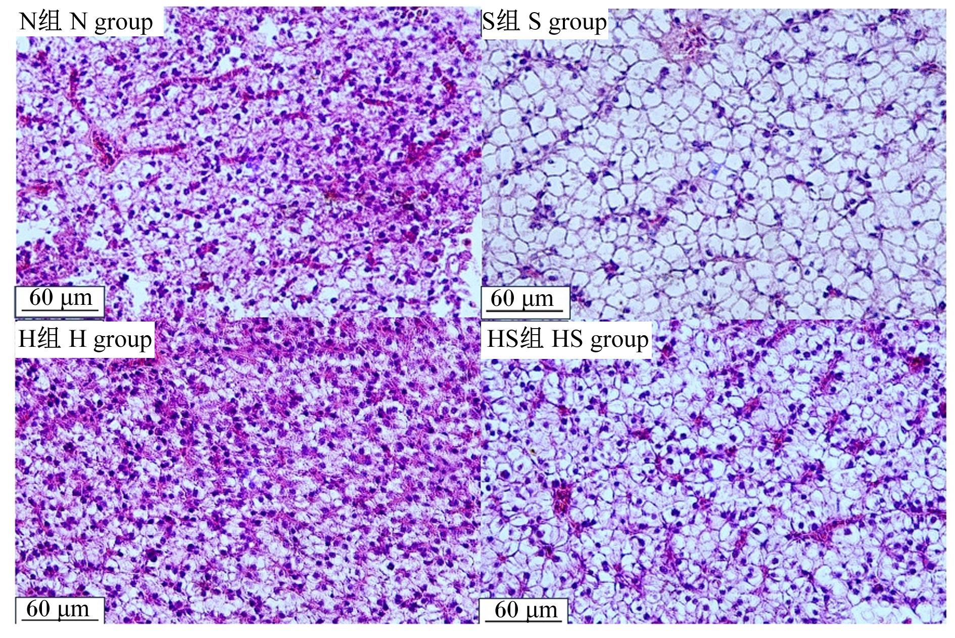
图2可知,S组、HS组大口黑鲈的肝细胞肿胀、细胞核偏移、伴随空泡出现,N组、H组的肝细胞结构相对完整,核较圆润清晰且处于细胞中心。
图2 大口黑鲈肝脏组织切片

Fig.2 Liver tissue sections of largemouth bass

2.4 脂脏和血清脂代谢相关指标

饲料糖水平和水体DO浓度对大口黑鲈肝脏和血清脂代谢相关指标的影响见表4。饲料糖水平对肝脏TG含量和血清TG、T-CHO含量有显著影响(P<0.05),对肝脏T-CHO含量和血清TG、HDL-C、LDL-C含量无显著影响(P>0.05)。高糖组肝脏中TG含量和血清中TG、T-CHO含量显著高于常糖组(P<0.05)。水体DO浓度对肝脏TG含量和血清TG、T-CHO、HDL-C、LDL-C含量有显著影响(P<0.05),对肝脏T-CHO含量无显著影响(P>0.05)。与常氧组相比,缺氧组肝脏和血清TG含量显著降低(P<0.05),而血清T-CHO、HDL-C、LDL-C含量显著升高(P<0.05)。饲料糖水平与水体DO浓度的交互作用显著影响肝脏TG含量和血清TG、HDL-C含量(P<0.05)。与N组相比,S组和HS组肝脏TG和血清TG、HDL-C含量显著升高(P<0.05);H组肝脏TG含量无显著变化(P>0.05),血清TG含量显著降低(P<0.05),血清HDL-C含量显著升高(P<0.05)。
表4 饲料糖水平和水体DO浓度对大口黑鲈肝脏和血清脂代谢相关指标的影响

Table 4 Effects of dietary sugar level and water DO concentration on lipid metabolism-related indicators in liver and serum of largemouth bass

项目
Items
组别Groups 双因素方差分析P
P-value of two-way ANOVA
N S H HS A B A×B
肝脏Liver/(mmol/g prot)
甘油三酯TG 0.21±0.02c 0.56±0.04a 0.19±0.02c 0.33±0.01b <0.001 <0.001 <0.001
总胆固醇T-CHO 0.08±0.02 0.11±0.03 0.10±0.02 0.11±0.02 0.229 0.328 0.312
血清Serum/(mmol/L)
甘油三酯TG 6.07±0.24c 7.09±0.21b 4.28±0.23d 7.78±0.46a <0.001 0.014 <0.001
总胆固醇T-CHO 8.41±0.33 10.74±0.93 10.71±1.42 11.21±0.97 0.038 0.042 0.148
高密度脂蛋白胆固醇
HDL-C
3.03±0.26c 3.81±0.30b 4.66±0.45a 3.86±0.53b 0.949 0.007 0.009
低密度脂蛋白胆固醇
LDL-C
7.27±0.33 7.28±0.19 9.42±0.35 8.37±0.92 0.124 0.001 0.122

2.5 肝脏抗氧化指标

饲料糖水平和水体DO浓度对大口黑鲈肝脏抗氧化指标的影响见表5。饲料糖水平对肝脏T-AOC、CAT和GSH-Px活性有显著影响(P<0.05),对SOD活性和MDA含量无显著影响(P>0.05)。与常糖组相比,高糖组肝脏T-AOC、CAT活性显著降低(P<0.05),而GSH-Px活性显著升高(P<0.05)。水体DO浓度显著影响肝脏T-AOC,SOD、CAT、GSH-Px活性和MDA含量(P<0.05)。与常氧组相比,缺氧组肝脏T-AOC、SOD和GSH-Px活性以及MDA含量显著下降(P<0.05),而CAT活性显著升高(P<0.05)。饲料糖水平与水体DO浓度的交互作用显著影响肝脏T-AOC与CAT、SOD、GSH-Px活性(P<0.05)。与N组相比,S组、H组、HS组肝脏T-AOC和SOD活性均显著降低(P<0.05),而GSH-Px活性均显著升高(P<0.05);H组肝脏CAT活性显著高于N组(P<0.05),而S组、HS组则与N组无显著差异(P>0.05)。
表5 饲料糖水平和水体DO浓度对大口黑鲈肝脏抗氧化指标的影响

Table 5 Effects of dietary sugar level and water DO concentration on liver antioxidant indicators of largemouth bass

项目
Items
组别Groups 双因素方差分析P
P-value of two-way ANOVA
N S H HS A B A×B
总抗氧化能力
T-AOC/(U/mg prot)
4.89±0.17a 3.02±0.13b 2.82±0.13b 2.13±1.08c <0.001 <0.001 <0.001
超氧化物歧化酶
SOD/(U/mg prot)
82.56±1.29a 79.13±0.65b 72.32±0.59c 73.11±1.49c 0.067 <0.001 0.010
过氧化氢酶
CAT/(U/mg prot)
12.07±1.57bc 17.15±2.15b 58.15±5.62a 10.25±1.74c <0.001 <0.001 <0.001
谷胱甘肽过氧化物酶
GSH-Px/(U/mg prot)
46.01±3.28d 116.67±6.59a 78.21±4.11b 59.85±6.25c <0.001 0.004 <0.001
丙二醛
MDA/(nmol/mg prot)
2.98±0.34 2.63±0.31 2.00±0.33 1.85±0.18 0.188 0.001 0.595

3 讨论

3.1 高糖饲料和慢性缺氧胁迫对大口黑鲈生长性能的影响

已有研究表明,慢性缺氧可显著降低大口黑鲈的增重率、特定生长率和摄食量[14];虹鳟鱼(Oncorhynchus mykiss)的存活率、增重和特定生长率在缺氧应激28 d后出现显著降低[15]。在本研究中,慢性缺氧胁迫下的大口黑鲈的末重、增重率、特定生长率显著降低,这可能归因于大口黑鲈在缺氧条件下无法获得足够的氧气,导致能量代谢减慢,生理功能受到限制,从而减慢生长速度[16]。在一定范围内限制氧气的可用性会限制鱼类对营养物质和能量的利用,从而影响生长。缺氧导致摄食量减少是鱼类应对缺氧胁迫的策略之一,这可能是由于代谢范围(MS)的受限所致。Chabot等[17]研究发现,缺氧相关的MS降低可能会影响欧洲海鲈(Dicentrarchus labrax)的生长。有研究认为,缺氧时食欲和生长率的降低是一种适应性行为反应,旨在保护有氧活动范围[18]。然而,除严重环境胁迫外,摄食量的减少并不一定会改变饲料效率。本试验中,缺氧状态下大口黑鲈的摄食量显著降低,但饲料系数无显著变化。类似结果在杂交条纹鲈幼鱼(Morone chrysops×M. saxatilis)[19]和尼罗罗非鱼(Oreochromis niloticus)[20]中亦有报道。

3.2 高糖饲料和慢性缺氧胁迫对大口黑鲈体成分和肝脏组织结构的影响

鱼类的体成分可间接反映营养物质的沉积量。本研究中,高糖饲料与慢性缺氧胁迫对大口黑鲈的部分体成分指标具有显著影响。与常氧环境相比,缺氧环境下全鱼水分含量升高,而粗脂肪含量降低。这一变化可能源于缺氧环境下鱼体新陈代谢与呼吸功能受限,脂质代谢活性下降,同时代谢废物与代谢物排泄减缓,进而影响水分平衡与脂质沉积[21]。研究表明,高糖饲料抑制了花鲈(Lateolabrax japonicus)体内蛋白质与灰分的沉积[22],提示高糖饲料可能干扰机体对糖、蛋白质等营养物质的正常代谢[23]。Zhang等[10]报道,高糖饲料会损伤鲈鱼的肠道,阻碍营养物质吸收,从而减少营养物质沉积。进一步的研究发现,高糖可抑制糖酵解,阻碍鲈鱼对葡萄糖的利用,减少能量供应[24]。由于营养物质的吸收与沉积需消耗能量,能量不足可能是限制营养物质沉积的重要原因[25]。本研究中,高糖饲料显著降低了大口黑鲈全鱼粗灰分与粗蛋白质含量,表明高糖饲料改变了鱼体中灰分与蛋白质的沉积模式;此外,高糖饲料与慢性缺氧胁迫对全鱼粗脂肪含量存在显著的交互作用。这可能是因为高糖饲料改变了大口黑鲈的新陈代谢效率,而缺氧则限制了氧气供应与能量转换,导致大口黑鲈无法获得足够能量以维持正常生理功能,进而影响营养物质的吸收与沉积。
研究表明,高淀粉饲料可诱导大口黑鲈[12]和花鲈[22]的脏体比和肝体比升高。组织学观察进一步证实,饲料中玉米淀粉水平为15%时,鲤(Cyprinus carpio)的肝细胞出现水样变性及轻微脂肪变性;玉米淀粉水平升至30%时,则表现为严重脂肪变性[26-27]。这表明高糖饲料可导致鱼类肝脏肿大、空泡化及脂肪样病变,损害肝脏健康[7]。本研究结果显示,与常糖组相比,高糖组大口黑鲈的脏体比、肝体比以及肝脏TG含量与血清TG、T-CHO含量均显著升高,肝脏组织学观察可见明显脂肪空泡,证实高糖饲料诱导了肝脏脂肪沉积。已有研究表明,与草食性或杂食性鱼类相比,肉食性鱼类利用淀粉获取能量的能力较差,其原因主要包括:葡萄糖感知与摄食的神经元调控体系不完善、葡萄糖转运能力差、胰岛素受体数量及亲和力不足[28]。过量的淀粉可诱发高血糖、糖原与脂质积累、慢性炎症及免疫功能与抗氧化能力下降,进而导致大口黑鲈出现代谢性肝病[29]

3.3 高糖饲料和慢性缺氧胁迫对大口黑鲈肝脏抗氧化能力的影响

缺氧可导致生物体氧化与抗氧化系统失衡,从而诱导鱼类氧化应激及过量ROS的产生。抗氧化酶系统(包括SOD、CAT和GSH-Px)在清除ROS中发挥关键作用[6]。本研究中,缺氧状态下大口黑鲈肝脏T-AOC、SOD和GSH-Px活性及MDA含量均显著下降,而CAT活性升高。这可能是由于机体通过抗氧化机制及时清除过量ROS,从而减少了脂质过氧化产物。MDA是细胞膜脂质过氧化的产物,其含量反映细胞所受氧化应激的程度;T-AOC可作为ROS清除能力及总抗氧化状态的综合指标,反映机体抵御氧化损害的能力[30];SOD是抵御氧化应激的第1道防线,主要将代谢或外界压力产生的ROS转化为过氧化氢,通过降解超氧自由基、减少细胞损伤发挥抗氧化保护作用[31]。类似研究表明,团头鲂(Megalobrama amblycephala)新品种在缺氧后肝脏和肠道MDA含量下降,而肠道CAT活性及肝脏GSH-Px活性升高,表明氧化应激诱导了这些酶活性的上调[32]。可口革囊星虫(Phascolosoma esculenta)体腔液中MDA含量在缺氧24、48、96 h时显著升高,但在复氧后显著下降[33]。为避免代谢应激(如高碳水化合物可能引起的代谢应激),生物体依赖抗氧化防御系统防止氧化损伤。本研究发现,高糖饲料未改变大口黑鲈肝脏MDA含量,但抑制了T-AOC和CAT活性,同时增强了GSH-Px活性。以往研究表明,较高的抗氧化酶活性与人类及水生动物更好的健康状况和代谢稳态相关。CAT作为过氧化氢清除剂,将SOD转化产生的过氧化氢还原为水和氧气;而GSH-Px则可清除细胞内的过氧化氢[31,34]。据报道,饲料中碳水化合物水平为47%时,团头鲂肝脏SOD活性和T-AOC呈下降趋势[35]。以上结果表明,高糖饲料诱导大口黑鲈肝脏发生氧化应激,机体通过激活抗氧化防御系统以应对潜在损害。值得注意的是,CAT和GSH-Px活性在常氧高糖组(S组)和缺氧常糖组(H组)中明显升高,而在缺氧高糖组(HS组)中呈现相反结果。这可能是因为这2种抗氧化酶随ROS水平变化处于合成与消耗的动态平衡状态:在高糖与缺氧共同作用下,大口黑鲈体内ROS积累达到峰值,超过CAT和GSH-Px的合成速率,导致其活性下降[36]。已有研究表明,某些抗氧化酶的活性在强烈氧化应激下会降低,而在中度压力下则升高[37]。团头鲂新品种[32]及银曼龙(Trichogaster microlepis)[38]在缺氧导致的强烈氧化应激下也表现出类似结果。
核因子E2相关因子2(Nrf2)是机体核转录因子家族成员之一,在调控氧化应激防御中发挥重要作用[39]。Nrf2的稳定性主要受Kelch样ECH关联蛋白1(Keap1)调控。当机体遭受氧化应激时,Nrf2从Keap1上解离并迅速转位进入细胞核,进而转录激活其下游抗氧化基因的表达[40]。研究发现,在温度胁迫或缺氧胁迫下,团头鲂的Nrf2及抗氧化相关基因表达水平上调;而在温度和缺氧联合胁迫下,这些基因的表达水平随时间呈先升高后下降的趋势,同时Keap1表达水平呈现相反的变化趋势[41]。缺氧与氨氮联合胁迫下的大口黑鲈也表现出类似结果[42]。以往研究表明,Nrf2/Keap1复合物可作为细胞响应氧化损伤的重要传感器,在调节抗氧化能力中发挥关键作用[39]。上述结果提示,短期胁迫可增强机体抗氧化能力,而长期联合胁迫则可能削弱这一能力。据此推测,与高糖或缺氧单一胁迫相比,高糖与缺氧的联合胁迫可能存在协同效应,从而加剧大口黑鲈的氧化应激,其具体作用机制有待进一步研究。

4 结论

① 慢性缺氧胁迫会降低大口黑鲈的生长性能,阻碍营养物质的吸收与沉积,并诱发氧化应激。
② 高糖饲料会导致大口黑鲈肝脏脂肪积累和氧化应激,从而损害肝脏健康。
③ 相较于单一胁迫因素,高糖饲料与慢性缺氧联合胁迫可能产生协同效应,加剧对大口黑鲈的负面影响。
[1]
MAHFOUZ M E, HEGAZI M M, EL-MAGD M A, et al. Metabolic and molecular responses in Nile tilapia,Oreochromis niloticus during short and prolonged hypoxia[J]. Marine and Freshwater Behaviour and Physiology, 2015, 48(5):319-340.

DOI

[2]
SAHA N, KONER D, SHARMA R. Environmental hypoxia:a threat to the gonadal development and reproduction in bony fishes[J]. Aquaculture and Fisheries, 2022, 7(5):572-582.

DOI

[3]
朱健. 不同投喂模式对大口黑鲈生长、饲料利用和肝脏健康的影响探究[D]. 硕士学位论文. 重庆: 西南大学, 2022.

ZHU J.Effect of different feeding strategies on growth,feed utilization and liver health of largemouth bass Micropterus salmoides[D]. Master’s Thesis. Chongqing: Southwest University, 2022. (in Chinese)

[4]
SUN J L, ZHAO L L, WU H, et al. Acute hypoxia changes the mode of glucose and lipid utilization in the liver of the largemouth bass (Micropterus salmoides)[J]. Science of the Total Environment, 2020, 713:135157.

DOI

[5]
DING Y, SHA W B, SUN Y F, et al. Respiratory metabolism and metabolomics of red swamp crayfish Procambarus clarkii under low temperature stress[J]. Journal of Experimental Zoology Part A:Ecological and Integrative Physiology, 2025, 343(5):578-589.

DOI

[6]
ALI S S, AHSAN H, ZIA M K, et al. Understanding oxidants and antioxidants:classical team with new players[J]. Journal of Food Biochemistry, 2020, 44(3):e13145.

[7]
LIN S M, SHI C M, MU M M, et al. Effect of high dietary starch levels on growth,hepatic glucose metabolism,oxidative status and immune response of juvenile largemouth bass,Micropterus salmoides[J]. Fish & Shellfish Immunology, 2018, 78:121-126.

[8]
ZHANG Y P, LIANG X F, HE S, et al. Effects of high carbohydrate diet-modulated microbiota on gut health in Chinese perch[J]. Frontiers in Microbiology, 2020, 11:575102.

DOI

[9]
XUE M Y, YAO T, XUE M, et al. Mechanism analysis of metabolic fatty liver on largemouth bass (Micropterus salmoides) based on integrated lipidomics and proteomics[J]. Metabolites, 2022, 12(8):759.

DOI

[10]
ZHANG Y M, XIE S W, WEI H L, et al. High dietary starch impaired growth performance,liver histology and hepatic glucose metabolism of juvenile largemouth bass,Micropterus salmoides[J]. Aquaculture Nutrition, 2020, 26(4):1083-1095.

DOI

[11]
GAO B, ZHANG X Y, ZHANG Y, et al. Effects of dietary carbohydrate levels on the growth,glycometabolism,antioxidant capacity and metabolome of largemouth bass (Micropterus salmoides)[J]. Aquaculture Research, 2022, 53(10):3748-3758.

DOI

[12]
LIANG X F, CHEN P, WU X L, et al. Effects of high starch and supplementation of an olive extract on the growth performance,hepatic antioxidant capacity and lipid metabolism of largemouth bass (Micropterus salmoides)[J]. Antioxidants, 2022, 11(3):577.

DOI

[13]
中华人民共和国农业农村部渔业渔政管理局, 全国水产技术推广总站, 中国水产学会. 2025中国渔业统计年鉴[M]. 北京: 中国农业出版社, 2025.

Bureau of Fisheries,Ministry of Agriculture and Rural Affairs of the People’s Republic of China,National Fisheries Technology Extension Center,China Society of Fisheries. 2025 China fishery statistical yearbook[M]. Beijing: China Agriculture Press,2025. (in Chinese)

[14]
HE Y, YU H D, ZHANG Z Y, et al. Effects of chronic hypoxia on growth performance,antioxidant capacity and protein turnover of largemouth bass (Micropterus salmoides)[J]. Aquaculture, 2022, 561:738673.

DOI

[15]
AKSAKAL E, EKINCI D. Effects of hypoxia and hyperoxia on growth parameters and transcription levels of growth,immune system and stress related genes in rainbow trout[J]. Comparative Biochemistry and Physiology Part A:Molecular & Integrative Physiology, 2021, 262:111060.

[16]
HE Y, ZHAO T, LV W H, et al. Interactive effects of dietary zinc and water temperature on growth performance,Zn transport,heat shock response and lipid metabolism in the livers of juvenile yellow catfish Pelteobagrus fulvidraco[J]. Aquaculture, 2023, 562:738795.

DOI

[17]
CHABOT D, CLAIREAUX G. Environmental hypoxia as a metabolic constraint on fish:the case of Atlantic cod,Gadus morhua[J]. Marine Pollution Bulletin, 2008, 57(6/12):287-294.

DOI

[18]
CLAIREAUX G, LEFRANÇOIS C. Linking environmental variability and fish performance:integration through the concept of scope for activity[J]. Philosophical Transactions of the Royal Society of Royal Series B:Biological Sciences, 2007, 362(1487):2031-2041.

[19]
GREEN B W, RAWLES S D, FULLER S A, et al. Hypoxia affects performance traits and body composition of juvenile hybrid striped bass (Morone chrysops×M. saxatilis)[J]. Aquaculture Research, 2016, 47(7):2266-2275.

DOI

[20]
OBIRIKORANG K A, ACHEAMPONG J N, DUODU C P, et al. Growth,metabolism and respiration in Nile tilapia (Oreochromis niloticus) exposed to chronic or periodic hypoxia[J]. Comparative Biochemistry and Physiology Part A:Molecular & Integrative Physiology, 2020, 248:110768.

[21]
HOU Z S, WEN H S, LI J F, et al. Environmental hypoxia causes growth retardation,osteoclast differentiation and calcium dyshomeostasis in juvenile rainbow trout (Oncorhynchus mykiss)[J]. Science of the Total Environment, 2020, 705:135272.

DOI

[22]
孙文豪, 邱建强, 田仁红, 等. 高糖饲料及添加缩合单宁对花鲈生长性能、体成分、血清代谢物、肠道短链脂肪酸含量及激素基因表达的影响[J]. 动物营养学报, 2024, 36(8):5254-5268.

DOI

SUN W H, QIU J Q, TIAN R H, et al. Effects of high sugar diet and condensed tannin supplementation on growth performance,body composition,serum metabolites,intestinal short-chain fatty acid contents and hormone gene expression of sea bass (Lateolabrax maculatus)[J]. Chinese Journal of Animal Nutrition, 2024, 36(8):5254-5268. (in Chinese)

[23]
PENG K, WANG G X, WANG Y X, et al. Condensed tannins enhanced antioxidant capacity and hypoxic stress survivability but not growth performance and fatty acid profile of juvenile Japanese seabass (Lateolabrax japonicus)[J]. Animal Feed Science and Technology, 2020, 269:114671.

DOI

[24]
ZHANG Y M, XIE S W, GUO T Y, et al. High dietary starch inclusion impairs growth and antioxidant status,and alters liver organization and intestinal microbiota in largemouth bass Micropterus salmoides[J]. Aquaculture Nutrition, 2020, 26(5):1806-1821.

DOI

[25]
KAYASTHA T B, KNOWAR B K, DUTTA S. Effect of energy level of feeds on feed intake,nutrient utilization and growth performance of indigenous pullets of Assam[J]. Indian Journal of Animal Nutrition, 2013, 30(2):175-178.

[26]
李静辉. 饲料不同糖水平对鲤生长、肝胰脏组织结构及代谢的影响[D]. 硕士学位论文. 天津: 天津农学院, 2017.

LI J H. Effects of different dietary carbohydrate levels on growth,hepatopancreas structure and metabolism of common carp[D]. Master’s Thesis. Tianjin: Tianjin Agricultural University, 2017. (in Chinese)

[27]
范泽, 吴迪, 李晨辉, 等. 苦瓜皂苷对饲喂高糖饲料松浦镜鲤幼鱼生长、消化及肝脏健康的影响[J]. 水产学杂志, 2023, 36(3):29-37.

FAN Z, WU D, LI C H, et al. Effects of bitter melon Momordica charantia saponins on growth,digestion and hepatopancreas health of juvenile Songpu mirror carp (Cyprinus carpio Songpu) fed high-carbohydrate diets[J]. Chinese Journal of Fisheries, 2023, 36(3):29-37. (in Chinese)

[28]
MA H J, MOU M M, PU D C, et al. Effect of dietary starch level on growth,metabolism enzyme and oxidative status of juvenile largemouth bass,Micropterus salmoides[J]. Aquaculture, 2019, 498:482-487.

DOI

[29]
YU H H, ZHANG L L, CHEN P, et al. Dietary bile acids enhance growth,and alleviate hepatic fibrosis induced by a high starch diet via AKT/FOXO1 and cAMP/AMPK/SREBP1 pathway in Micropterus salmoides[J]. Frontiers in Physiology, 2019, 10:1430.

DOI

[30]
CHENG C H, MA H L, DENG Y Q, et al. Oxidative stress,cell cycle arrest,DNA damage and apoptosis in the mud crab (Scylla paramamosain) induced by cadmium exposure[J]. Chemosphere, 2021, 263:128277.

DOI

[31]
IGHODARO O M, AKINLOYE O A. First line defence antioxidants-superoxide dismutase (SOD),catalase (CAT) and glutathione peroxidase (GPX):their fundamental role in the entire antioxidant defence grid[J]. Alexandria Journal of Medicine, 2018, 54(4):287-293.

DOI

[32]
SHUANG L, CHEN S L, REN C, et al. Effects of hypoxia and reoxygenation on oxidative stress,histological structure,and apoptosis in a new hypoxia-tolerant variety of blunt snout bream (Megalobrama amblycephala)[J]. Comparative Biochemistry and Physiology Part A:Molecular & Integrative Physiology, 2023, 278:111358.

[33]
XING J M, ZOU J, LIU X D, et al. Effects of acute hypoxia and reoxygenation on the coelomic fluid of Phascolosoma esculenta:oxidative stress and transcriptome analysis[J]. Aquaculture Reports, 2024, 39:102424.

DOI

[34]
OGUEJI E, NWANI C, MBAH C, et al. Oxidative stress,biochemical,lipid peroxidation,and antioxidant responses in Clarias gariepinus exposed to acute concentrations of ivermectin[J]. Environmental Science and Pollution Research, 2020, 27(14):16806-16815.

DOI

[35]
ZHOU C, LIU B, GE X, et al. Effect of dietary carbohydrate on the growth performance,immune response,hepatic antioxidant abilities and heat shock protein 70 expression of Wuchang bream,Megalobrama amblycephala[J]. Journal of Applied Ichthyology, 2013, 29(6):1348-1356.

DOI

[36]
WANG Q F, SHEN W L, HOU C C, et al. Physiological responses and changes in gene expression in the large yellow croaker Larimichthys crocea following exposure to hypoxia[J]. Chemosphere, 2017, 169:418-427.

DOI

[37]
RODRIGUEZ C, MAYO J C, SAINZ R M, et al. Regulation of antioxidant enzymes:a significant role for melatonin[J]. Journal of Pineal Research, 2004, 36(1):1-9.

DOI

[38]
HUANG C Y, LIN H C, LIN C H. Effects of hypoxia on ionic regulation,glycogen utilization and antioxidative ability in the gills and liver of the aquatic air-breathing fish Trichogaster microlepis[J]. Comparative Biochemistry and Physiology Part A:Molecular & Integrative Physiology, 2015, 179:25-34.

[39]
MA Q. Role of Nrf2 in oxidative stress and toxicity[J]. Annual Review of Pharmacology and Toxicology, 2013, 53(1):401-426.

DOI

[40]
HO C Y, CHENG Y T, CHAU C F, et al. Effect of diallyl sulfide on in vitro and in vivo Nrf2-mediated pulmonic antioxidant enzyme expression via activation ERK/p38 signaling pathway[J]. Journal of Agricultural and Food Chemistry, 2012, 60(1):100-107.

DOI

[41]
CHEN K, JIA Y H, HE Z, et al. Regulation mechanism of oxidative status,immunity and apoptosis induced by hypoxia and heat exposure via PI3K/Akt signaling pathway in Megalobrama amblycephala[J]. Fish & Shellfish Immunology, 2024, 155:110027.

[42]
ZHAO L L, CUI C, LIU Q, et al. Combined exposure to hypoxia and ammonia aggravated biological effects on glucose metabolism,oxidative stress,inflammation and apoptosis in largemouth bass (Micropterus salmoides)[J]. Aquatic Toxicology, 2020, 224:105514.

DOI

Outlines

/