

1 材料与方法
1.1 试剂与仪器
1.1.1 主要试剂
1.1.2 主要仪器
1.2 试验设计
1.3 指标检测
1.3.1 肠道组织形态学观察
1.3.2 肠道黏膜屏障相关蛋白表达检测
1.3.3 肠道促炎因子及炎症信号通路相关蛋白表达检测
1.3.4 肠道细胞焦亡及坏死性凋亡相关蛋白表达检测
1.3.5 肠道PXR表达检测
1.4 数据统计分析
2 结果与分析
2.1 PWD对仔猪小肠各段组织结构的影响
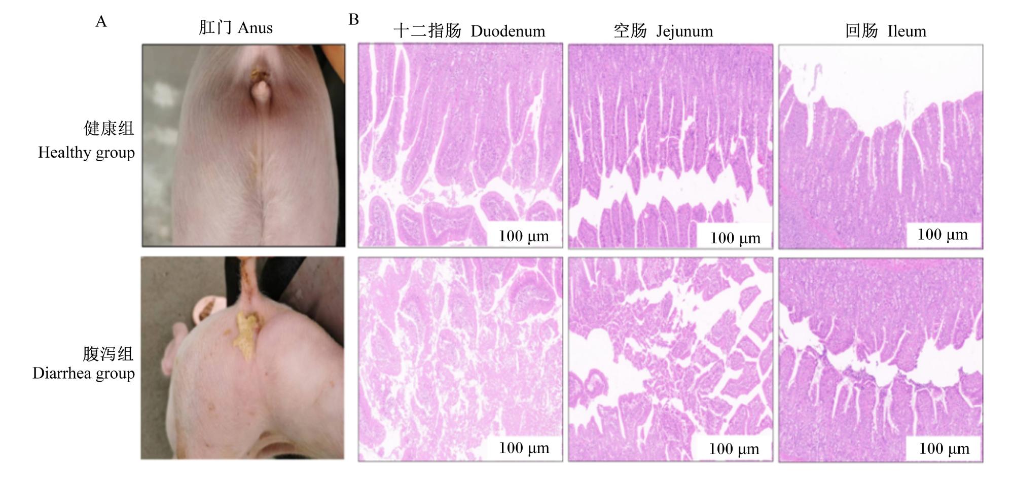
2.2 PWD对仔猪肠道黏膜屏障的影响
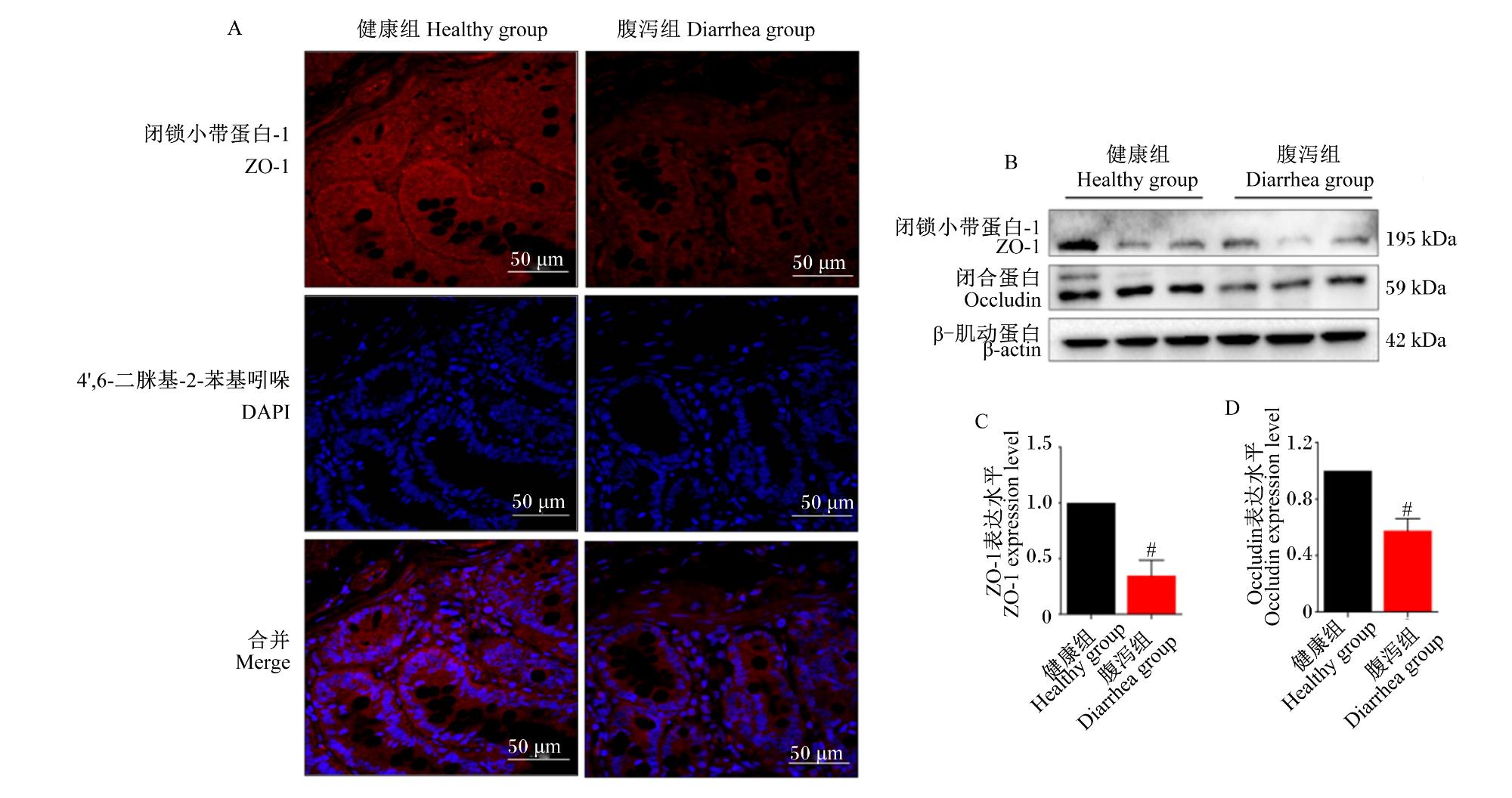
图2 健康组与腹泻组仔猪空肠组织中黏膜屏障相关蛋白的表达情况A:免疫荧光检测ZO-1的定位分布;B:蛋白质印迹检测ZO-1与Occludin的表达;C:ZO-1表达的定量分析;D:Occludin表达的定量分析。目的蛋白表达水平以内参β-肌动蛋白校正后,将健康组的表达水平设为1,健康组的数据以健康组的倍数表示。腹泻组数据柱标注“#”表示与健康组相比差异显著(P<0.05),标注“##”表示与健康组相比差异极显著(P<0.01)。下图同。 Fig.2 Expression of mucosal barrier-associated proteins in jejunal tissues of piglets in healthy group and A: localization and distribution of ZO-1 detected by IF; B: expression of ZO-1 and Occludin detected by WB; C: quantitative analysis of ZO-1 expression; D: quantitative analysis of Occludin expression. The expression level of the target protein was normalized to the internal control β-actin. The expression level in the healthy group was set to 1, and data from the diarrhea group were expressed as fold change relative to the healthy group. Compare with the healthy group, data columns of diarrhea group with “#” indicated significant difference (P<0.05), and with “##” indicated extremely significant difference (P<0.01). The same as below. diarrhea group (n=6) |
2.3 PWD对仔猪肠道促炎因子分泌的影响
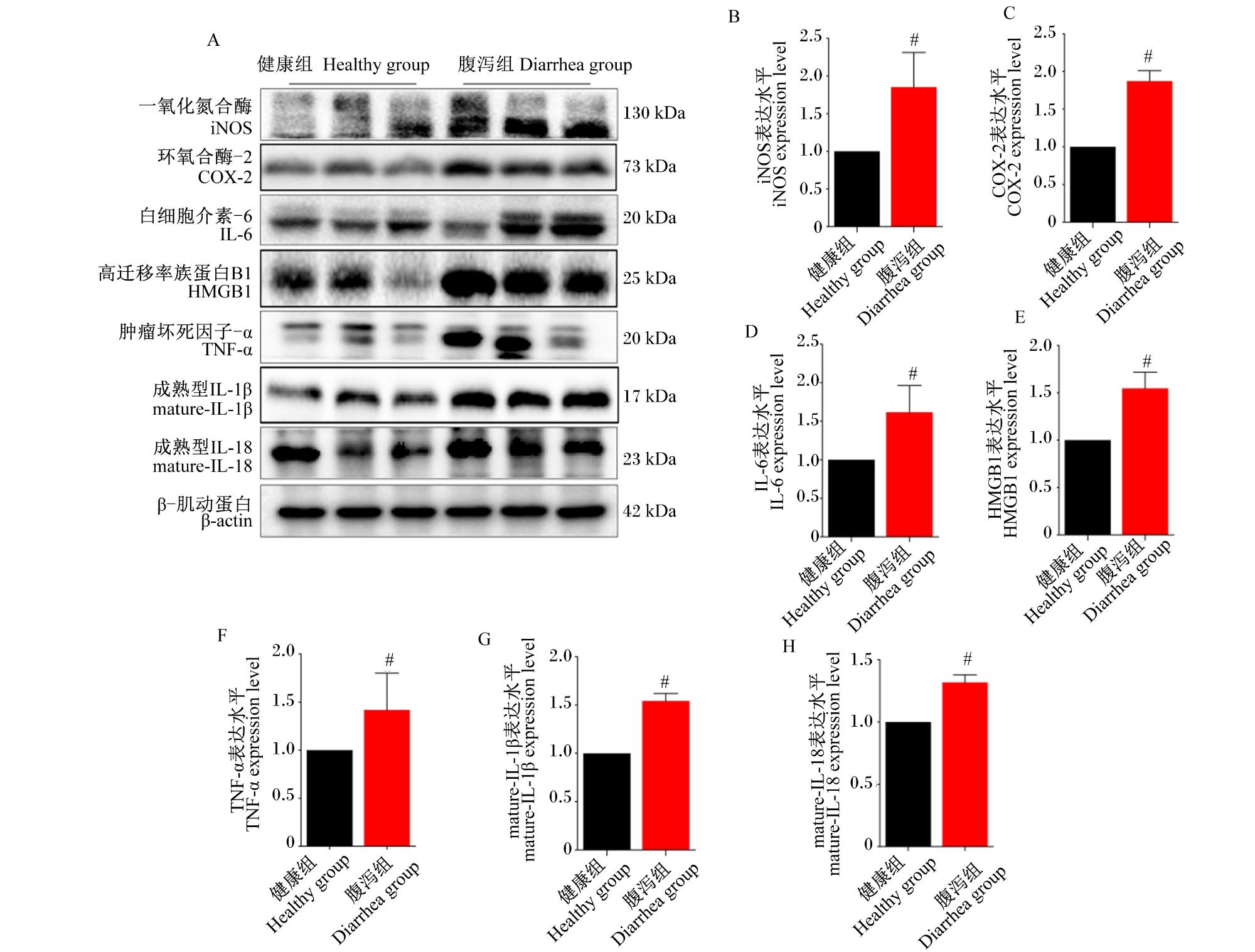
图3 健康组与腹泻组仔猪空肠组织中促炎因子的表达情况A:蛋白质印迹检测iNOS、COX-2、IL-6、HMGB1、TNF-α、成熟型IL-1β、成熟型IL-18的表达;B:iNOS表达的定量分析;C:COX-2表达的定量分析;D:IL-6表达的定量分析;E:HMGB1表达的定量分析;F:TNF-α表达的定量分析;G:成熟型IL-1β表达的定量分析;H:成熟型IL-18表达的定量分析。 Fig.3 Expression of pro-inflammatory cytokines in jejunal tissues of piglets in healthy group and diarrhea group (n=6) A: expression of iNOS, COX-2, IL-6, HMGB1, TNF-α, mature-IL-1β and mature-IL-18 detected by WB; B: quantitative analysis of iNOS expression; C: quantitative analysis of COX-2 expression; D: quantitative analysis of IL-6 expression; E: quantitative analysis of HMGB1 expression; F: quantitative analysis of TNF-α expression; G: quantitative analysis of mature-IL-1β expression; H: quantitative analysis of mature-IL-18 expression. |
2.4 PWD对仔猪肠道炎症信号通路的影响
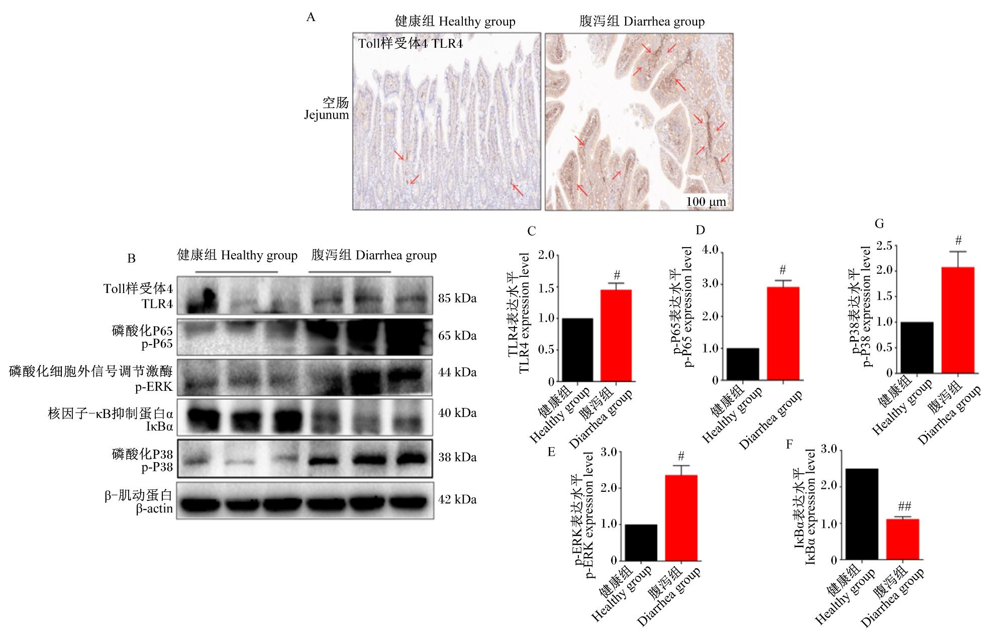
图4 健康组与腹泻组仔猪空肠组织中炎症信号通路相关蛋白的表达情况A:免疫组化检测TLR4的定位分布;B:蛋白质印迹检测TLR4、p-P65、p-ERK、IκBα及p-P38的表达;C:TLR4表达的定量分析;D:p-P65表达的定量分析;E:p-ERK表达的定量分析;F:IκBα表达的定量分析;G:p-P38表达的定量分析。 Fig.4 Expression of pro-inflammatory cytokines in jejunal tissues of piglets in healthy group and diarrheal group (n=6) A: localization and distribution of TLR4 detected by IHC; B: expression of TLR4, p-P65, p-ERK, IκBα and p-P38 detected by WB; C: quantitative analysis of TLR4 expression; D: quantitative analysis of p-P65 expression; E: quantitative analysis of p-ERK expression; F: quantitative analysis of IκBα expression; G: quantitative analysis of p-P38 expression. |
2.5 PWD对仔猪肠道细胞焦亡的影响
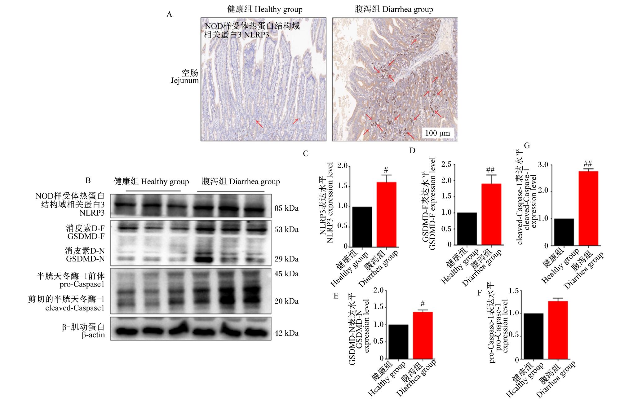
图5 健康组与腹泻组仔猪空肠组织中细胞焦亡相关蛋白的表达情况A:免疫组化检测NLRP3的定位分布;B:蛋白质印迹检测NLRP3、GSDMD-F、GSDMD-N、pro-Caspase-1 和cleaved-Caspase-1的表达;C:NLRP3表达的定量分析;D:GSDMD-N表达的定量分析;E:GSDMD-N表达的定量分析;F:pro-Caspase-1表达的定量分析;G:cleaved-Caspase-1表达的定量分析。 Fig.5 Expression of pyroptosis-related proteins in jejunal tissues of piglets in healthy group and diarrheal group (n=6) A: localization and distribution of NLRP3 detected by IHC; B: expression of NLRP3, GSDMD-F, GSDMD-N, pro-Caspase-1 and cleaved-Caspase-1 detected by WB; C: quantitative analysis of NLRP3 expression; D: quantitative analysis of GSDMD-N expression; E: quantitative analysis of GSDMD-N expression; F: quantitative analysis of pro-Caspase-1 expression; G: quantitative analysis of cleaved-Caspase-1 expression. |
2.6 PWD对仔猪肠道坏死性凋亡的影响
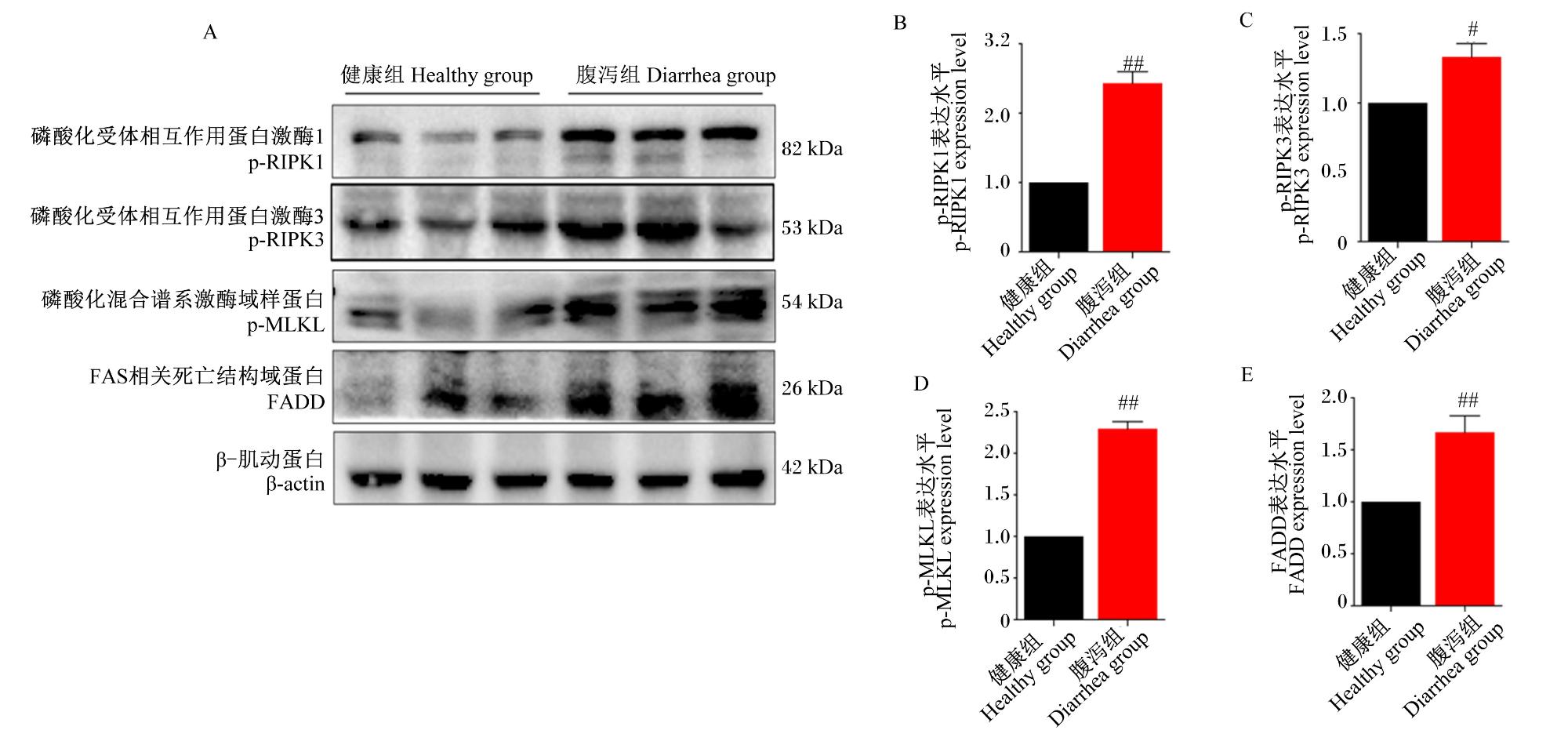
图6 健康组与腹泻组仔猪空肠组织中坏死性凋亡相关蛋白的表达情况A:蛋白质印迹检测p-RIPK1、p-RIPK3、p-MLKL和FADD的表达;B:p-RIPK1表达的定量分析;C:p-RIPK3表达的定量分析;D:p-MLKL表达的定量分析;E:FADD表达的定量分析。 Fig.6 Expression of necroptosis-related proteins in jejunal tissues of piglets in healthy group and diarrheal group (n=6) A: expression of p-RIPK1, p-RIPK3, p-MLKL and FADD detected by WB; B: quantitative analysis of p-RIPK1 expression; C: quantitative analysis of p-RIPK3 expression; D: quantitative analysis of p-MLKL expression; E: quantitative analysis of FADD expression. |
2.7 PWD对仔猪肠道PXR表达的影响
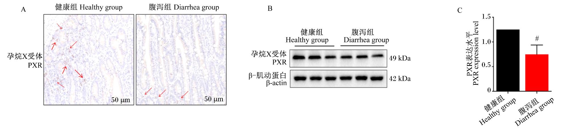


图7 健康组与腹泻组仔猪空肠组织中PXR的表达情况A:免疫组化检测PXR的定位发布;B:蛋白质印迹检测PXR的表达;C:PXR表达的定量分析。 Fig.7 Expression of PXR in jejunal tissues of piglets in healthy group and diarrheal group (n=6) A: localization and distribution of PXR detected by IHC; B: expression of PXR detected by WB; C: quantitative analysis of PXR expression. |