

1 材料与方法
1.1 BLF的制备及超高效液相色谱-四极杆飞行时间质谱(UHPLC-QTOF-MS)分析
1.2 BLF活性成分及其抑制氧化应激靶点的筛选
1.3 活性成分-交集靶点网络和蛋白质-蛋白质相互作用(PPI)网络构建
1.4 基因本体(GO)功能和京都基因与基因组百科全书(KEGG)通路富集分析及相互作用网络构建
1.5 分子对接和分子动力学模拟
1.6 免疫荧光检测
1.7 Western blot检测
2 结果与分析
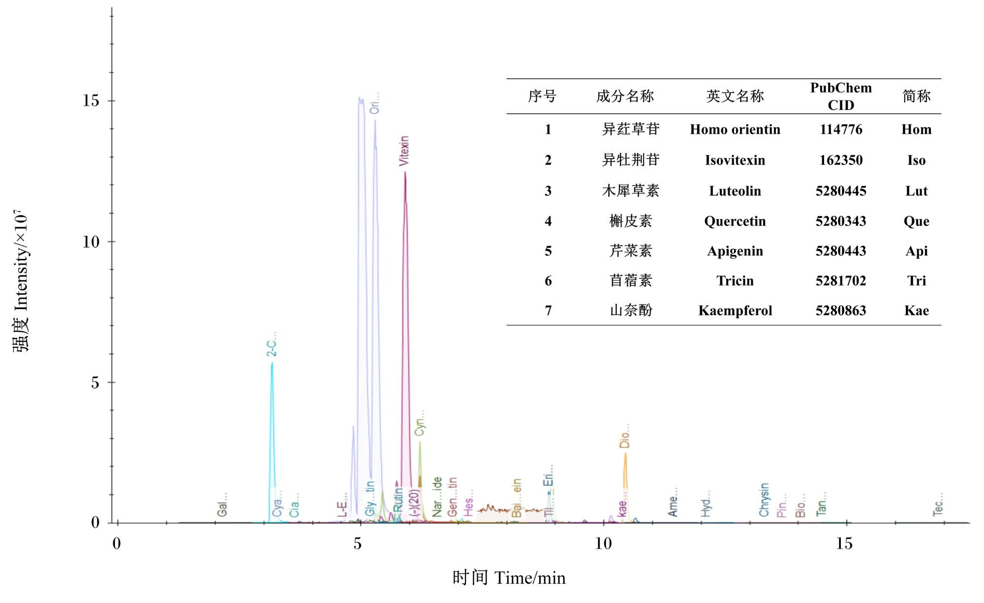
2.1 通过UHPLC-QTOF-MS鉴定BLF提取物中的化学成分以及潜在靶点筛选
2.2 BLF抑制氧化应激靶点获取结果及PPI网络
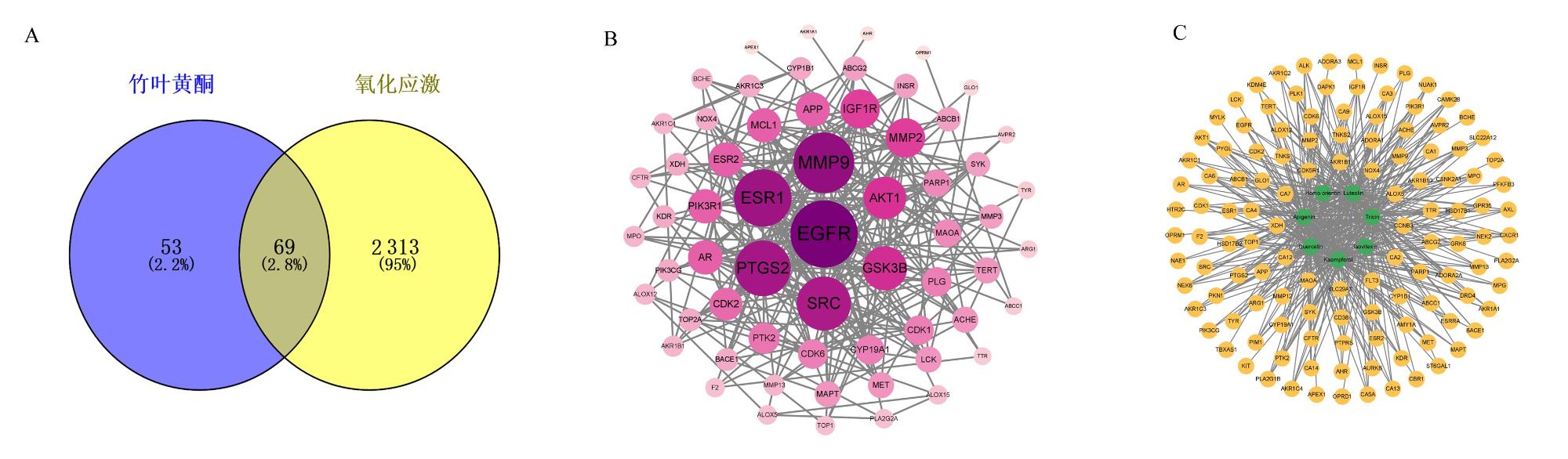
图2 BLF抑制氧化应激潜在作用靶点和PPI网络图A:BLF与氧化应激交集靶点韦恩图;B:PPI网络图;C:活性成分-交集靶点网络图。A: Venn diagram of intersection targets of BLF and oxidative stress; B: PPI network diagram; C: active component-intersection target network diagram. Fig.2 Potential targets of BLF in inhibiting oxidative stress and PPI network diagram |
表1 度值排名前20的关键靶点Table 1 Top 20 key targets in degree value |
| 关键靶点 Key targets | 中文名称 Chinese name | 度值 Degree value | 关键靶点 Key targets | 中文名称 Chinese name | 度值 Degree value | |
|---|---|---|---|---|---|---|
| EGFR | 表皮生长因子受体 | 33 | AR | 雄激素受体 | 13 | |
| ESR1 | 雌激素受体1 | 29 | ESR2 | 雌激素受体2 | 13 | |
| MMP9 | 基质金属蛋白酶9 | 27 | MCL1 | 髓系细胞白血病1 | 13 | |
| PTGS2 | 前列腺素内过氧化物合酶2 | 26 | PIK3R1 | 磷脂酰肌醇3-激酶调节亚基1 | 13 | |
| SRC | SRC原癌基因蛋白 | 25 | CDK2 | 细胞周期蛋白依赖性激酶2 | 12 | |
| GSK3B | 糖原合成酶激酶3β | 19 | PTK2 | 蛋白酪氨酸激酶2 | 11 | |
| AKT1 | 蛋白激酶B1 | 18 | CDK1 | 细胞周期蛋白依赖性激酶1 | 10 | |
| IGF1R | 胰岛素样生长因子1受体 | 16 | CDK6 | 细胞周期蛋白依赖性激酶6 | 10 | |
| MMP2 | 基质金属蛋白酶2 | 16 | CYP19A1 | 细胞色素P450家族19亚家族A成员1 | 10 | |
| APP | β-淀粉样前体蛋白 | 13 | PLG | 纤溶酶原 | 10 |
2.3 BLF抑制氧化应激靶点的GO功能和KEGG通路富集分析及相互作用网络
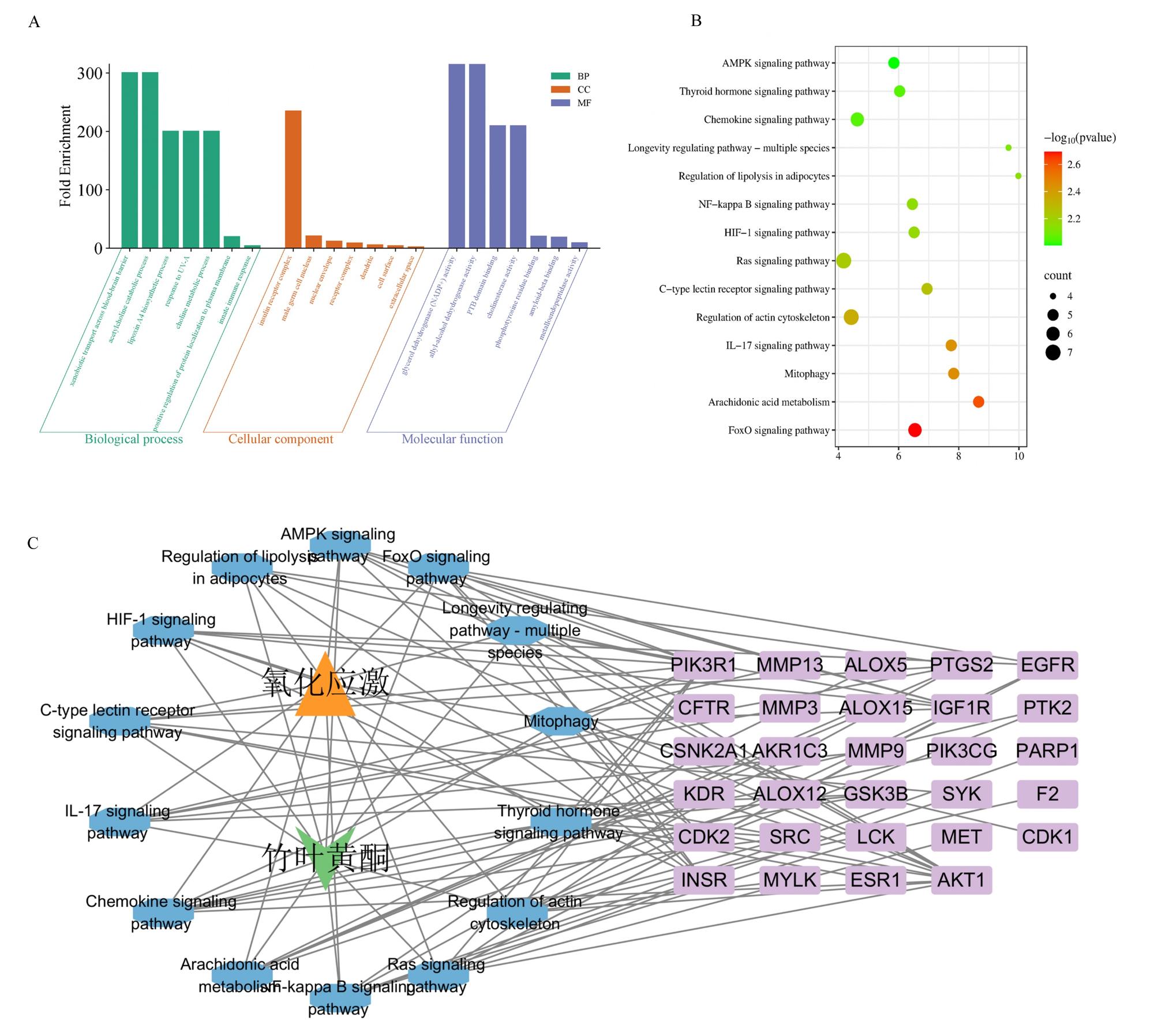
图3 BLF抑制氧化应激靶点的GO功能和KEGG通路富集分析及相互作用网络A:GO功能富集分析 GO function enrichment analysis;B:KEGG通路富集分析 KEGG pathway enrichment analysis;C:BLF-关键靶点-关键通路-氧化应激相互作用网络图 BLF-key target-key pathway-oxidative stress interaction network diagram。 Fig.3 Enrichment analysis of GO function and KEGG pathway of BLF in inhibiting oxidative stress targets and interaction network |
2.4 分子对接和分子动力学模拟验证
表2 BLF核心活性成分与关键靶点分子对接结合能Table 2 Binding energy of core active components of BLF to key target molecules kcal/mol |
| 活性成分 Active components | EGFR结合能 EGFR binding energy | ESR1结合能 ESR1 binding energy | MMP9结合能 MMP9 binding energy |
|---|---|---|---|
| 异荭草苷Homo orientin | -7.9 | -7.3 | -7.0 |
| 异牡荆苷Isovitexin | -8.2 | -7.3 | -6.5 |
| 木犀草素Luteolin | -7.1 | -7.3 | -6.4 |
| 槲皮素Quercetin | -7.0 | -7.1 | -6.3 |
| 芹菜素Apigenin | -7.2 | -6.8 | -6.2 |
| 苜蓿素Tricin | -7.2 | -6.8 | -6.0 |
| 山奈酚Kaempferol | -7.1 | -6.8 | -6.1 |
1 kcal/mol≈4.184 kJ/mol。 |
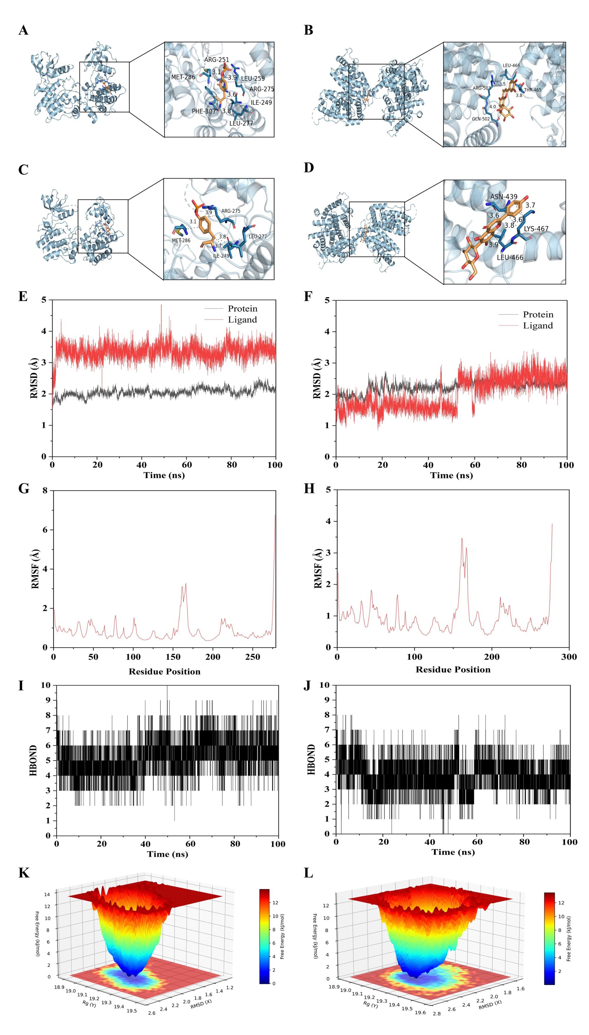
图4 分子对接和分子动力学模拟结果A和B分别表示异荭草苷与EGFR和ESR1的结合,C和D分别表示异牡荆苷与EGFR和ESR1的结合;E和F分别表示异荭草苷和异牡荆苷与EGFR的RMSD图;G和H分别表示异荭草苷和异牡荆苷与EGFR的RMSF图;I和J分别表示异荭草苷和异牡荆苷与EGFR的氢键数量图;K和L分别表示异荭草苷和异牡荆苷与EGFR的自由能形貌图。 Fig.4 Molecular docking and molecular dynamics simulation results |
2.5 BLF对H2O2诱导奶牛乳腺上皮细胞屏障功能及PINK1/P62介导的线粒体自噬的影响
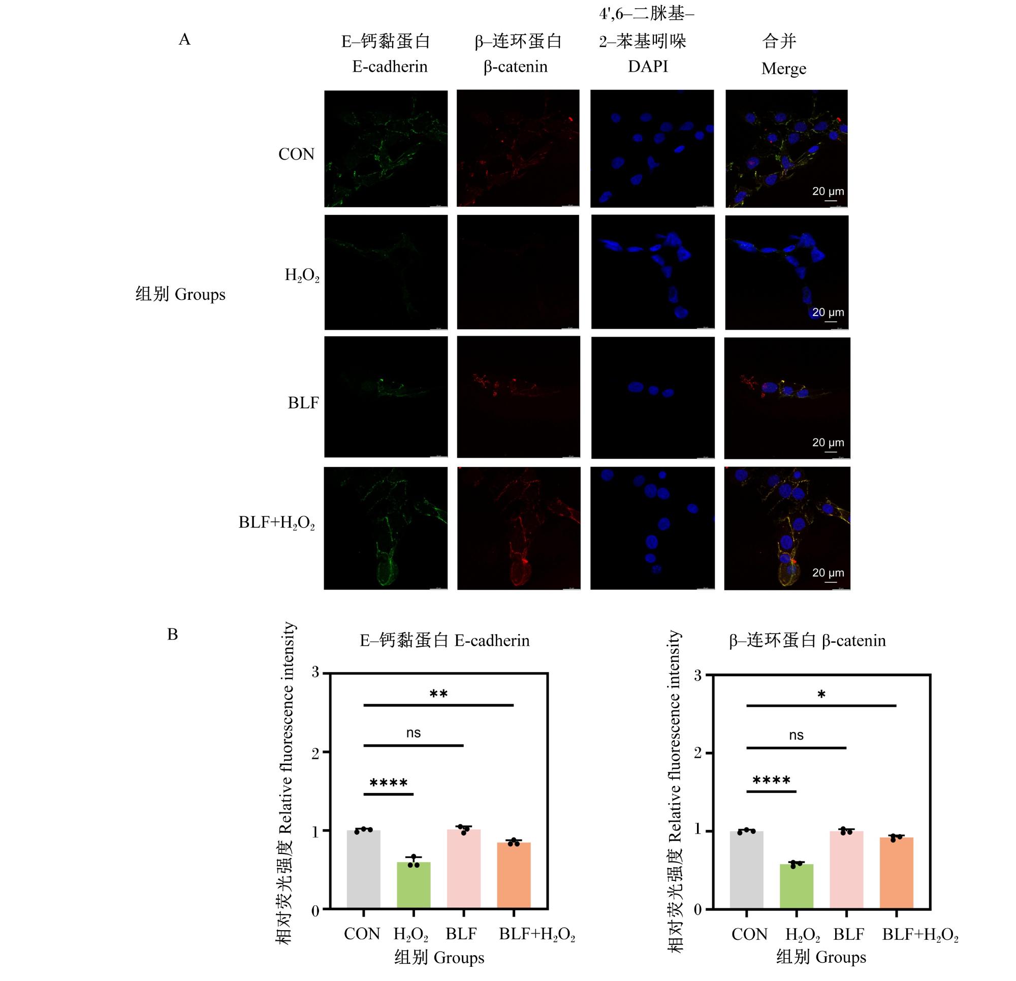
图5 BLF对H2O2诱导奶牛乳腺上皮细胞连接蛋白分布和表达的影响A:激光扫描共聚焦显微镜观察到的细胞中E-钙黏蛋白和β-连环蛋白的荧光图像 fluorescence images of E-cadherin and β-catenin in cells visualized by laser scanning confocal microscopy;B:细胞中E-钙黏蛋白和β-连环蛋白的相对荧光强度 relative fluorescence intensity of E-cadherin and β-catenin in cells。 Fig.5 Effects of BLF on distribution and expression of junction proteins in bovine mammary epithelial cells stimulated with H2O2 Data were presented as mean±SD (n=3); **** indicated P<0.000 1, ** indicated P<0.01, * indicated P<0.05, and ns indicated P>0.05. The same as Fig.6. |
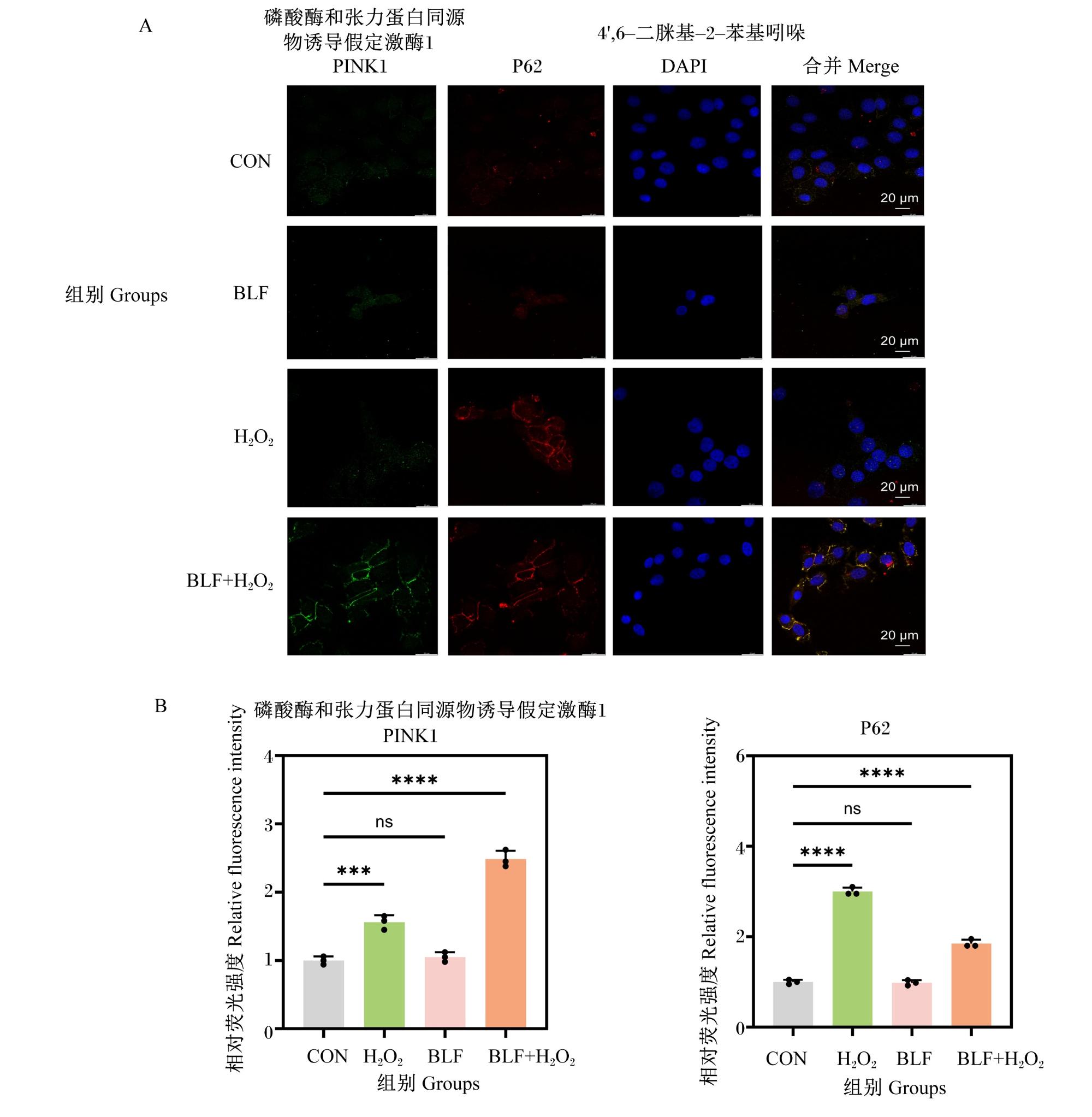
图6 BLF对H2O2诱导奶牛乳腺上皮细胞PINK1和P62分布和表达的影响A:激光扫描共聚焦显微镜观察到的细胞中PINK1和P62的荧光图像 fluorescence images of PINK1 and P62 in cells visualized by laser scanning confocal microscopy;B:细胞中PINK1和P62的相对荧光强度 relative fluorescence intensity of PINK1 and P62 in cells。 Fig.6 Effects of BLF on distribution and expression of PINK1 and P62 in bovine mammary epithelial cells stimulated with H2O2 |
2.6 BLF对H2O2诱导奶牛乳腺上皮细胞线粒体自噬相关蛋白表达的影响
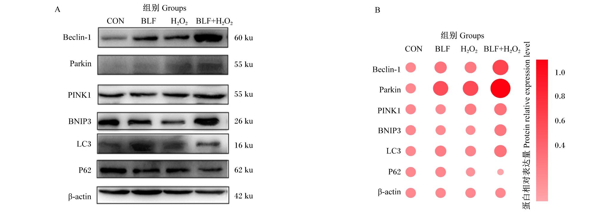


图7 BLF对H2O2诱导奶牛乳腺上皮细胞线粒体自噬相关蛋白表达的影响A:线粒体自噬相关关键蛋白Western blot代表性条带图;B:线粒体自噬相关蛋白相对表达量的气泡图分析,气泡颜色越深代表蛋白相对表达量越高。 Fig.7 Effects of BLF on expression of mitophagy-related proteins in bovine mammary epithelial cells stimulated with H2O2 A: representative Western blot bands of key mitophagy-related proteins; B: bubble plot analysis of relative expression levels of mitophagy-related proteins, where darker bubble colors indicated higher protein relative expression levels. |