REVIEW

Effect of Selenium on Programmed Cell Death in Animals Induced by Cadmium Exposure and Its Mechanism

  • FANG Manxin , 1, 2, 3 ,
  • LIU Ben 1, 2, 3 ,
  • HU Wei 1, 2, 3 ,
  • ZHENG Lucheng 1, 2, 3
Expand
  • 1 School of Life Sciences and Environmental Resources, Yichun University, Yichun 336000, China
  • 2 Yichun University Research Center for Traditional Chinese Veterinary Medicine and Animal Embryo Engineering Technology, Yichun 336000, China
  • 3 Engineering Technology Research Center of Jiangxi Universities and Colleges for Selenium Agriculture, Yichun 336000, China

FANG Manxin,associate professor,E-mail:

Received date: 2025-12-23

  Online published: 2026-08-13

Abstract

As a typical toxic heavy metal, cadmium has extremely strong accumulative properties. It can trigger acute toxic reactions in livestock and poultry, and is more likely to cause long-term potential chronic toxic damage, posing a severe and persistent threat to the growth, development and physiological functions of livestock and poultry. Though the trace element selenium is present in extremely low concentrations in animal bodies, it plays an irreplaceable physiological role and is crucial for maintaining the normal physiological metabolism of the organism and ensuring healthy growth. In recent years, numerous studies have shown that selenium can antagonize the toxicity of cadmium. This paper mainly summarized the research reports on cadmium-induced programmed cell death in animals and the mechanism of selenium’s antagonistic effect, with the aim of providing references for the prevention and control of cadmium pollution and the research and application of selenium.

Cite this article

FANG Manxin , LIU Ben , HU Wei , ZHENG Lucheng . Effect of Selenium on Programmed Cell Death in Animals Induced by Cadmium Exposure and Its Mechanism[J]. Chinese Journal of Animal Nutrition, 2026 , 38(8) : 5531 -5542 . DOI: 10.12418/CJAN2026.441

镉作为环境中广泛存在的重金属元素,被动物摄入后在体内半衰期长且易蓄积,难以代谢排出,对动物生长性能、生理功能、繁殖性能、免疫性能等层面造成严重影响[1-4],给养殖业带来经济损失。硒是动物和人体不可或缺的微量矿物元素,可激活谷胱甘肽过氧化物酶(glutathione peroxidase,GPX)等抗氧化酶,清除镉诱导产生的过量活性氧(reactive oxygen species,ROS),减少脂质过氧化,保护细胞免受损伤等[5]。本文综述了硒对镉暴露致动物细胞程序性死亡的影响及其作用机制,旨在为镉污染的防控治理与硒的应用提供参考。

1 硒的生物学功能

硒是动物机体必需的微量矿物元素,其生物学功能主要通过以硒代半胱氨酸为活性中心的各类硒蛋白,在抗氧化、抗炎、免疫调节及甲状腺激素代谢等关键生理过程中发挥不可替代的作用。在抗氧化功能方面,机体内近半数硒蛋白,如GPX家族、硫氧还蛋白还原酶(thioredoxin reductase,TrxR)家族、硒蛋白P、硒蛋白M、硒蛋白H、硒蛋白O、硒蛋白V等参与氧化还原稳态调控。硒作为GPX的核心成分,可催化谷胱甘肽(glutathione,GSH)还原反应,将有毒过氧化物降解为无毒羟基化合物,有效清除细胞代谢产生的过量ROS,缓解氧化应激造成的脂质过氧化、蛋白质羰基化与DNA损伤,保护细胞膜、蛋白质及遗传物质稳定[5-7];在抗炎功能方面,硒通过多途径调控炎症信号通路与炎症介质表达,维持机体炎症平衡。硒可抑制核因子-κB(nuclear factor-κB,NF-κB)的过度磷酸化与激活,减少肿瘤坏死因子-α(tumor necrosis factor-α,TNF-α)、白细胞介素-1β(interleukin-1β,IL-1β)等促炎细胞因子的合成与释放,同时可促进白细胞介素-10(interleukin-10,IL-10)、转化生长因子-β(transforming growth factor-β,TGF-β)等抗炎因子生成。此外,抑制巨噬细胞、树突状细胞促炎活性及T细胞过度增殖,协同调控趋化因子、黏附分子与炎症相关脂质介质代谢,避免过度炎症对组织造成损伤[8-10];在免疫调节功能方面,硒可同时提升动物非特异性与特异性免疫水平。对于非特异性免疫,硒能够激活巨噬细胞并通过抗氧化作用减轻其在吞噬病原体时受到的氧化损伤,增强吞噬与清除能力。对于特异性免疫,硒在胸腺、淋巴结、脾脏等免疫器官中分布丰富,可保护胸腺结构、维持淋巴细胞活性,促进B细胞增殖分化与抗体及免疫球蛋白合成,同时增强T细胞增殖与细胞毒性功能,全面提升机体抗感染能力[3,11-12];在甲状腺激素代谢方面,硒作为碘甲腺原氨酸脱碘酶的必需组分,参与甲状腺激素的合成、活化与利用;硒可维持甲状腺细胞内氧化还原稳态与谷胱甘肽还原状态,保障碘摄取、酪氨酸碘化及激素释放过程正常进行,碘甲腺原氨酸脱碘酶各亚型均为含硒代半胱氨酸的硒蛋白;硒可催化无活性的四碘甲状腺原氨酸转化为具有生理活性的三碘甲状腺原氨酸,硒代半胱氨酸是其脱碘活性的核心保障,进而保证甲状腺激素对动物生长发育与物质代谢的调控作用[13]。综上可知,硒通过多种硒蛋白参与机体氧化还原、炎症反应、免疫功能及内分泌调控,对维持动物正常生理功能与健康状态具有重要意义。

2 硒缓解镉暴露致动物细胞程序性死亡的作用机制

2.1 硒缓解镉暴露致动物细胞焦亡的作用机制

细胞焦亡作为一种依赖半胱氨酸天冬氨酸蛋白酶(cysteine-aspartic protease,Caspase)的促炎型程序性细胞死亡,镉诱导细胞焦亡的核心途径是激活NOD样受体蛋白3(NOD-like receptor protein 3,NLRP3)炎症小体,通过诱导氧化应激产生过量ROS作为激活信号,直接刺激NLRP3炎症小体组装(模式识别受体识别内源性危险信号),进而招募并激活Caspase-1[14]。Zhou等[15]报道,镉(5 μg/mL)通过诱导氧化应激和NLRP3炎症小体激活诱导小鼠睾丸细胞焦亡。另外,NLRP3炎症小体介导的猪细胞焦亡通过IL-1β/核因子-κB抑制蛋白α(inhibitor α of nuclear factor-κB,IκBα)-NF-κB-NLRP3反馈回路参与镉诱导的神经炎症[16]。镉(10.0 μmol/L)通过ROS/NLRP3/Caspase-1信号通路诱导鸭肾小管上皮细胞焦亡,而抑制Caspase-1依赖性焦亡可减轻镉诱导的细胞凋亡[17]。此外,核因子E2相关因子2(nuclear factor E2-related factor 2,Nrf2)是细胞抗氧化应激的核心转录因子,可抑制NLRP3炎症小体激活,镉能抑制Nrf2通路活性,解除其对NLRP3的负调控,导致Nrf2/NLRP3通路调控失衡,从而促进细胞焦亡。Hu等[18]研究表明,钼和镉可抑制Nrf2介导的鸭脑细胞抗氧化防御反应,共同诱导细胞焦亡。镉诱导线粒体损伤和抑制Nrf2活性导致ROS的产生,促进NLRP3炎症小体的激活,而镉诱导的鸡肝细胞焦亡受Nrf2/ROS/NLRP3轴的调节[19]
硒通过抑制细胞焦亡拮抗镉毒性。Sun等[20]研究表明,镉(140 mg/kg)可诱导肝细胞焦亡及炎症小体活化,使炎症小体释放到星形胶质细胞中并激活;而纳米硒(0.6 mg/kg)能拮抗镉诱导的肝脏毒性及纤维化改变。Wang等[21]研究表明,硒(0.34 mg/kg BW)对谷胱甘肽过氧化物酶4(glutathione peroxidase 4,GPX4)的利用是缓解镉(1 mg/kg BW)诱导的绵羊肾脏细胞焦亡所必需的,硒增加了GPX4和转录因子激活增强子结合蛋白2c(transcription factor activating enhancer binding protein 2c,TFAP2c)、特异性蛋白1(specificity protein 1,SP1)的表达水平,并改善了镉诱导的细胞焦亡相关蛋白NLRP3、凋亡相关点样蛋白(apoptosis associated speck-like protein containing a CARD domain,ASC)、Caspase-1、成孔蛋白消皮素D(gasdermin D,GSDMD)、IL-1β、白细胞介素-18(interleukin-18,IL-18)表达。酵母硒(0.5 mg/kg)通过调节硒蛋白水平发挥抗氧化作用,镉(150 mg/kg)暴露诱导肝脏氧化应激,使硒蛋白的表达水平降低,硒和镉的联合处理可使抗氧化水平和硒蛋白表达水平恢复,肝脏细胞焦亡减少[7]。此外,硒可通过维持线粒体膜电位、抑制线粒体损伤介导的炎症小体组装。硫酸化壳寡糖硒通过恢复免疫平衡和抑制线粒体-焦亡串扰减轻镉(75 mg/L饮水)诱导的氧化应激、炎症和免疫毒性[22]。硒(0.5 mg/kg BW)通过干扰心脏磷脂酰肌醇3-激酶(phosphatidylinositol 3-kinase,PI3K)/蛋白激酶B(protein kinase B,AKT)/磷酸酶及张力蛋白同源物(phosphatase and tensin homolog,PTEN)信号传导来保护心脏免受镉(1 mg/kg BW)诱导的细胞焦亡,使NLRP3、IL-18、IL-1βCaspase-1的表达水平显著下调[23]。综上可知,氧化应激通过ROS-炎症小体-焦亡蛋白级联反应、线粒体功能损伤介导的信号放大、多通路交叉调控等多重机制,成为镉驱动细胞焦亡发生的核心因素。硒通过增强硒蛋白活性,高效清除镉诱导产生的过量ROS,从源头减轻氧化应激损伤,阻断NLRP3炎症小体激活的启动环节以及保护线粒体等机制,有效抑制细胞焦亡的发生与发展。

2.2 硒缓解镉暴露致动物细胞铁死亡的作用机制

铁死亡是一种具有独特机制的程序性细胞死亡类型,镉诱导铁死亡可通过耗竭谷胱甘肽(glutathione,GSH)抑制GPX4活性,镉可与GSH的巯基结合形成复合物,导致细胞内GSH水平急剧下降,同时,镉竞争性结合GPX4的活性中心硒代半胱氨酸(selenocysteine,Sec),直接抑制GPX4的催化功能,双重阻断抗氧化防线,引发脂质过氧化爆发。另外,镉通过诱导铁过载,强化芬顿反应,生成大量脂质过氧化物(lipid peroxide,LPO)以启动铁死亡。镉调控脂质代谢,激活乙酰辅酶A羧化酶和脂肪酸合成酶,促进多不饱和脂肪酸的合成与膜磷脂整合,引发脂质过氧化链式反应,生成丙二醛等毒性产物,破坏细胞膜完整性,最终触发铁死亡[24-26]。研究表明,镉暴露引起小鼠肝脏氧化应激,诱导铁自噬降解铁蛋白和分解血红素的方式造成细胞内铁过载,促进脂质过氧化物的产生和积聚,同时通过下调GPX4等蛋白的表达,降低细胞抗氧化能力,导致肝细胞铁死亡发生[27];镉暴露损伤颗粒细胞结构并通过小鼠细胞内铁超载和氧化应激引起铁死亡,阻碍卵母细胞成熟[28];青春期接触镉,会通过细胞内铁超载和氧化应激引起铁死亡,损害小鼠睾丸发育和精子发生[29];镉通过靶向长链酰基辅酶A合成酶家族成员4(acyl-CoA synthetase long-chain family member 4,ACSL4)/NF-κB轴诱导绵羊肠道细胞铁死亡[30];镉还通过Nrf2/血红素加氧酶-1(heme oxygenase-1,HO-1)通路诱导猪肾脏PK-15细胞铁死亡[31];镉还导致肝组织氧化应激,引起肝脏内脂质过氧化物蓄积、铁代谢紊乱,进而诱发鸡肝细胞铁死亡[32]。此外,在鱼类中,镉激活尼罗罗非鱼肝细胞铁死亡和中性粒细胞细胞外陷阱的形成,加重肝脏损伤[33],镉还通过溶血和炎症扰乱全身铁稳态,导致鲤鱼肝细胞铁死亡[34]
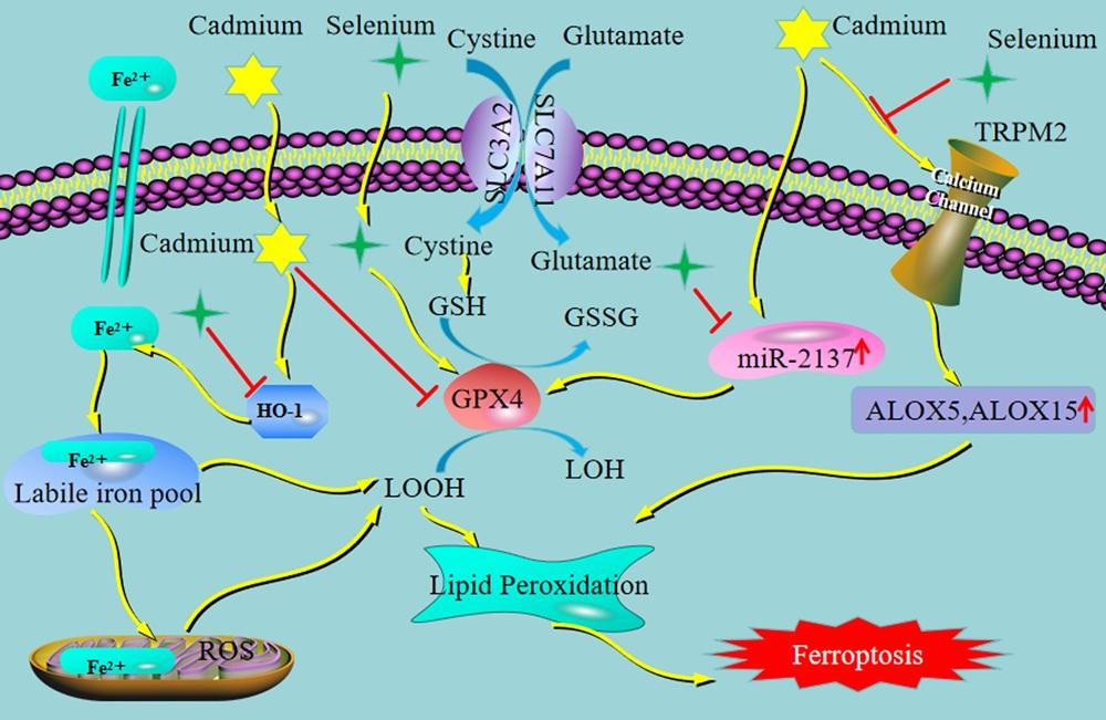
基于硒的功能特性,硒(0.3 mg/kg)减轻了镉(0.05 mg/L)对黄颡鱼肝细胞铁死亡的影响[35]。Wen等[36]研究表明,来自镉(1.25 mg/kg,注射)处理的肝细胞外泌体miR-2137通过GPX4抑制驱动肾脏细胞铁死亡,并被硒(2 mg/kg)缓解,与铁死亡抗性有关的基因包括溶质载体家族3成员2(solute carrier family 3 member 2,SLC3A2)、溶质载体家族7成员11(solute carrier family 7 member 11,SLC7A11)、溶质载体家族40成员1(solute carrier family 40 member 1,SLC40A1)、铁蛋白重链1(ferritin heavy chain 1,FTH1)和谷氨酸-半胱氨酸连接酶催化亚基(glutamate-cysteine ligase catalytic subunit,GCLC)在镉处理中显著下调,硒处理减弱了这种影响。纳米硒在体内和体外均是通过抑制铁死亡和氧化应激来减轻镉诱导的肝脏毒性[37]。此外,研究表明,瞬时受体电位M型-2(transient receptor potential melastatin-2,TRPM2)是一种非选择性阳离子通道,能够介导钙离子(Ca2+)内流,同时作为氧化应激传感器响应ROS,纳米硒[0.6 mg/(kg BW·d)]通过调节TRPM2通道缓解镉[140 mg/(kg BW·d)]诱导的鸡肝脏细胞铁死亡[26]。综上可知,镉可诱导小鼠、羊、猪、鱼等多种动物的不同类型细胞发生铁死亡,硒对镉诱导细胞铁死亡的拮抗机制与焦亡存在部分重叠,但也有针对性靶点,主要有增强GPX4活性,促进脂质过氧化物清除,阻断铁死亡核心通路,调节铁代谢等(图1)。
图1 硒对镉诱导的铁死亡保护作用机制示意图

Cadmium:镉;Selenium:硒;Cystine:胱氨酸;Glutamate:谷氨酸;SLC3A2:溶质载体家族3成员2 solute carrier family 3 member 2;SLC7A11:溶质载体家族7成员11 solute carrier family 7 member 11;Fe2+:亚铁离子 ferrous ion;GSH:谷胱甘肽 glutathione;GSSG:氧化型谷胱甘肽 oxidized glutathione;GPX4:谷胱甘肽过氧化物酶4 glutathione peroxidase 4;HO-1:血红素加氧酶-1 heme oxygenase-1;LOOH:脂质氢过氧化物 lipid hydroperoxide;LOH:脂质醇 lipid alcohol;Lipid Peroxidation:脂质过氧化;Labile iron pool:不稳定铁池;ROS:活性氧 reactive oxygen species;TRPM2:瞬时受体电位M型-2 transient receptor potential melastatin-2;ALOX5:花生四烯酸-5-脂加氧酶 arachidonate-5-lipoxygenase;ALOX15:花生四烯酸-15-脂加氧酶 arachidonate-15-lipoxygenase;Ferroptosis:铁死亡。黄色箭头表示激活,红色箭头表示抑制 yellow arrow indicates activation, and red arrow indicates inhibition。

Fig.1 Schematic diagram of protective mechanism of selenium against cadmium-induced ferroptosis

2.3 硒缓解镉暴露致动物细胞自噬的作用机制

细胞自噬又称Ⅱ型细胞程序性死亡,是一种溶酶体依赖的受控降解过程,有助于维持细胞稳态、应对应激、调控细胞存活与死亡(适度自噬促进细胞存活,过度自噬诱导自噬性死亡)[38]。高剂量镉长期暴露诱发过度自噬或自噬流受阻,最终促进细胞死亡。ROS是自噬调控的核心上游信号,可直接损伤线粒体、DNA等,同时通过激活AMP活化蛋白激酶(AMP-activated protein kinase,AMPK)/哺乳动物雷帕霉素靶蛋白(mammalian target of rapamycin,mTOR)通路等,诱导自噬过度启动。此外,镉还会破坏自噬体与溶酶体的融合过程或抑制溶酶体水解酶的活性,导致自噬体无法正常降解(自噬流阻滞)。镉还可以直接结合自噬相关蛋白以破坏其功能,同时干扰自噬信号通路平衡,导致自噬启动与终止机制失衡,形成持续性异常自噬[39-40]。Shi等[41]研究表明,镉触发miR-30a-葡萄糖调节蛋白78(glucose-regulated protein 78,GRP78)信号轴紊乱,诱导内质网应激并激活肌醇需求酶1(inositol-requiring enzyme 1,IRE1)-c-Jun氨基末端激酶(c-Jun N-terminal kinase,JNK)通路,促进鸡肾脏细胞自噬。镉诱导鲤鱼氧化-抗氧化失衡,引发氧化应激并导致能量障碍,进而通过AMPK/mTOR/Unc-51样激酶1(Unc-51 like kinase 1,ULK1)通路参与氧化应激介导的细胞自噬[42]。镉诱导鸭肾小管上皮细胞通过磷脂酶C(phospholipase C,PLC)-肌醇-1,4,5-三磷酸(inositol-1,4,5-trisphosphate,IP3)/肌醇-1,4,5-三磷酸受体(inositol-1,4,5-trisphosphate receptor,IP3R)信号通路自噬[43]。镉暴露可诱导猪心脏毒性,其机制与溶酶体功能障碍介导的自噬通量抑制密切相关[44]。AMPK/过氧化物酶体增殖物激活受体-γ(peroxisome proliferator-activated receptor-γ,PPAR-γ)/NF-κB轴参与镉诱导猪肝脏中ROS介导的细胞凋亡与自噬过程[45]。此外,适度的自噬对镉的毒性损伤发挥了一定的保护作用,mTOR介导的自噬对镉暴露诱导的NLRP3炎症小体依赖性肾脏损伤有显著保护作用[46],自噬通过减轻氧化应激和内质网应激保护原代鸡肝细胞免受镉诱导的细胞损伤[47]。共济失调毛细血管扩张突变蛋白(ataxia telangiectasia mutated protein,ATM)/AMPK/mTOR轴的激活促进自噬,以应对钼和镉诱导的鸭睾丸氧化应激介导的DNA损伤[48]
研究表明,酵母硒(0.4 mg/kg基础饲粮)通过减少颗粒细胞的自噬来保护蛋鸡免受镉(25 mg/kg)诱导的卵泡闭锁[49]。硒通过抑制过量G蛋白质偶联雌激素受体1(G protein-coupled estrogen receptor 1,GPER1)介导的自噬激活,恢复自噬通量,从而减轻镉诱导的肝脏毒性,硒与镉联合处理显著降低了镉诱导的自噬,微管相关蛋白1轻链3A(microtubule-associated protein 1 light chain 3 alpha,LC3A)、微管相关蛋白1轻链3B(microtubule-associated protein 1 light chain 3 beta,LC3B)、自噬相关蛋白4B(autophagy-related 4B,ATG4B)、自噬相关蛋白5(autophagy-related 5,ATG5)、自噬相关蛋白7(autophagy-related 7,ATG7)、自噬相关蛋白6(autophagy-related 6,ATG6或Beclin-1)、PTEN诱导激酶1(PTEN-induced kinase 1,PINK1)和E3泛素连接酶帕金蛋白(Parkin)的表达水平增加,同时显著提高了线粒体外膜转位酶2(translocase of the outer membrane of mitochondria 20,TOMM20)的表达水平,增加了镉诱导的线粒体融合蛋白1(mitofusin 1,MFN1)和线粒体融合蛋白2(mitofusin 2,MFN2)相对水平的下降,缓解镉诱导的肝细胞过度自噬激活[50]。硒通过FUN14结构域包含蛋白1(FUN14 domain containing 1,FUNDC1)介导的绵羊肺部细胞线粒体质量控制途径缓解镉诱导的线粒体自噬[51]。纳米硒通过抑制ROS和恢复自噬流,减轻镉诱导的小鼠睾丸间质细胞损伤[52]。硒通过PI3K/AKT/PTEN信号通路抑制氧化应激和程序性细胞死亡(焦亡、自噬和凋亡),缓解镉引起的心脏损伤[23]。硒(0.5 mg/kg BW)触发Nrf2-AMPK串扰,缓解了镉(1 mg/kg BW)诱导的兔大脑自噬[53]。硒通过调节兔肝细胞核外源性受体反应和氧化应激来保护兔免受镉诱导的自噬[54]。综上可知,镉可通过氧化应激、钙稳态失衡、自噬流阻断及信号通路紊乱等多种机制诱导细胞自噬异常,而硒通过抑制镉诱导的过度自噬/自噬流受阻以缓解镉毒性,硒调控自噬的核心机制包括抑制氧化应激介导的自噬激活,调控转录因子(如Nrf2)或自噬相关基因表达,对线粒体自噬、脂自噬等选择性自噬具有特异性调控作用,改善线粒体功能,促进受损线粒体的自噬降解(线粒体自噬),维持线粒体功能稳态等。

2.4 硒缓解镉暴露致动物细胞坏死性凋亡的作用机制

坏死性凋亡也称为程序性坏死,是一种受基因严密调控、不依赖Caspase的程序性细胞坏死方式,兼具坏死的形态学特征(细胞膜破裂)和凋亡的调控特征(信号通路介导)[55]。镉对细胞坏死性凋亡的调控主要通过非经典通路且与氧化应激、钙稳态失衡等上游信号密切关联,过量ROS可直接氧化修饰受体相互作用蛋白激酶1(receptor-interacting protein kinase 1,RIPK1)的丝氨酸/苏氨酸残基,促进RIPK1去泛素化并与受体相互作用蛋白激酶(receptor-interacting protein kinase 3,RIPK3)结合,形成功能性坏死小体,启动坏死性凋亡。同时,ROS还能损伤线粒体,释放细胞色素c(cytochrome c,Cyt-c)等信号分子,进一步增强坏死性凋亡的激活,激活的RIPK3进一步促进线粒体ROS释放,形成“ROS-RIPK3-ROS”正反馈循环,加速混合谱系激酶结构域样蛋白(mixed lineage kinase domain-like protein,MLKL)磷酸化[56]。此外,镉通过MAPK、NF-κB等通路,干扰坏死性凋亡信号平衡,进一步放大坏死性凋亡信号。镉激活的p38 MAPK可磷酸化RIPK3,增强其活性,NF-κB通路的激活则会促进炎症因子(TNF-α、IL-1β)释放,TNF-α作为坏死性凋亡的经典起始配体,会形成炎症-坏死性凋亡恶性循环[57-58]。研究表明,镉通过氧化应激和TNF-α/TNFR1信号传导引起的坏死性凋亡诱导小鼠睾丸间质细胞损伤[59];镉通过TLR4/NF-κB信号通路诱导氧化应激介导的猪附睾细胞坏死性凋亡[60];镉引发氧化应激、坏死性凋亡、Th1/Th2失衡并通过TNF-α/NF-κB信号通路引起猪小肠炎症[61];此外,镉暴露显著增加了下丘脑中镉的积累,抑制了PI3K/AKT信号通路,导致能量代谢紊乱、线粒体动力学失衡和离子稳态失衡,抑制B细胞淋巴瘤-2(B-cell lymphoma-2,Bcl-2)和RIPK1的高水平表达,最终引发细胞凋亡和程序性坏死,导致猪下丘脑损伤[62];镉通过激活TLR4/MAPK/NF-κB信号通路诱导睾丸间质细胞发生坏死性凋亡,进而抑制睾酮合成,最终导致雄性仔猪性行为能力下降[63]
研究表明,酵母硒(0.5 mg/kg)通过抑制氧化应激和MAPK通路对镉(150 mg/kg)诱导的鸡肝坏死性凋亡发挥保护作用[64]。与亚硒酸钠(Na2SeO3)相比,酵母硒(0.5 mg/kg)对镉(150 mg/kg)所致鸡肾脏坏死性凋亡的保护作用更为显著,其机制具有多靶点特征:通过激活miR-26a-5p表达并下调PTEN的表达,进而上调PI3K/AKT信号通路;同时,酵母硒能更有效地阻断镉在肾脏中的蓄积,并显著抑制RIPK1、RIPK3和MLKL的表达,以及降低热休克蛋白60(heat shock protein,HSP)60、HSP70和HSP90的水平[65]。Hou等[66]和Wang等[67]的研究均表明,镉暴露导致氧化应激和内质网应激,引发鸡睾丸细胞程序性坏死,硒显著拮抗了镉的毒性损伤。综上可知,镉诱导动物细胞坏死性凋亡的核心分子机制包括对RIPK3/MLKL信号通路的核心调控、线粒体氧化应激的介导作用、炎症反应的级联放大效应及其他通路的协同调控等。硒对细胞坏死性凋亡的调控核心是抑制异常激活、维持通路稳态,其一方面通过抗氧化作用减少ROS对RIPK1、RIPK3的氧化修饰,切断ROS-RIPK3-ROS正反馈循环;另一方面直接调控坏死性凋亡核心分子,抑制坏死性凋亡的激活。

2.5 硒缓解镉暴露致动物细胞凋亡的作用机制

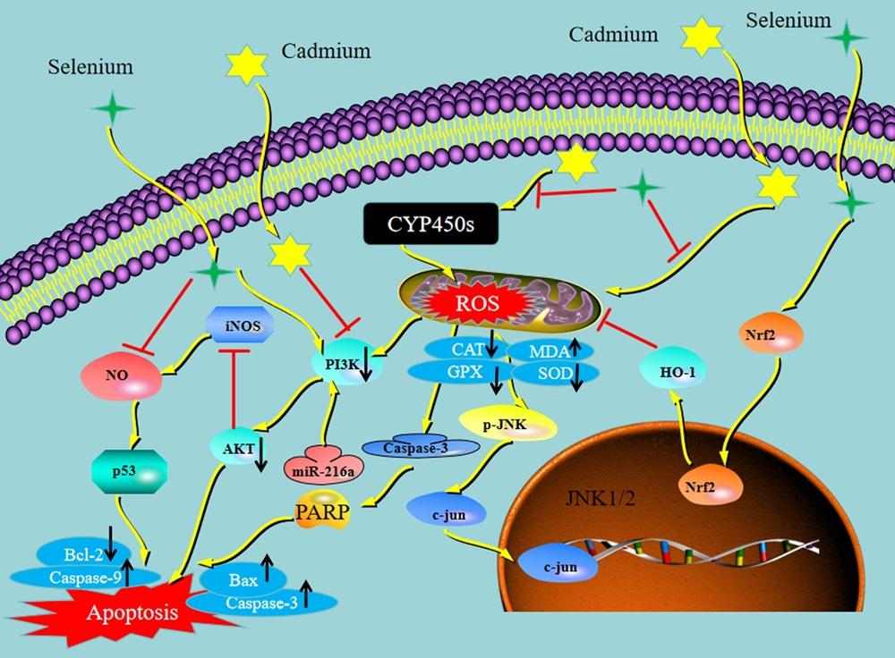
细胞凋亡是由基因调控的主动程序性细胞死亡,按激活信号不同分为外源性和内源性途径,后者包括线粒体和内质网途径,最终均汇聚于Caspase激活,诱导细胞凋亡[68]。镉诱导细胞凋亡核心环节与氧化应激、线粒体损伤、钙稳态失衡密切相关。过量ROS攻击线粒体,破坏线粒体通路,导致线粒体膜电位下降、通透性增加,释放Cyt-c等促凋亡因子,激活Caspase-9、Caspase-3,诱导细胞凋亡[69-70]。镉显著上调促凋亡蛋白p53、Bcl-2相关X蛋白(Bcl-2-associated X protein,Bax)等的表达,并下调抑凋亡蛋白(Bcl-2等)的表达,打破细胞存活与死亡的平衡,推动凋亡进程[71]。针对不同动物的研究表明,即使在低浓度下,镉也会引起大口黑鲈肝细胞线粒体损伤,加剧氧化应激,导致细胞炎症和凋亡[72];镉还诱导小鼠体外骨骼肌细胞的氧化损伤和凋亡[73]。此外,镉激活氧化应激和内质网应激途径促进鲤鱼脑细胞凋亡[74]。MicroRNA-129-1-3p通过下调线粒体钙单向转运蛋白(mitochondrial calcium uniporter,MCU)介导的Ca2+信号通路,保护鸡颗粒细胞免受镉诱导的凋亡[75]。MAPK、AKT/叉头框蛋白O3a(forkhead box protein O3a,FoxO3a)和mTOR通路参与镉调节鸡卵泡颗粒细胞凋亡[76]。PI3K/AKT通路是细胞内关键的信号转导通路,主要通过磷酸化修饰下游靶分子,从多个层面抑制凋亡、促进细胞存活,具体机制包括调控Bcl-2家族蛋白平衡、抑制Caspase家族活性、调控转录因子与凋亡相关基因表达。在猪上的研究表明,镉可通过靶向PTEN的miR-9-5p调节PI3K/AKT通路诱导肾上腺细胞凋亡[77];通过下调PI3K/AKT通路诱导小肠细胞凋亡[78];AMPK/PPAR-γ/NF-κB轴参与镉在肝脏中引起的ROS介导的凋亡[45];亚急性镉暴露通过调节miR-369-TNFα轴影响Th1极化,引发内质网应激并诱导肝细胞凋亡[79];此外,镉可激活快速加速纤维肉瘤激酶1(rapidly accelerated fibrosarcoma 1,RAF1)/丝裂原活化蛋白激酶激酶(mitogen-activated protein kinase kinase,MEK)/细胞外信号调节激酶(extracellular signal-regulated kinase,ERK)和NF-κB通路诱导附睾炎症和凋亡[80],并通过PI3K/AKT/缺氧诱导因子-1α(hypoxia-inducible factor-1α,HIF-1α)通路诱导淋巴结凋亡[81]。对鸡的研究表明,硒(1.0 mg/kg)可通过激活Nrf2/HO-1信号通路抑制氧化应激,减轻镉(150.00 mg/kg)诱导的鸡睾丸细胞凋亡和炎症,有效抑制镉诱导的ROS、丙二醛(malondialdehyde,MDA)和过氧化氢(hydrogen peroxide,H2O2)水平升高,并阻止过氧化氢酶(catalase,CAT)活性、GSH含量和总抗氧化能力(total antioxidant capacity,T-AOC)降低;同时,硒可改善镉诱导的Nrf2、HO-1和Bcl-2表达水平的降低,拮抗Caspase-3、BaxCyt-cCaspase-9、TNF-α、白细胞介素-2(interleukin-2,IL-2)、白细胞介素-6(interleukin-6,IL-6)和IL-1β的过表达[82]。纳米硒可通过调节氧化应激、减少细胞凋亡减轻镉引起的雄性Wistar大鼠生殖毒性[83]。硒可缓解镉诱导的草鱼肝脏细胞氧化应激、内质网应激和凋亡[84]。在鲤鱼淋巴细胞中,镉(4×10⁻⁵ mol/L)诱导的氧化应激通过调节miR-216a-PI3K/AKT轴促进细胞凋亡,该效应可被硒(10-6 mol/L)拮抗,镉暴露导致淋巴细胞凋亡和坏死性凋亡,增加细胞色素P450酶系(cytochrome P450 enzymes,CYP450s)和糖代谢相关酶的表达及ROS的产生,同时刺激氧化应激,上调miR-216a表达并降低PI3K水平,而硒镉联合组中这些变化均得到明显缓解[85]。此外,硒可通过抑制ROS/JNK/c-Jun信号通路改善镉诱导的小鼠睾丸间质细胞凋亡[86];硒蛋氨酸可通过调节PI3K/AKT通路改善镉诱导的鸡肝细胞凋亡[87]。综上可知,镉诱导动物细胞凋亡整体呈现出以氧化应激为核心、多信号通路交叉调控的特征,涉及线粒体损伤、死亡受体激活和炎症反应等多个层面。硒通过强力清除过量ROS、保护线粒体膜完整性,阻断线粒体凋亡通路的激活;同时通过激活PI3K/AKT等信号通路,上调Bcl-2等抗凋亡蛋白的表达,抑制Bax、p53等促凋亡蛋白的激活,恢复细胞内凋亡平衡(图2)。
图2 硒对镉诱导的细胞凋亡保护作用机制示意图

Cadmium:镉;Selenium:硒;ROS:活性氧 reactive oxygen species;Nrf2:核因子E2相关因子2 nuclear factor erythroid 2-related factor 2;HO-1:血红素加氧酶1 heme oxygenase-1;JNK1/2:c-Jun氨基末端激酶1/2 c-Jun N-terminal kinase 1/2;CYP450s:细胞色素P450酶系 cytochrome P450 enzymes;PI3K:磷酸肌醇3-激酶 phosphoinositide 3-kinase;AKT:蛋白激酶B protein kinase B;iNOS:诱导型一氧化氮合酶 inducible nitric oxide synthase;NO:一氧化氮 nitric oxide;Caspase-3:半胱氨酸天冬氨酸蛋白酶-3 cysteine-aspartic protease-3;Bcl-2:B-cell lymphoma-2 B淋巴细胞瘤-2;Bax:Bcl-2相关蛋白X Bcl-2-associated X protein;Apoptosis:凋亡;p-JNK:磷酸化c-Jun氨基末端激酶 phosphorylated c-Jun N-terminal kinase;CAT:过氧化氢酶 catalase;GPX:谷胱甘肽过氧化物酶 glutathione peroxidase;MDA:丙二醛 malondialdehyde;SOD:超氧化物歧化酶 superoxide dismutase;PARP:聚腺苷二磷酸核糖聚合酶 poly(ADP-ribose) polymerase。黄色箭头表示激活,红色箭头表示抑制 yellow arrow indicates activation, and red arrow indicates inhibition。

Fig.2 Schematic diagram of protective mechanism of selenium on cadmium induced cell apoptosis

3 小结

镉严重威胁畜禽的生长性能、繁殖能力及产品安全性。细胞程序性死亡本是畜禽细胞应对损伤的重要防御机制,但镉可扭曲并利用这一机制,而硒则能有效缓解甚至逆转镉的毒性损伤。未来应基于硒的拮抗作用机制,结合转录组、蛋白质组、代谢组与单细胞测序等多组学技术,系统解析硒与镉调控细胞程序性死亡的全局网络,挖掘新的调控靶点与信号通路,尤其是参与镉暴露下程序性死亡进程的硒蛋白种类及其精细调控机制。
[1]
XING L, ZHANG R, GONG R, et al. Ameliorative effects of dietary selenium against cadmium toxicity on production performance and egg quality in laying hens[J]. Ecotoxicology and Environmental Safety, 2022, 248:114317.

[2]
RAFIEIAN-NAEINI H R, ZHANDI M, SADEGHI M, et al. Effects of coenzyme Q10 on reproductive performance of laying Japanese quail (Coturnix japonica) under cadmium challenge[J]. Poultry Science, 2021, 100(11):101418.

[3]
SUN Y, ZHOU C, WANG Z, et al. Selenium alleviates cadmium-induced immune dysfunction in M1 macrophages by modulating energy metabolism[J]. Journal of Trace Elements in Medicine and Biology, 2026, 95:127902.

[4]
RADWAN M, AL MALKI, WAHEED R M, et al. Mitigation exposed to acute cadmium stress in Nile Tilapia by dietary quercetin:exploring the growth performance,intestinal health,stress biomarkers,and immune responses[J]. Biological Trace Element Research, 2026, 204(1):368-384.

[5]
YAZĞAN Y, BAYIR M H, et al. Selenium reduces cadmium-induced cardiotoxicity by modulating oxidative stress and the ROS/PARP-1/TRPM2 signalling pathway in rats[J]. Toxics. 2025, 13(8):611.

[6]
LONG C, ZHU G Y, SHENG X H, et al. Dietary supplementation with selenomethionine enhances antioxidant capacity and selenoprotein gene expression in layer breeder roosters[J]. Poultry Science, 2022, 101(11):102113.

[7]
XING P P, SHI X, CUI W, et al. Yeast selenium exerts an antioxidant effect by regulating the level of selenoprotein to antagonize Cd-induced pyroptosis of chicken liver[J]. Biological Trace Element Research, 2022, 200(9):4079-4088.

[8]
CUI L, ZHANG M, ZHENG F, et al. Selenium elicited an enhanced anti-inflammatory effect in primary bovine endometrial stromal cells with high cortisol background[J]. BMC Veterinary Research, 2024, 20(1):383.

[9]
BI C L, ZHANG S J, SHEN Y Z, et al. Selenium plays an anti-inflammatory role by regulation NLRP3 inflammasome in Staphylococcus aureus-infected mouse mammary gland[J]. Biological Trace Element Research, 2021, 199(2):604-610.

[10]
SWETHA K, INDUMATHI M C, KISHAN R, et al. Selenium mitigates caerulein and LPS-induced severe acute pancreatitis by inhibiting MAPK,NF-κB,and STAT3 signaling via the Nrf2/HO-1 pathway[J]. Biological Trace Element Research, 2025, 203(9):4728-4750.

[11]
DING D, MOU D, ZHAO L, et al. Maternal organic selenium supplementation alleviates LPS induced inflammation,autophagy and ER stress in the thymus and spleen of offspring piglets by improving the expression of selenoproteins[J]. Food & Function, 2021, 12(22):11214-11228.

[12]
ROKADE A, THORAT S T, CHANDRAMORE K, et al. Integrating immunity,antioxidative status, and gene regulation against nickel and high-temperature stress in fish:selenium nanoparticles for mitigation[J]. Environmental Science and Pollution Research, 2025, 32(7):3987-4003.

[13]
MA P, HU Z, LI L, et al. Dietary selenium promotes the growth performance through growth hormone-insulin-like growth factor and hypothalamic-pituitary-thyroid axes in grass carp (Ctenopharyngodon idella)[J]. Fish Physiology and Biochemistry, 2021, 47(4):1313-1327.

[14]
ZHANG Y, LIU Q, YIN H, et al. Cadmium exposure induces pyroptosis of lymphocytes in carp pronephros and spleens by activating NLRP3[J]. Ecotoxicology and Environmental Safety, 2020, 202:110903.

[15]
ZHOU J Z, ZHANG Y W, ZENG L, et al. Cadmium exposure induces pyroptosis of TM4 cells through oxidative stress damage and inflammasome activation[J]. Ecotoxicology and Environmental Safety, 2024, 270:115930.

[16]
CAI J Z, GUAN H Y, JIAO X, et al. NLRP3 inflammasome mediated pyroptosis is involved in cadmium exposure-induced neuroinflammation through the IL-1β/IκB-α-NF-κB-NLRP3 feedback loop in swine[J]. Toxicology, 2021, 453:152720.

[17]
WEI Z J, NIE G H, YANG F, et al. Inhibition of ROS/NLRP3/Caspase-1 mediated pyroptosis attenuates cadmium-induced apoptosis in duck renal tubular epithelial cells[J]. Environmental Pollution, 2021, 273:115919.

[18]
HU Z S, NIE G H, LUO J R, et al. Molybdenum and cadmium co-induce pyroptosis via inhibiting Nrf2-mediated antioxidant defense response in the brain of ducks[J]. Biological Trace Element Research, 2023, 201(2):874-887.

[19]
LI L, XU H, WANG Y, et al. From inflammation to pyroptosis:understanding the consequences of cadmium exposure in chicken liver cells[J]. Ecotoxicology and Environmental Safety, 2024, 272:116004.

[20]
SUN X H, LV M W, ZHAO Y X, et al. Nano-selenium antagonized cadmium-induced liver fibrosis in chicken[J]. Journal of Agricultural and Food Chemistry, 2023, 71(1):846-856.

[21]
WANG L, YANG F, HU M W, et al. GPX4 utilization by selenium is required to alleviate cadmium-induced ferroptosis and pyroptosis in sheep kidney[J]. Environmental Toxicology, 2023, 38(4):962-974.

[22]
ZHENG H, DONG W X, QIU H L, et al. S-COS-Se mitigates cadmium-induced oxidative stress,inflammation,and immunotoxicity by restoring immune balance and inhibiting mitochondria-pyroptosis crosstalk[J]. Ecotoxicology and Environmental Safety, 2025, 300:118462.

[23]
FENG J P, YANG F, WU H S, et al. Selenium protects against cadmium-induced cardiac injury by attenuating programmed cell death via PI3K/AKT/PTEN signaling[J]. Environmental Toxicology, 2022, 37(5):1185-1197.

[24]
HONG H, LIN X, XU Y, et al. Cadmium induces ferroptosis mediated inflammation by activating Gpx4/Ager/p65 axis in pancreatic β-cells[J]. Science of the Total Environment, 2022, 849:157819.

[25]
LUO T, SONG S, WANG S, et al. Mechanistic insights into cadmium-induced nephrotoxicity:NRF2-driven HO-1 activation promotes ferroptosis via iron overload and oxidative stress in vitro[J]. Free Radical Biology and Medicine, 2025, 235:162-175.

[26]
WANG Z J, LIANG Y S, REN Y F, et al. Nano-selenium alleviates cadmium-induced ferroptosis in chicken liver by modulating the TRPM2 channel[J]. Free Radical Biology & Medicine, 2026, 242:495-507.

[27]
郁孝强, 吴亚, 罗通旺, 等. 镉暴露诱导小鼠肝脏铁死亡的作用机制[J]. 中国兽医学报, 2024, 44(3):543-549.

YU X Q, WU Y, LUO T W, et al. Mechanism of ferroptosis in mice liver induced by cadmium exposure[J]. Chinese Journal of Veterinary Science, 2024, 44(3):543-549. (in Chinese)

[28]
WU M Q, LI W Q, HE M R, et al. Acute cadmium exposure damaged granulosa cell structure and blocked oocyte maturation via ferroptosis caused by intracellular iron overload and oxidative stress in mice[J]. Ecotoxicology and Environmental Safety, 2025, 303:118851.

[29]
WANG Y, WU J, ZHANG M M, et al. Cadmium exposure during puberty damages testicular development and spermatogenesis via ferroptosis caused by intracellular iron overload and oxidative stress in mice[J]. Environmental Pollution, 2023, 325:121434.

[30]
MA Z M, YAN S, ZHANG H M, et al. Cadmium induced ferroptosis and inflammation in sheep via targeting ACSL4/NF-κB axis[J]. Frontiers in Veterinary Science, 2025, 12:1617190.

[31]
宋圣哲, 罗通旺, 沈灵俊, 等. 镉通过Nrf2/HO-1通路诱导猪肾PK-15细胞铁死亡[J]. 中国兽医学报, 2024, 44(9):1992-1998.

SONG S Z, LUO T W, SHEN L J, et al. Cadmium induces ferroptosis by modulating Nrf2/HO-1 pathway in PK-15 cells[J]. Chinese Journal of Veterinary Science, 2024, 44(9):1992-1998. (in Chinese)

[32]
陈敬宜, 于淼, 张金洋, 等. 铁死亡参与镉暴露鸡肝损伤的研究[J]. 畜牧兽医学报, 2023, 54(2):787-802.

CHEN J Y, YU M, ZHANG J Y, et al. Study on the involvement of ferroptosis in liver injury of cadmium-exposed chickens[J]. Acta Veterinaria et Zootechnica Sinica, 2023, 54(2):787-802. (in Chinese)

[33]
WANG J J, CHEN Y C, WANG X, et al. Cadmium aggravates liver injury by activating ferroptosis and neutrophil extracellular traps formation in Nile tilapia (Oreochromis niloticus)[J]. Environmental Toxicology, 2024, 39(7):4047-4057.

[34]
GUO W L, ZHANG J J, ZHANG X Q, et al. Environmental cadmium exposure perturbs systemic iron homeostasis via hemolysis and inflammation,leading to hepatic ferroptosis in common carp (Cyprinus carpio L.)[J]. Ecotoxicology and Environmental Safety, 2024, 275:116246.

[35]
XU Q, TANG Q, YI L Y, et al. Selenium alleviates the effects of cadmium on growth performance, oxidative stress,cholesterol synthesis and ferroptosis of hybrid yellow catfish,Pelteobagrus fulvidraco ♀×Pelteobagrus vachelli ♂[J]. Comparative Biochemistry and Physiology Part C:Toxicology & Pharmacology, 2025, 298:110290.

[36]
WEN Q, QI M Y, WU Y J, et al. Exosomal miR-2137 from cadmium-treated hepatocytes drives renal ferroptosis via GPX4 suppression and is alleviated by selenium[J]. Frontiers in Cell and Developmental Biology, 2025, 13:1585106.

[37]
SHAO C Y, LUO T W, WANG S J, et al. Selenium nanoparticles alleviates cadmium induced hepatotoxicity by inhibiting ferroptosis and oxidative stress in vivo and in vitro[J]. Chemosphere, 2024, 364:143004.

[38]
LEVINE B, KROEMER G. Autophagy in the pathogenesis of disease[J]. Cell, 2008; 132:27-42.

[39]
MIREAULT M, XIAO Y, BARBEAU B, et al. Cadmium affects autophagy in the human intestinal cells Caco-2 through ROS-mediated ERK activation[J]. Cell Biology and Toxicology, 2023, 39(3):945-966.

[40]
ZHOU L, CHEN Y, SUN Y, et al. Cadmium induces apoptosis of mouse spermatocytes through JNK activation and disruption of autophagic flux[J]. Ecotoxicology and Environmental Safety, 2023, 265:115505.

[41]
SHI Q X, JIN X, FAN R F, et al. Cadmium-mediated miR-30a-GRP78 leads to JNK-dependent autophagy in chicken kidney[J]. Chemosphere, 2019, 215:710-715.

[42]
LIU Y H, LIN X, HAO Z Y, et al. Cadmium exposure caused cardiotoxicity in common carps (Cyprinus carpio L.):miR-9-5p,oxidative stress,energetic impairment,mitochondrial division/fusion imbalance,inflammation,and autophagy[J]. Fish & Shellfish Immunology, 2023, 138:108853.

[43]
GUO H L, HUANG B Y, CUI T, et al. Cadmium exposure induces autophagy via PLC-IP3-IP3R signaling pathway in duck renal tubular epithelial cells[J]. Environmental Toxicology, 2022, 37(11):2660-2672.

[44]
ZHAO X, SHI X, YAO Y J, et al. Autophagy flux inhibition mediated by lysosomal dysfunction participates in the cadmium exposure-induced cardiotoxicity in swine[J]. BioFactors, 2022, 48(4):946-958.

[45]
WANG H, WANG A Q, WANG X Q, et al. AMPK/PPAR-γ/NF-κB axis participates in ROS-mediated apoptosis and autophagy caused by cadmium in pig liver[J]. Environmental Pollution, 2022, 294:118659.

[46]
HU Y, WANG K, XU J, et al. mTOR-mediated autophagy regulates cadmium-induced kidney injury via pyroptosis[J]. International Journal of Molecular Sciences, 2025, 26(6):2589.

[47]
LI N, YI B J, SALEEM M A U, et al. Autophagy protects against Cd-induced cell damage in primary chicken hepatocytes via mitigation of oxidative stress and endoplasmic reticulum stress[J]. Ecotoxicology and Environmental Safety, 2023, 259:115056.

[48]
PU W J, CHU X S, GUO H L, et al. The activated ATM/AMPK/mTOR axis promotes autophagy in response to oxidative stress-mediated DNA damage co-induced by molybdenum and cadmium in duck testes[J]. Environmental Pollution, 2023, 316(Pt 2):120574.

[49]
WU C M, JIANG Y X, ZHOU Z Y, et al. Selenized yeast protects against cadmium-induced follicular atresia in laying hens by reducing autophagy in granulosa cells[J]. Current Issues in Molecular Biology, 2024, 46(11):13119-13130.

[50]
ZHANG H L, YAN J, XIE D N, et al. Selenium restored mitophagic flux to alleviate cadmium-induced hepatotoxicity by inhibiting excessive GPER1-mediated mitophagy activation[J]. Journal of Hazardous Materials, 2024, 475:134855.

[51]
LIN S X, YANG F, HU M W, et al. Selenium alleviates cadmium-induced mitophagy through FUNDC1-mediated mitochondrial quality control pathway in the lungs of sheep[J]. Environmental Pollution, 2023, 319:120954.

[52]
HU X D, LIN R, ZHANG C Q, et al. Nano-selenium alleviates cadmium-induced mouse Leydig cell injury,via the inhibition of reactive oxygen species and the restoration of autophagic flux[J]. Reproductive Sciences, 2023, 30(6):1808-1822.

[53]
XUE H T, CAO H B, XING C H, et al. Selenium triggers Nrf2-AMPK crosstalk to alleviate cadmium-induced autophagy in rabbit cerebrum[J]. Toxicology, 2021, 459:152855.

[54]
ZHANG L W, YANG F, LI Y, et al. The protection of selenium against cadmium-induced mitophagy via modulating nuclear xenobiotic receptors response and oxidative stress in the liver of rabbits[J]. Environmental Pollution, 2021, 285:117301.

[55]
RODRIGUEZ D A, WEINLICH R, BROWN S, et al. Characterization of RIPK3-mediated phosphorylation of the activation loop of MLKL during necroptosis[J]. Cell Death and Differentiation, 2016, 23(1):76-88.

[56]
XIA Y, ZHANG Y M, ZHANG J T, et al. Cadmium exposure induces necroptosis of porcine spleen via ROS-mediated activation of STAT1/RIPK3 signaling pathway[J]. Environmental and Molecular Mutagenesis, 2023, 64(7):382-392.

[57]
ZHAO X, WANG S C, LI X J, et al. Cadmium exposure induces TNF-α-mediated necroptosis via FPR2/TGF-β/NF-κB pathway in swine myocardium[J]. Toxicology, 2021, 453:152733.

[58]
QI X, ZHANG Y, LIU H G, et al. Cadmium exposure induces inflammation and necroptosis in porcine adrenal gland via activating NF-κB/MAPK pathway[J]. Journal of Inorganic Biochemistry, 2021, 223:111516.

[59]
ZHENG X P, SUN Y H, WANG J H, et al. Cadmium exposure induces Leydig cell injury via necroptosis caused by oxidative stress and TNF-α/TNFR1 signaling[J]. Biochemical and Biophysical Research Communications, 2025, 761:151717.

[60]
LI Y L, WANG S, WANG Y F. Cadmium exposure induces oxidative stress-mediated necroptosis via TLR4/NF-κB signaling pathway in pig epididymis[J]. Environmental Pollution, 2025, 366:125514.

[61]
CHEN X M, BI M Y, YANG J, et al. Cadmium exposure triggers oxidative stress,necroptosis,Th1/Th2 imbalance and promotes inflammation through the TNF-α/NF-κB pathway in swine small intestine[J]. Journal of Hazardous Materials, 2022, 421:126704.

[62]
CHEN D, YAO Y J, SHI X, et al. Cadmium exposure causes mitochondrial fission and fusion disorder in the pig hypothalamus via the PI3K/AKT pathway[J]. Ecotoxicology and Environmental Safety, 2022, 242:113880.

[63]
LI Y L, ZHANG Y, FENG R, et al. Cadmium induces testosterone synthesis disorder by testicular cell damage via TLR4/MAPK/NF-κB signaling pathway leading to reduced sexual behavior in piglets[J]. Ecotoxicology and Environmental Safety, 2022, 233:113345.

[64]
WANG Y, CHEN H W, CHANG W H, et al. Protective effects of selenium yeast against cadmium-induced necroptosis via inhibition of oxidative stress and MAPK pathway in chicken liver[J]. Ecotoxicology and Environmental Safety, 2020, 206:111329.

[65]
CHEN H J, LI P, SHEN Z Q, et al. Protective effects of selenium yeast against cadmium-induced necroptosis through miR-26a-5p/PTEN/PI3K/AKT signaling pathway in chicken kidney[J]. Ecotoxicology and Environmental Safety, 2021, 220:112387.

[66]
HOU L L, WANG S Z, WANG Y Y, et al. Antagonistic effect of selenium on programmed necrosis of testicular Leydig cells caused by cadmium through endoplasmic reticulum stress in chicken[J]. Environmental Science and Pollution Research, 2023, 30(52):112517-112535.

[67]
WANG M, WANG Y Y, WANG S Z, et al. Selenium alleviates cadmium-induced oxidative stress,endoplasmic reticulum stress and programmed necrosis in chicken testes[J]. Science of the Total Environment, 2023, 863:160601.

[68]
MUSTAFA M, AHMAD R, TANTRY I Q, et al. Apoptosis:a comprehensive overview of signaling pathways,morphological changes,and physiological significance and therapeutic implications[J]. Cells, 2024, 13(22):1838.

[69]
ZHANG Y M, LIU Z Y, LAN J, et al. Cadmium induces the thymus apoptosis of pigs through ROS-dependent PTEN/PI3K/AKT signaling pathway[J]. Environmental Science and Pollution Research International, 2021, 28(29):39982-39992.

[70]
YI L N, SHANG X J, LV L L, et al. Cadmium-induced apoptosis of Leydig cells is mediated by excessive mitochondrial fission and inhibition of mitophagy[J]. Cell Death & Disease, 2022, 13(11):928.

[71]
LI W Q, WU M Q, SHI H T, et al. Cadmium-induced apoptosis of granulosa cells is mediated by excessive activation of mitophagy and ROS-mediated mitochondrial dysfunction[J]. Toxicology and Applied Pharmacology, 2025, 503:117462.

[72]
DUAN Y P, LIU H, ZHANG W C, et al. Novel insights into cadmium-induced liver toxicity in largemouth bass (Micropterus salmoides):oxidative stress,inflammatory response,and mitochondria-mediated apoptosis pathway[J]. Biological Trace Element Research, 2026, 204(3):1901-1914.

[73]
LI W H, JIE Y, YUAN Z, et al. Cadmium treatment induces oxidative damage and apoptosis in vitro skeletal muscle cells[J]. Toxicology, 2025, 515:154139.

[74]
LIU J, LU W, GUO Z Y, et al. Cadmium activated oxidative stress and endoplasmic reticulum stress pathway promote apoptosis in the common carp (Cyprinus carpio L.) brain[J]. Fish & Shellfish Immunology, 2025, 165:110538.

[75]
ZHU M K, YAN M, MUSA M, et al. MicroRNA-129-1-3p protects chicken granulosa cells from cadmium-induced apoptosis by down-regulating the MCU-mediated Ca2+ signaling pathway[J]. Ecotoxicology and Environmental Safety, 2024, 269:115906.

[76]
ZHU M K, MIAO S S, ZHOU W T, et al.MAPK, AKT/FoxO3a and mTOR pathways are involved in cadmium regulating the cell cycle,proliferation and apoptosis of chicken follicular granulosa cells[J]. Ecotoxicology and Environmental Safety, 2021, 214:112091.

[77]
QI X, REN Z H, CUI Y, et al. Cadmium induces apoptosis by miR-9-5p targeting PTEN and regulates the PI3K/AKT pathway in the piglet adrenal gland[J]. Environmental Science and Pollution Research International, 2022, 29(48):73001-73010.

[78]
ZHANG H R, HUANG J Q, YANG J, et al. Cadmium induces apoptosis and autophagy in swine small intestine by downregulating the PI3K/Akt pathway[J]. Environmental Science and Pollution Research International, 2022, 29(27):41207-41218.

[79]
HAN D X, SONG N, WANG W, et al. Subacute cadmium exposure modulates Th1 polarization to trigger ER stress-induced porcine hepatocyte apoptosis via regulation of miR-369-TNFα axis[J]. Environmental Science and Pollution Research International, 2022, 29(11):16576-16587.

[80]
ZHANG Y, LI Y L, ZHANG J X, et al. Cadmium induced inflammation and apoptosis of porcine epididymis via activating RAF1/MEK/ERK and NF-κB pathways[J]. Toxicology and Applied Pharmacology, 2021, 415:115449.

[81]
LU Y M, HAN Y F, YIN H, et al. Cadmium induces apoptosis of pig lymph nodes by regulating the PI3K/AKT/HIF-1α pathway[J]. Toxicology, 2021, 451:152694.

[82]
LI Y L, WANG S, FENG R. Dietary selenium mitigates cadmium-induced apoptosis and inflammation in chicken testicles by inhibiting oxidative stress through the activation of the Nrf2/HO-1 signaling pathway[J]. Poultry Science, 2025, 104(4):104990.

[83]
TAROMSARI S, EIDI A, MORTAZAVI P, et al. Effect of nano-selenium on cadmium chloride-induced infertility in male Wistar rats[J]. Journal of Biochemical and Molecular Toxicology, 2025, 39(2):e70179.

[84]
ZHANG T T, SUN S Y, GAVRILOVIĆ A, et al. Selenium alleviates cadmium-induced oxidative stress,endoplasmic reticulum stress,and apoptosis in L8824 cells[J]. Ecotoxicology and Environmental Safety, 2023, 262:115337.

[85]
ZHANG J Q, ZHENG S F, WANG S C, et al. Cadmium-induced oxidative stress promotes apoptosis and necrosis through the regulation of the miR-216a-PI3K/AKT axis in common carp lymphocytes and antagonized by selenium[J]. Chemosphere, 2020, 258:127341.

[86]
REN X M, WANG S S, ZHANG C Q, et al. Selenium ameliorates cadmium-induced mouse Leydig TM3 cell apoptosis via inhibiting the ROS/JNK/c-Jun signaling pathway[J]. Ecotoxicology and Environmental Safety, 2020, 192:110266.

[87]
XIONG X Y, ZHANG Y, XING H J, et al. Ameliorative effect of selenomethionine on cadmium-induced hepatocyte apoptosis via regulating PI3K/AKT pathway in chickens[J]. Biological Trace Element Research, 2020, 195(2):559-568.

Outlines

/